Genetic Chimerism: Marmosets contain multitudes
Single-cell RNA sequencing reveals the extent to which marmosets carry genetically distinct cells from their siblings.
- Center for Evolution and Medicine, School of Life Sciences, Arizona State University, United States
Marmosets almost always produce non-identical twins which, unusually, share much more than just a womb. The same circulatory system connects the siblings during pregnancy, allowing two genetically distinct individuals to freely exchange the hematopoietic stem cells that give rise to all blood and immune cells (Gengozian et al., 1969; Wislocki, 1939; Hill, 1932). As a result, a drop of blood from this tiny South American primate reveals a mixture of cells originating from each twin (Benirschike and Brownhill, 1962). This is known as sibling chimerism – from Chimera, the monster from Greek mythology that is a hybrid of a lion, a goat and other creatures.
This quirk of nature has been known for decades (Benirschke et al., 1962). Yet, while studies have detected genetic information from the other twin in certain organs, it has remained difficult to precisely test whether sibling chimerism is limited to blood-related cells or extends to other cell types (Sweeney et al., 2012; Ross et al., 2007). Answering this question would help to uncover how cellular exchange takes place between marmoset twins, while also allowing researchers to investigate how genetic variations affect cell function in vivo. However, this requires technology that has only become recently available, and which makes it possible to isolate and sequence the genetic material of individual cells in a single tissue. If different genomes are identified in a heart sample, for example, these newer approaches can now distinguish whether they originate from the muscle cells of the heart, the blood cells pumped through it, or the resident immune cells that protect it. Now, in eLife, Ricardo del Rosario, Steven McCarroll and colleagues at Harvard Medical School, the Broad Institute and MIT report having addressed this knowledge gap by applying single-cell sequencing to various marmoset organs (del Rosario et al., 2024).
Rather than focusing on DNA, the team took advantage of the thousands of genetic variants transcribed into RNAs and opted instead for single-nucleus RNA-sequencing. Being able to capture all the genes being transcribed in an individual cell did double duty. del Rosario et al. could identify which tissues contained cells from the other twin; and they could also examine the impact of these genetic differences on gene expression patterns and cell function.
To investigate the first question, the team tested for sibling chimerism in the blood, liver, kidney and brain – all tissues which contain varying amounts of cell types deriving from hematopoietic stem cells. This confirmed that chimerism is prevalent and widespread in marmosets, while also allowing del Rosario et al. to precisely identify which cells were from the sampled individual, and which were from its twin. In doing so, they demonstrated that sibling chimerism is limited to cells from the two lineages (myeloid and lymphoid) that hematopoietic stem cells give rise to. Across the organs studied, all the other cell types examined originated from the twin being sampled (Figure 1).
Figure 1
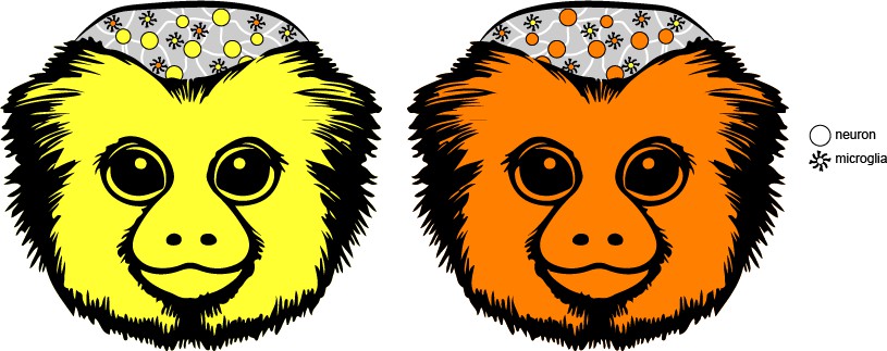
Investigating sibling chimerism in marmosets.
Marmosets almost always produce non-identical twins (also known as fraternal or dizygotic twins). However, these siblings are more alike than expected because they share a circulatory system in the womb, which allows them to exchange hematopoietic stem cells. As a result, the tissues and organs of the adult twins can contain cells carrying the genetic information of their sibling (orange cells in the yellow individual, and vice versa). Single-nucleus RNA sequencing approaches allowed del Rosario et al. to investigate which tissues contained cells from both twins, and in which proportion. This showed that only cells derived from hematopoietic stem cells display such ‘sibling chimerism’. In the brain, for example, the team found evidence for this phenomenon primarily in just one type of cell – immune cells known as microglia – with other cell types (such as neurons) being solely from the individual sampled.
The team then turned its attention to microglia, the primary immune cells of the brain, using a single-nucleus RNA-sequencing dataset of more than 2.2 million cells from various brain regions of 11 marmosets (Krienen et al., 2023). Consistent with the findings for non-brain tissues, this analysis first revealed that the proportion of sibling-derived microglia varies between the brain areas of an individual. del Rosario et al. suggest two possible explanations for this finding: (1) Such variations may be due to cell migration and proliferation taking place in a random manner, with the level of sibling-derived cells reflecting which cells arrived first and multiplied the most in a tissue. (2) Alternatively, cells from the twin may be recruited and proliferate differently across brain tissues and even local environments due to their genetic background.
To then test if genetic variations between cells could indeed impact their expression patterns, del Rosario et al. directly compared how microglia in the same brain region expressed their genes depending on whether they were from the sampled animal or its twin. When assessing if cells facing the same constraints respond differently because of variations in their genomes, this is as environmentally controlled a study can get in a living animal – nature’s very own common garden experiment, where scientists examine how populations of different genetic origins fare when facing the same conditions. The results show that the local environment, such as which brain region the cells were in, had a much larger impact in shaping gene expression than the genetic background. This does not indicate that genetic variation is unimportant, but it does highlight how cells – no matter who they come from – are very good at doing their job when they find themselves in the right place at the right time.
All told, this thoughtful and thorough study accomplishes two important goals. First, it all but closes a previously open question on the extent and cell origins of sibling chimerism. Second, it sets the stage for using this unique model system to examine, in a natural context, how genetic variation in microglia may impact brain development, function, and disease.
References
-
Further observations on marrow chimerism in marmosetsCytogenetics 1:245–257.https://doi.org/10.1159/000129734
-
The developmental history of the primatesPhilosophical Transactions of the Royal Society of London. Series B 221:45–178.https://doi.org/10.1098/rstb.1932.0002
-
Observations on twinning in marmosetsAmerican Journal of Anatomy 64:445–483.https://doi.org/10.1002/aja.1000640305
Article and author information
Author details
Noah Snyder-Mackler

Publication history
- Version of Record published: April 25, 2024 (version 1)
Copyright
© 2024, Chiou and Snyder-Mackler
This article is distributed under the terms of the Creative Commons Attribution License, which permits unrestricted use and redistribution provided that the original author and source are credited.
Metrics
-
- 291
- views
-
- 24
- downloads
-
- 0
- citations
Views, downloads and citations are aggregated across all versions of this paper published by eLife.
Download links
A two-part list of links to download the article, or parts of the article, in various formats.
Downloads (link to download the article as PDF)
Open citations (links to open the citations from this article in various online reference manager services)
Cite this article (links to download the citations from this article in formats compatible with various reference manager tools)
Genetic Chimerism: Marmosets contain multitudes
eLife 13:e97866.
https://doi.org/10.7554/eLife.97866
Further reading
-
- Genetics and Genomics
- Microbiology and Infectious Disease
Despite over a century of observations, the obligate insect parasites within the order Entomophthorales remain poorly characterized at the genetic level. In this manuscript, we present a genome for a laboratory-tractable Entomophthora muscae isolate that infects fruit flies. Our E. muscae assembly is 1.03 Gb, consists of 7810 contigs and contains 81.3% complete fungal BUSCOs. Using a comparative approach with recent datasets from entomophthoralean fungi, we show that giant genomes are the norm within Entomophthoraceae owing to extensive, but not recent, Ty3 retrotransposon activity. In addition, we find that E. muscae and its closest allies possess genes that are likely homologs to the blue-light sensor white-collar 1, a Neurospora crassa gene that has a well-established role in maintaining circadian rhythms. We uncover evidence that E. muscae diverged from other entomophthoralean fungi by expansion of existing families, rather than loss of particular domains, and possesses a potentially unique suite of secreted catabolic enzymes, consistent with E. muscae’s species-specific, biotrophic lifestyle. Finally, we offer a head-to-head comparison of morphological and molecular data for species within the E. muscae species complex that support the need for taxonomic revision within this group. Altogether, we provide a genetic and molecular foundation that we hope will provide a platform for the continued study of the unique biology of entomophthoralean fungi.
-
- Evolutionary Biology
- Genetics and Genomics
A protein’s genetic architecture – the set of causal rules by which its sequence produces its functions – also determines its possible evolutionary trajectories. Prior research has proposed that the genetic architecture of proteins is very complex, with pervasive epistatic interactions that constrain evolution and make function difficult to predict from sequence. Most of this work has analyzed only the direct paths between two proteins of interest – excluding the vast majority of possible genotypes and evolutionary trajectories – and has considered only a single protein function, leaving unaddressed the genetic architecture of functional specificity and its impact on the evolution of new functions. Here, we develop a new method based on ordinal logistic regression to directly characterize the global genetic determinants of multiple protein functions from 20-state combinatorial deep mutational scanning (DMS) experiments. We use it to dissect the genetic architecture and evolution of a transcription factor’s specificity for DNA, using data from a combinatorial DMS of an ancient steroid hormone receptor’s capacity to activate transcription from two biologically relevant DNA elements. We show that the genetic architecture of DNA recognition consists of a dense set of main and pairwise effects that involve virtually every possible amino acid state in the protein-DNA interface, but higher-order epistasis plays only a tiny role. Pairwise interactions enlarge the set of functional sequences and are the primary determinants of specificity for different DNA elements. They also massively expand the number of opportunities for single-residue mutations to switch specificity from one DNA target to another. By bringing variants with different functions close together in sequence space, pairwise epistasis therefore facilitates rather than constrains the evolution of new functions.


