Chromatin Mapping: A cut above
A new technique called CUT&RUN can map the distribution of proteins on the genome with higher resolution and accuracy than existing approaches.
- Perelman School of Medicine, University of Pennsylvania, United States
The genome is regulated by thousands of proteins and mapping the interactions between these proteins and the genome is crucial in many areas of biology. However, the mapping process is complicated by the fact that eukaryotic genomes are normally packaged inside a macromolecular complex called chromatin, which is found in the cell nucleus. The basic building block of chromatin is a structure called a nucleosome, which consists of about 150 base pairs of DNA wrapped around proteins called histones (Campos and Reinberg, 2009). The most popular technique for mapping protein–DNA interactions is called ChIP, which is short for chromatin immunoprecipitation.
Although a number of variations on ChIP have been developed over the years (Zentner and Henikoff, 2014), the original method – called crosslinked ChIP or X-ChIP – is still the most widely used. In X-ChIP the protein-DNA interactions are frozen in place (by using formaldehyde to form crosslinks between them; Figure 1A) and the chromatin is fragmented by sonication and made soluble. Antibodies are then used to physically separate the chromatin fragments that contain the protein of interest from those that do not, so that the DNA in these fragments can be identified. In native ChIP, the freezing-in-place (crosslinking) step is skipped and the chromatin is fragmented by an enzyme called micrococcal nuclease (MNase; Figure 1B). The DNA obtained by X-ChIP or native ChIP can be identified using microarrays (ChIP-chip) or deep sequencing techniques (ChIP-seq; Johnson et al., 2007). The final results are maps spanning the entire genome and studded with peaks and valleys that represent the presence and absence of a given protein at each genomic locus.
Figure 1
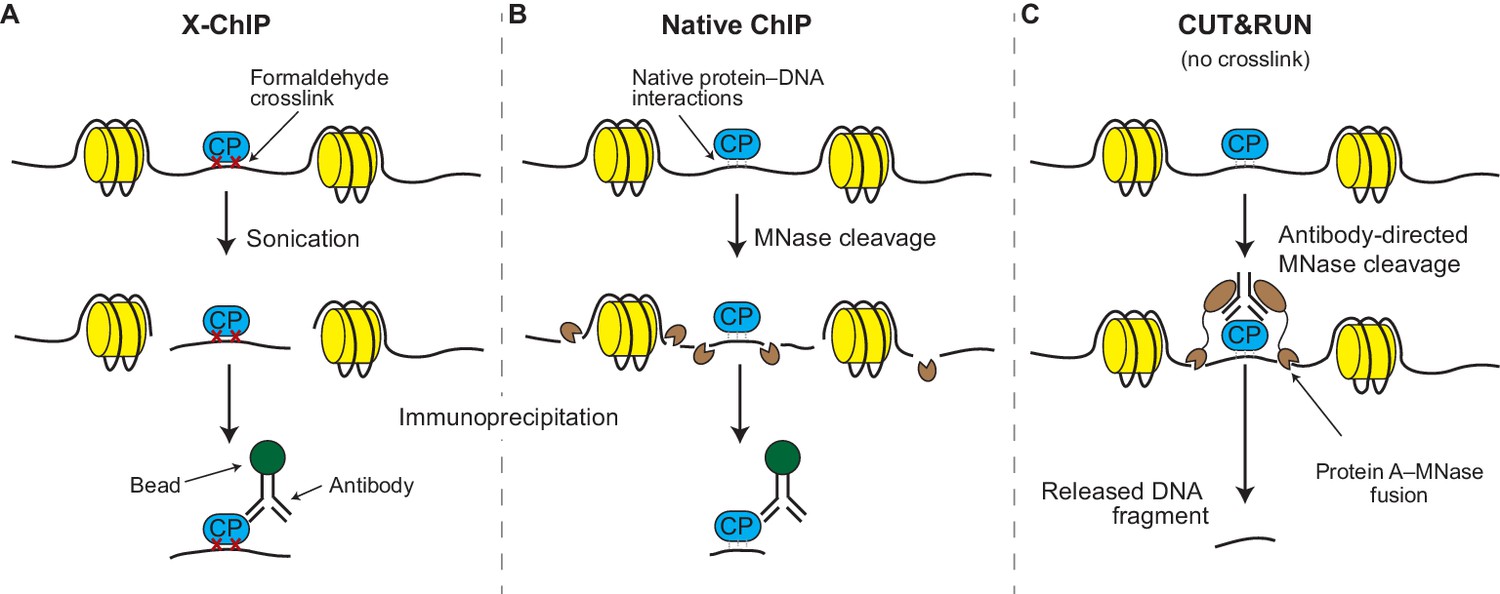
X-ChIP, native ChIP, and CUT&RUN.
(A) In X-ChIP, cells are first crosslinked (red crosses) with formaldehyde to freeze the interactions between the DNA (black line) and a chromatin-binding protein of interest (CP; blue). Sonication fragments the chromatin and makes it soluble. Antibodies are used to recognize the protein–DNA fragments, which are then ‘pulled’ out of the solution using antibody-binding beads, in a process called immunoprecipitation. The histones are shown in yellow. (B) In native ChIP, chromatin is fragmented and solubilized by treating cells with an enzyme called micrococcal nuclease (MNase; small brown shapes). The natural affinity of the protein for its DNA target keep them together during the immunoprecipitation process. (C) In CUT&RUN, antibodies direct the activity of the MNase enzyme to ensure that chromatin cleavage happens close to the protein of interest. A protein called protein A (brown ovals) binds the MNase enzyme to the antibody. The resulting small DNA fragments can be isolated as they diffuse out of the nuclei.
Crosslinking-based ChIP techniques, especially ChIP-seq, are now widely used to study gene regulation and they have been employed in a number of large-scale studies, such as the ENCODE project (ENCODE Consortium, 2012) and the NIH Roadmap Epigenomics project (Roadmap Epigenomics Consortium, 2015). Thousands of ChIP-seq datasets are available in public databases and there are very few papers in the fields of transcription, gene regulation, and epigenetics that do not contain at least one ChIP-seq experiment.
However, ChIP-seq suffers from a number of limitations, including poor resolution, suboptimal signal-to-noise ratio, and a tendency for false positives. These problems are caused by the chemicals used to crosslink the chromatin, the strong forces used to fragment it during sonication, and the detergents used to make the fragments soluble (Jain et al., 2015; Teytelman et al., 2013). Now, in eLife, Peter Skene and Steven Henikoff of the Fred Hutchinson Cancer Research Center report that they have developed a new technique called ‘cleavage under targets and release using nuclease’ (CUT&RUN) that has the potential to overcome the limitations of X-ChIP (Skene and Henikoff, 2017).
CUT&RUN is similar to native ChIP in that it uses the same enzyme, MNase, to fragment the DNA (Figure 1C). However, unlike native ChIP, the antibodies are not used to physically separate wanted from unwanted chromatin fragments (which is one of the sources of noise in ChIP); instead, CUT&RUN uses the antibodies to guide the cutting activity of the MNase enzyme to the protein of interest while the latter is still bound to intact chromatin. This means that only nearby DNA is cut into small fragments; because of their size, these fragments of DNA float out of the nuclei (leaving the rest of the uncut genome behind) and can be identified by deep sequencing. Therefore, a significant advantage of CUT&RUN is that it does not require the use of strong chemicals or sonication and that the nuclease only cuts the DNA in the region of interest, thus minimizing the noise from unwanted chromatin regions. Skene and Henikoff borrowed this idea from an older technique called chromatin immunocleavage (ChIC; Schmid et al., 2004) but adapted it to native nuclei and modernized it for the sequencing age.
To test the performance of CUT&RUN, Skene and Henikoff created maps of well-known proteins that bind the genome in yeast and human cells and compared these to maps produced by ChIP-seq. The maps obtained through CUT&RUN had a higher dynamic range and recovered a larger fraction of ‘true positives’ (sites containing short DNA sequences recognized by each protein). Skene and Henikoff were also able to modify CUT&RUN to study poorly soluble complexes by extracting all the DNA from the nuclei and only sequencing fragments below a certain size limit, which were specifically generated by the antibody-guided MNase enzyme. Due to lower sample and sequencing requirements, CUT&RUN might be applicable to a number of real-world situations, where cost and sample availability are often an issue.
As a new technique, CUT&RUN will require further evaluation to determine whether it could replace (or at least complement) ChIP-seq and its variants at the same scale. For example, it is not clear if CUT&RUN can be applied to samples in which it is not possible to isolate the nuclei of the cells; this could include some fresh tissue samples and many samples that have been fixed and then frozen. Moreover, like X-ChIP, CUT&RUN has the disadvantage that in addition to cleaving DNA in the target region, the nuclease also cleaves DNA that is far away from the target region in sequence space, but close in real space because of the three-dimensional structure of the DNA. To distinguish between these direct and indirect targets, Skene and Henikoff compared CUT&RUN maps with those obtained with their native ChIP protocol (Kasinathan et al., 2014), which only detects direct targets. However, this two-pronged approach can be labor intensive and CUT&RUN would be even more useful if it could be modified to only detect DNA directly bound to the protein of interest.
Nevertheless, CUT&RUN is a welcome addition to the arsenal of techniques in the fast-evolving field of functional genomics and we look forward to the new insights that it might facilitate in the future.
References
-
Histones: annotating chromatinAnnual Review of Genetics 43:559–599.https://doi.org/10.1146/annurev.genet.032608.103928
-
Active promoters give rise to false positive 'Phantom Peaks' in ChIP-seq experimentsNucleic Acids Research 43:6959–6968.https://doi.org/10.1093/nar/gkv637
-
ChIC and ChEC; genomic mapping of chromatin proteinsMolecular Cell 16:147–157.https://doi.org/10.1016/j.molcel.2004.09.007
-
High-resolution digital profiling of the epigenomeNature Reviews Genetics 15:814–827.https://doi.org/10.1038/nrg3798
Article and author information
Author details
Publication history
- Version of Record published: February 15, 2017 (version 1)
Copyright
© 2017, He et al.
This article is distributed under the terms of the Creative Commons Attribution License, which permits unrestricted use and redistribution provided that the original author and source are credited.
Metrics
-
- 4,573
- views
-
- 483
- downloads
-
- 1
- citations
Views, downloads and citations are aggregated across all versions of this paper published by eLife.
Download links
A two-part list of links to download the article, or parts of the article, in various formats.
Downloads (link to download the article as PDF)
Open citations (links to open the citations from this article in various online reference manager services)
Cite this article (links to download the citations from this article in formats compatible with various reference manager tools)
Chromatin Mapping: A cut above
eLife 6:e25000.
https://doi.org/10.7554/eLife.25000
Further reading
-
- Chromosomes and Gene Expression
- Immunology and Inflammation
Ikaros is a transcriptional factor required for conventional T cell development, differentiation, and anergy. While the related factors Helios and Eos have defined roles in regulatory T cells (Treg), a role for Ikaros has not been established. To determine the function of Ikaros in the Treg lineage, we generated mice with Treg-specific deletion of the Ikaros gene (Ikzf1). We find that Ikaros cooperates with Foxp3 to establish a major portion of the Treg epigenome and transcriptome. Ikaros-deficient Treg exhibit Th1-like gene expression with abnormal production of IL-2, IFNg, TNFa, and factors involved in Wnt and Notch signaling. While Ikzf1-Treg-cko mice do not develop spontaneous autoimmunity, Ikaros-deficient Treg are unable to control conventional T cell-mediated immune pathology in response to TCR and inflammatory stimuli in models of IBD and organ transplantation. These studies establish Ikaros as a core factor required in Treg for tolerance and the control of inflammatory immune responses.
-
- Cell Biology
- Chromosomes and Gene Expression
Heat stress is a major threat to global crop production, and understanding its impact on plant fertility is crucial for developing climate-resilient crops. Despite the known negative effects of heat stress on plant reproduction, the underlying molecular mechanisms remain poorly understood. Here, we investigated the impact of elevated temperature on centromere structure and chromosome segregation during meiosis in Arabidopsis thaliana. Consistent with previous studies, heat stress leads to a decline in fertility and micronuclei formation in pollen mother cells. Our results reveal that elevated temperature causes a decrease in the amount of centromeric histone and the kinetochore protein BMF1 at meiotic centromeres with increasing temperature. Furthermore, we show that heat stress increases the duration of meiotic divisions and prolongs the activity of the spindle assembly checkpoint during meiosis I, indicating an impaired efficiency of the kinetochore attachments to spindle microtubules. Our analysis of mutants with reduced levels of centromeric histone suggests that weakened centromeres sensitize plants to elevated temperature, resulting in meiotic defects and reduced fertility even at moderate temperatures. These results indicate that the structure and functionality of meiotic centromeres in Arabidopsis are highly sensitive to heat stress, and suggest that centromeres and kinetochores may represent a critical bottleneck in plant adaptation to increasing temperatures.


