Cardiac Hypertrophy: A tail of translational regulation
An RNA-binding protein called PABPC1 has an important role in determining protein synthesis rates and hypertrophy in the heart.
- University of Edinburgh, United Kingdom
The mammalian heart is a muscular pump that handles about five litres of blood per minute in the average human. Muscle cells known as cardiomyocytes make up 25–35% of all cells in the heart, with the remainder largely being vascular endothelial cells and fibroblasts (Pinto et al., 2016). The proliferation of cardiomyocytes is key to the growth of the heart before birth, and also soon after birth, but most cardiomyocytes lose their capacity to proliferate after the early post-natal period in both mice (Soonpaa et al., 1996) and humans (Bergmann et al., 2015). Thereafter, cardiac growth is achieved mainly by increases in the size of cardiomyocytes in a process known as hypertrophy.
In adulthood cardiac hypertrophy can occur as a physiological adaptation to an increased need to pump blood following sustained athletic training or during pregnancy (reviewed in Maillet et al., 2013). Cardiac hypertrophy can also be pathological in nature – for example, when it is caused by long-term hypertension. Although cellular signalling pathways and gene transcription change in distinct ways during 'physiological' and 'pathological' cardiac hypertrophy, both require de novo protein synthesis in order to increase cardiomyocyte size (Figure 1; reviewed in Heineke and Molkentin, 2006). Now, in eLife, Auinash Kalsotra of the University of Illinois and co-workers – including Sandip Chorghade and Joseph Seimetz as joint first authors – reveal an unexpected role for an RNA-binding protein called PABPC1 in cardiac hypertrophy (Chorghade et al., 2017).
Figure 1
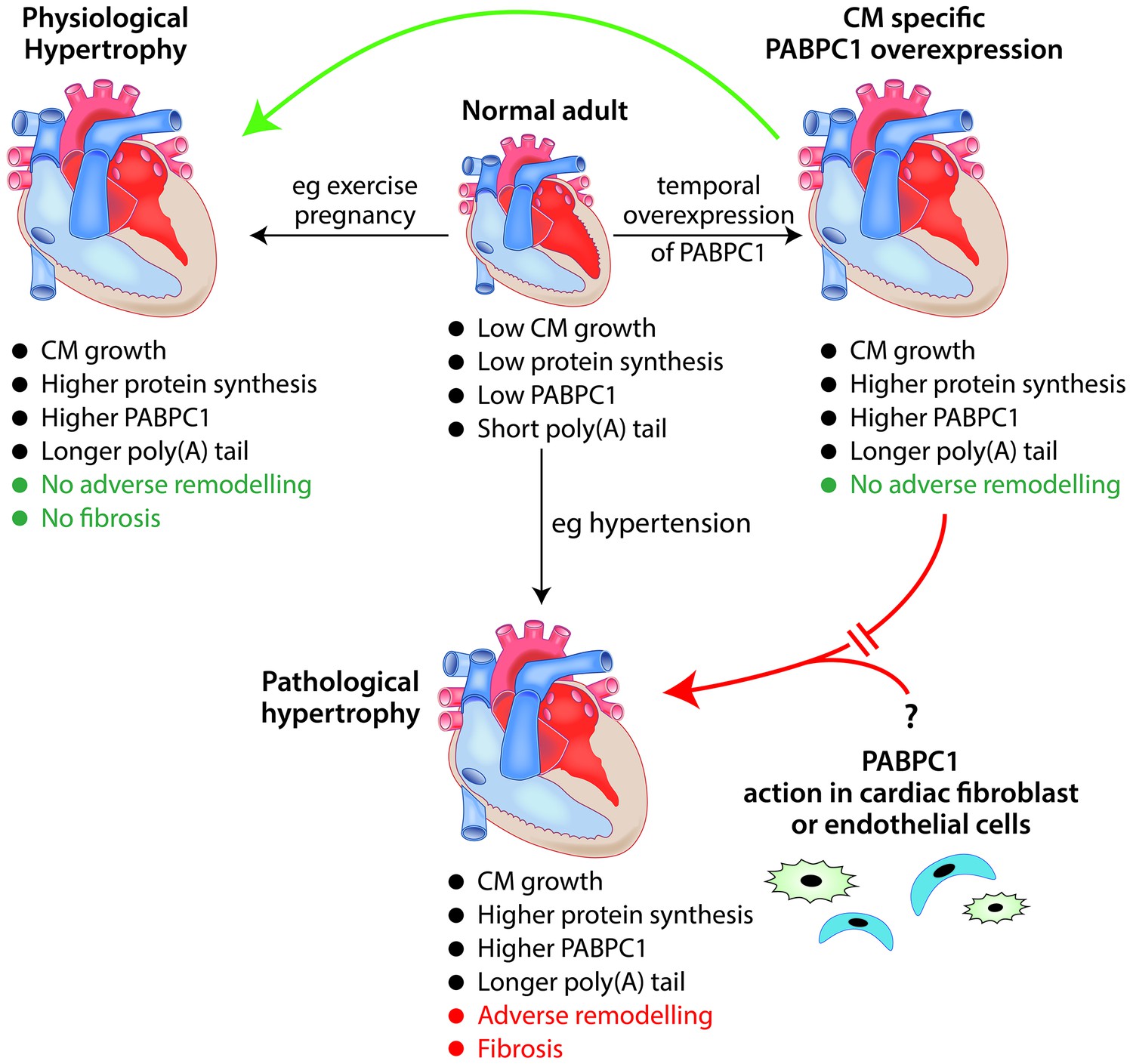
The role of the RNA-binding protein PABPC1 in cardiac hypertrophy.
In a normal adult heart, cardiomyocytes (CM) show little growth or turnover, and protein synthesis and PABPC1 levels are low. The temporal overexpression of PABPC1 in adult cardiomyocytes leads to a form of cardiac hypertrophy (top right) that resembles (green arrow) physiological cardiac hypertrophy (e.g. induced by pregnancy or endurance exercise) but not (red broken arrow) pathological cardiac hypertrophy (e.g. induced by long-term hypertension). However, a number of features (such as cardiomyocyte growth, higher protein synthesis and higher levels of PABPC1) are common to both physiological and pathological cardiac hypertrophy. This raises the question (bottom right) of whether PABPC1 function in other types of cardiac cells (such as endothelial cells or fibroblasts) may be important in the induction of pathological cardiac hypertrophy. During cardiac hypertrophy, longer poly(A) tails are associated with more efficient translation of the messenger RNA for PABPC1.
Illustration: Ronnie Grant
PABPC1 (commonly known as PABP1) is one of the 1000+ RNA-binding proteins found in humans, and has multiple functions in controlling the synthesis of new proteins (reviewed in Smith et al., 2014). However, it is best characterized as a factor that is required for efficient messenger RNA (mRNA) translation and to maintain mRNA stability, and it is generally considered to be an abundant and essential protein (Smith et al., 2014).
Protein synthesis is surprisingly low in the adult heart, perhaps reflecting the relatively low level of cell turnover there. Intriguingly, Chorghade et al. show that PABPC1 is abundant in the neonatal heart, but is only present at very low levels in the adult heart, and they go on to show that the low levels of PABPC1 in the adult heart are due to reduced translation of PABPC1 mRNA. Chorghade et al. also found that this reduction in translation coincided with a shortening of the 3’ poly(A) tail of PABPC1 mRNA: poly(A) tails are critical for efficient translation but their length can be exquisitely regulated by a number of mechanisms (reviewed in Charlesworth et al., 2013).
Intriguingly, the length of the poly(A) tail of PABPC1 mRNA and its translation were increased in experimental models of both physiological hypertrophy (induced by swimming) and pathological hypertrophy (induced by narrowing of the aorta to mimic high blood pressure), leading to an increase in PABPC1 levels.
It was already known that the translation of PABPC1 mRNA was controlled by two mechanisms (reviewed in Eliseeva et al., 2013), but uncovering that its poly(A) tail length is also regulated further underscores how important it is to control cellular levels of PABPC1. However, Chorghade et al. found that the 3’untranslated region of PABPC1 mRNA is not important for its regulated translation. This is surprising since studies in multiple cell types have shown that sequences within the 3’untranslated region normally regulate poly(A) tail length. Therefore, more work is needed to clarify the mechanism by which PABPC1 mRNA translation and poly(A) tail length is controlled in cardiomyocytes.
The altered levels of PABPC1 suggested that it may facilitate a dynamic response to an increased workload being placed on the heart. Importantly, Chorghade et al. probed the functional relevance of altered PABPC1 levels and in doing so uncovered new avenues for exploration. Reducing PABPC1 levels in cultured neonatal mouse cardiomyocytes blocked the normal increase in protein synthesis and cell size associated with stimulus-induced growth: moreover, this effect that could be rescued with wild-type PABPC1, but not with mutant PABPC1.
In transgenic mice, the overexpression of PABPC1 in adult cardiomyocytes increased protein synthesis and heart size without any loss of cardiac function, similar to what happens in physiological hypertrophy (Figure 1). The transcriptional changes that accompanied these increases were also similar to physiological rather than pathological hypertrophy. Therefore, despite the level of PABPC1 increasing in models of both forms of hypertrophy, increased PABPC1 levels in cardiomyocytes do not appear sufficient to drive pathological hypertrophy.
Other types of cardiac cells, including endothelial cells and fibroblasts, proliferate during pathological hypertrophy and can influence the behaviour of cardiomyocytes (Figure 1; Frieler and Mortensen, 2015). Future research is needed to determine the extent to which PABPC1 levels in other cardiac cell types may be involved in this process.
In addition to adding to the growing body of evidence that translational control has a role in cardiac hypertrophy, the work of Chorghade et al. also greatly extends our knowledge of the physiological roles of PABPC1. Previous whole-organism studies of PABPC1 function in vertebrates were confined to the frog X. laevis, where it is essential for early embryonic development (Gorgoni et al., 2011).
Since these new results also showed that PABPC1 levels were highest in fetal and neonatal hearts (prior to hypertrophic growth), this suggests that PABPC1 might have a critical role in cell proliferation and/or the growth of heart tissue during heart development and maturation, as well as in adult cardiac hypertrophy. Thus, ascertaining its function in fetal and neonatal hearts in mammals is an exciting question for the future.
Finally, there is significant interest in invoking pathways that promote physiological cardiac hypertrophy as an alternative to the detrimental structural and functional “remodelling” of the heart that is associated with heart failure. The work of Chorghade et al. has opened a new window on our understanding of cardiac hypertrophy, and future research should reveal whether or not PABPC1 has potential as a therapeutic target for cardiovascular disease.
References
-
Specificity factors in cytoplasmic polyadenylationWiley Interdisciplinary Reviews: RNA 4:437–461.https://doi.org/10.1002/wrna.1171
-
Regulation of cardiac hypertrophy by intracellular signalling pathwaysNature Reviews Molecular Cell Biology 7:589–600.https://doi.org/10.1038/nrm1983
-
Molecular basis of physiological heart growth: fundamental concepts and new playersNature Reviews Molecular Cell Biology 14:38–48.https://doi.org/10.1038/nrm3495
-
Revisiting cardiac cellular compositionCirculation Research 118:400–409.https://doi.org/10.1161/CIRCRESAHA.115.307778
-
Poly(A)-binding proteins are required for diverse biological processes in metazoansBiochemical Society Transactions 42:1229–1237.https://doi.org/10.1042/BST20140111
-
Cardiomyocyte DNA synthesis and binucleation during murine developmentAmerican Journal of Physiology 271:H2183–H2189.
Article and author information
Author details
Publication history
- Version of Record published: June 27, 2017 (version 1)
Copyright
© 2017, Gray et al.
This article is distributed under the terms of the Creative Commons Attribution License, which permits unrestricted use and redistribution provided that the original author and source are credited.
Metrics
-
- 2,655
- Page views
-
- 288
- Downloads
-
- 5
- Citations
Article citation count generated by polling the highest count across the following sources: Crossref, PubMed Central, Scopus.
Download links
A two-part list of links to download the article, or parts of the article, in various formats.
Downloads (link to download the article as PDF)
Open citations (links to open the citations from this article in various online reference manager services)
Cite this article (links to download the citations from this article in formats compatible with various reference manager tools)
Cardiac Hypertrophy: A tail of translational regulation
eLife 6:e29104.
https://doi.org/10.7554/eLife.29104
Further reading
-
- Biochemistry and Chemical Biology
- Plant Biology
Metabolism and biological functions of the nitrogen-rich compound guanidine have long been neglected. The discovery of four classes of guanidine-sensing riboswitches and two pathways for guanidine degradation in bacteria hint at widespread sources of unconjugated guanidine in nature. So far, only three enzymes from a narrow range of bacteria and fungi have been shown to produce guanidine, with the ethylene-forming enzyme (EFE) as the most prominent example. Here, we show that a related class of Fe2+- and 2-oxoglutarate-dependent dioxygenases (2-ODD-C23) highly conserved among plants and algae catalyze the hydroxylation of homoarginine at the C6-position. Spontaneous decay of 6-hydroxyhomoarginine yields guanidine and 2-aminoadipate-6-semialdehyde. The latter can be reduced to pipecolate by pyrroline-5-carboxylate reductase but more likely is oxidized to aminoadipate by aldehyde dehydrogenase ALDH7B in vivo. Arabidopsis has three 2-ODD-C23 isoforms, among which Din11 is unusual because it also accepted arginine as substrate, which was not the case for the other 2-ODD-C23 isoforms from Arabidopsis or other plants. In contrast to EFE, none of the three Arabidopsis enzymes produced ethylene. Guanidine contents were typically between 10 and 20 nmol*(g fresh weight)-1 in Arabidopsis but increased to 100 or 300 nmol*(g fresh weight)-1 after homoarginine feeding or treatment with Din11-inducing methyljasmonate, respectively. In 2-ODD-C23 triple mutants, the guanidine content was strongly reduced, whereas it increased in overexpression plants. We discuss the implications of the finding of widespread guanidine-producing enzymes in photosynthetic eukaryotes as a so far underestimated branch of the bio-geochemical nitrogen cycle and propose possible functions of natural guanidine production.
-
- Biochemistry and Chemical Biology
- Medicine
Type 2 diabetes (T2D) is associated with higher fracture risk, despite normal or high bone mineral density. We reported that bone formation genes (SOST and RUNX2) and advanced glycation end-products (AGEs) were impaired in T2D. We investigated Wnt signaling regulation and its association with AGEs accumulation and bone strength in T2D from bone tissue of 15 T2D and 21 non-diabetic postmenopausal women undergoing hip arthroplasty. Bone histomorphometry revealed a trend of low mineralized volume in T2D (T2D 0.249% [0.156–0.366]) vs non-diabetic subjects 0.352% [0.269–0.454]; p=0.053, as well as reduced bone strength (T2D 21.60 MPa [13.46–30.10] vs non-diabetic subjects 76.24 MPa [26.81–132.9]; p=0.002). We also showed that gene expression of Wnt agonists LEF-1 (p=0.0136) and WNT10B (p=0.0302) were lower in T2D. Conversely, gene expression of WNT5A (p=0.0232), SOST (p<0.0001), and GSK3B (p=0.0456) were higher, while collagen (COL1A1) was lower in T2D (p=0.0482). AGEs content was associated with SOST and WNT5A (r=0.9231, p<0.0001; r=0.6751, p=0.0322), but inversely correlated with LEF-1 and COL1A1 (r=–0.7500, p=0.0255; r=–0.9762, p=0.0004). SOST was associated with glycemic control and disease duration (r=0.4846, p=0.0043; r=0.7107, p=0.00174), whereas WNT5A and GSK3B were only correlated with glycemic control (r=0.5589, p=0.0037; r=0.4901, p=0.0051). Finally, Young’s modulus was negatively correlated with SOST (r=−0.5675, p=0.0011), AXIN2 (r=−0.5523, p=0.0042), and SFRP5 (r=−0.4442, p=0.0437), while positively correlated with LEF-1 (r=0.4116, p=0.0295) and WNT10B (r=0.6697, p=0.0001). These findings suggest that Wnt signaling and AGEs could be the main determinants of bone fragility in T2D.


