Vaccine Design: Chimeras could help in the fight against leptospirosis
Understanding the structure of an antigen can guide the design of improved antigen-based vaccines.
- Instituto Butantan, Brazil
Leptospirosis is a disease that affects humans and animals worldwide, with a high prevalence in tropical and subtropical regions. The Leptospira bacteria that cause the disease colonize the kidneys of wild and domestic animals, and humans come into contact with these bacteria via the urine of infected animals – primarily rats in urban areas (Bharti et al., 2003). Symptoms of leptospirosis range from a mild influenza-like illness to severe infections that are fatal in over half of cases (Marotto et al., 1999). The lack of an effective vaccine has hampered efforts to prevent and control the disease.
Vaccines contain substances that allow the host immune system to learn how to recognize a particular pathogen. The parts of the pathogen that are recognized by the immune system are known as antigens, and immune molecules called antibodies bind to these antigens as part of the immune response.
Current vaccines against leptospirosis consist of whole inactivated bacterial cells, which induce the host immune system to produce antibodies against lipopolysaccharide molecules in the outer membrane of the bacteria. However, these vaccines only provide short-term immunity against the specific varieties of bacteria that are included (in their inactivated form) in the vaccine (Adler and de la Peña Moctezuma, 2010). Moreover, there are more than 250 serum varieties of Leptospira, so developing a vaccine that is effective against all of them with this methodology is unrealistic.
Advances in recombinant DNA techniques, allied to whole-genome sequencing and bioinformatics technologies, have led to a new approach for the identification of vaccine candidates. This approach, known as reverse vaccinology, uses the genome of the pathogen to predict the exact part of an antigen that antibodies interact with (Rappuoli et al., 2014). However, despite these advances, only a few antigens that protect against leptospirosis have been identified. Now, in eLife, Yung-Fu Chang and Robert Oswald of Cornell University and colleagues – including Ching-Lin Hsieh and Christopher Ptak as joint first authors – report a valuable step forward in efforts to develop an effective vaccine against pathogenic Leptospira (Hsieh et al., 2017).
LigB is a protein found on the surface of pathogenic forms of Leptospira (Matsunaga et al., 2003). It contains a short N-terminal domain (which anchors it to the outer membrane of the bacterium), twelve consecutive immunoglobulin-like domains (called LigB1-12), and a large non-immunoglobulin-like domain at the C-terminal end. The 12 central domains can be divided into a conserved region (LigB1-7) and a more variable region (LigB7-12; Ptak et al., 2014). LigB has been considered the most promising target for an effective vaccine (Conrad et al., 2017), but controversial results suggested only a partial immunization (Yan et al., 2009; Silva et al., 2007) or did not confer sterilizing immunity (protection against infection as well as disease; Evangelista et al., 2017).
Hsieh et al. combined structural biology with immunoreactive assays to determine the region of the LigB protein that most strongly induces an immune response. They used small-angle X-ray scattering to determine the low-resolution structure of the LigB1-12 region by working their way along this region, imaging five of the domains at a time. The final structure demonstrated an extensive surface area that is present across almost all of the 12 domains. This provides a high degree of exposure to the host immune system.
To confirm the capability of the protein to induce a host immune response, Hsieh et al. used two truncated forms of LigB – one that consisted of LigB1-7, and one formed of LigB7-12 – to generate a library of anti-LigB monoclonal antibodies. The bactericidal activity of these antibodies was evaluated by measuring how they interacted with LigB and how well they adhered to the surface of pathogenic Leptospira. These interactions were then correlated with the ability of the monoclonal antibodies to kill the bacteria in the presence of innate immune proteins called serum complement. By blocking important domains of LigB, monoclonal antibodies render Leptospira susceptible to attack and killing by complement proteins.
Using a technique called nuclear magnetic resonance spectroscopy, Hsieh et al. worked out the structure of the monoclonal antibody domains that have bactericidal activity. These data helped them to build chimera proteins from selected domains that were then used to immunize hamsters against virulent Leptospira (Figure 1). One chimera containing just three domains – LigB10-B7-B7 – afforded better protection to hamsters than longer constructs, such as LigB7-12 (which contains six domains).
Figure 1
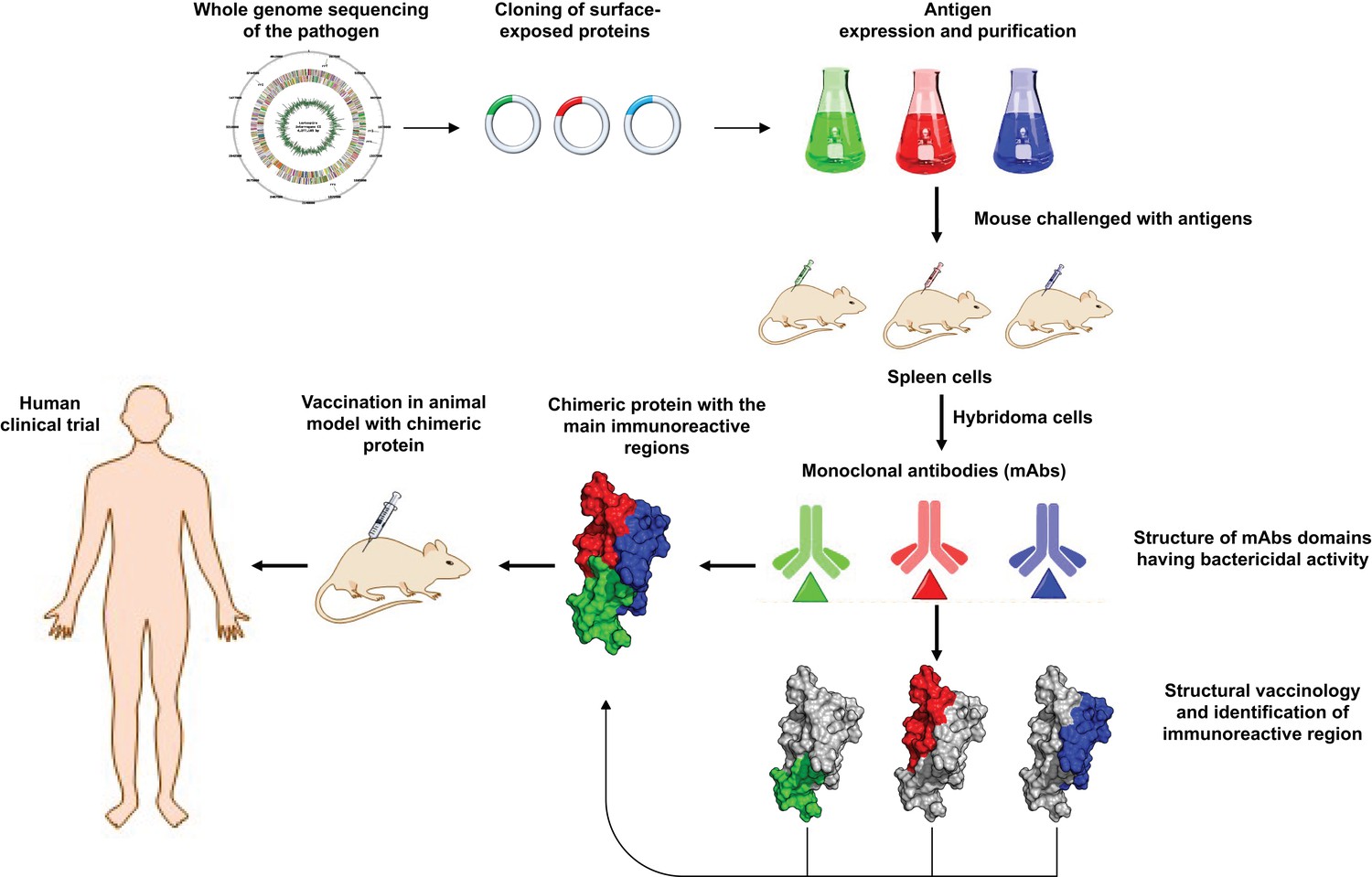
Schematic representation of how structural biology contributes to vaccine design.
Sequencing the genome of a pathogen (top left) makes it possible to clone protein-based antigens – the features of the pathogen that are detected by antibodies in the host immune system. When mice are injected with the purified antigens, cells in their spleen produce monoclonal antibodies (mAbs) via cells called hybridomas. By studying the structure of the antibodies, and identifying the regions that interact most strongly with the antigens, researchers can build chimeric proteins from these regions. The effectiveness of the chimera as a vaccine for the pathogen can then be tested in animal models and human clinical trials.
The results of Hsieh et al. reinforce previous work that showed that structural biology represents a powerful tool for structure-based vaccine design. Their findings have significantly advanced our knowledge of LigB and represent an important step toward an improved vaccine against leptospirosis.
References
-
Leptospira and leptospirosisVeterinary Microbiology 140:287–296.https://doi.org/10.1016/j.vetmic.2009.03.012
-
Leptospirosis: a zoonotic disease of global importanceThe Lancet Infectious Diseases 3:757–771.https://doi.org/10.1016/S1473-3099(03)00830-2
-
LigB subunit vaccine confers sterile immunity against challenge in the hamster model of leptospirosisPLOS Neglected Tropical Diseases 11:e0005441.https://doi.org/10.1371/journal.pntd.0005441
-
Acute lung injury in leptospirosis: clinical and laboratory features, outcome, and factors associated with mortalityClinical Infectious Diseases 29:1561–1563.https://doi.org/10.1086/313501
Article and author information
Author details
Jademilson C Santos

Ana Lucia TO Nascimento
Publication history
- Version of Record published: January 19, 2018 (version 1)
Copyright
© 2018, Santos et al.
This article is distributed under the terms of the Creative Commons Attribution License, which permits unrestricted use and redistribution provided that the original author and source are credited.
Metrics
-
- 1,334
- views
-
- 147
- downloads
-
- 2
- citations
Views, downloads and citations are aggregated across all versions of this paper published by eLife.
Download links
A two-part list of links to download the article, or parts of the article, in various formats.
Downloads (link to download the article as PDF)
Open citations (links to open the citations from this article in various online reference manager services)
Cite this article (links to download the citations from this article in formats compatible with various reference manager tools)
Vaccine Design: Chimeras could help in the fight against leptospirosis
eLife 7:e34087.
https://doi.org/10.7554/eLife.34087
Further reading
-
- Biochemistry and Chemical Biology
- Structural Biology and Molecular Biophysics
NADPH oxidases (NOX) are transmembrane proteins, widely spread in eukaryotes and prokaryotes, that produce reactive oxygen species (ROS). Eukaryotes use the ROS products for innate immune defense and signaling in critical (patho)physiological processes. Despite the recent structures of human NOX isoforms, the activation of electron transfer remains incompletely understood. SpNOX, a homolog from Streptococcus pneumoniae, can serves as a robust model for exploring electron transfers in the NOX family thanks to its constitutive activity. Crystal structures of SpNOX full-length and dehydrogenase (DH) domain constructs are revealed here. The isolated DH domain acts as a flavin reductase, and both constructs use either NADPH or NADH as substrate. Our findings suggest that hydride transfer from NAD(P)H to FAD is the rate-limiting step in electron transfer. We identify significance of F397 in nicotinamide access to flavin isoalloxazine and confirm flavin binding contributions from both DH and Transmembrane (TM) domains. Comparison with related enzymes suggests that distal access to heme may influence the final electron acceptor, while the relative position of DH and TM does not necessarily correlate with activity, contrary to previous suggestions. It rather suggests requirement of an internal rearrangement, within the DH domain, to switch from a resting to an active state. Thus, SpNOX appears to be a good model of active NOX2, which allows us to propose an explanation for NOX2’s requirement for activation.
-
- Cell Biology
- Structural Biology and Molecular Biophysics
Acetylation of α-tubulin at the lysine 40 residue (αK40) by αTAT1/MEC-17 acetyltransferase modulates microtubule properties and occurs in most eukaryotic cells. Previous literatures suggest that acetylated microtubules are more stable and damage resistant. αK40 acetylation is the only known microtubule luminal post-translational modification site. The luminal location suggests that the modification tunes the lateral interaction of protofilaments inside the microtubule. In this study, we examined the effect of tubulin acetylation on the doublet microtubule (DMT) in the cilia of Tetrahymena thermophila using a combination of cryo-electron microscopy, molecular dynamics, and mass spectrometry. We found that αK40 acetylation exerts a small-scale effect on the DMT structure and stability by influencing the lateral rotational angle. In addition, comparative mass spectrometry revealed a link between αK40 acetylation and phosphorylation in cilia.


