Antagonistic control of DDK binding to licensed replication origins by Mcm2 and Rad53
- Molecular Biology Program, Sloan Kettering Institute, Memorial Sloan Kettering Cancer Center, United States
- Weill-Cornell Graduate School of Medical Sciences, United States
Figures
Figure 1 with 1 supplement
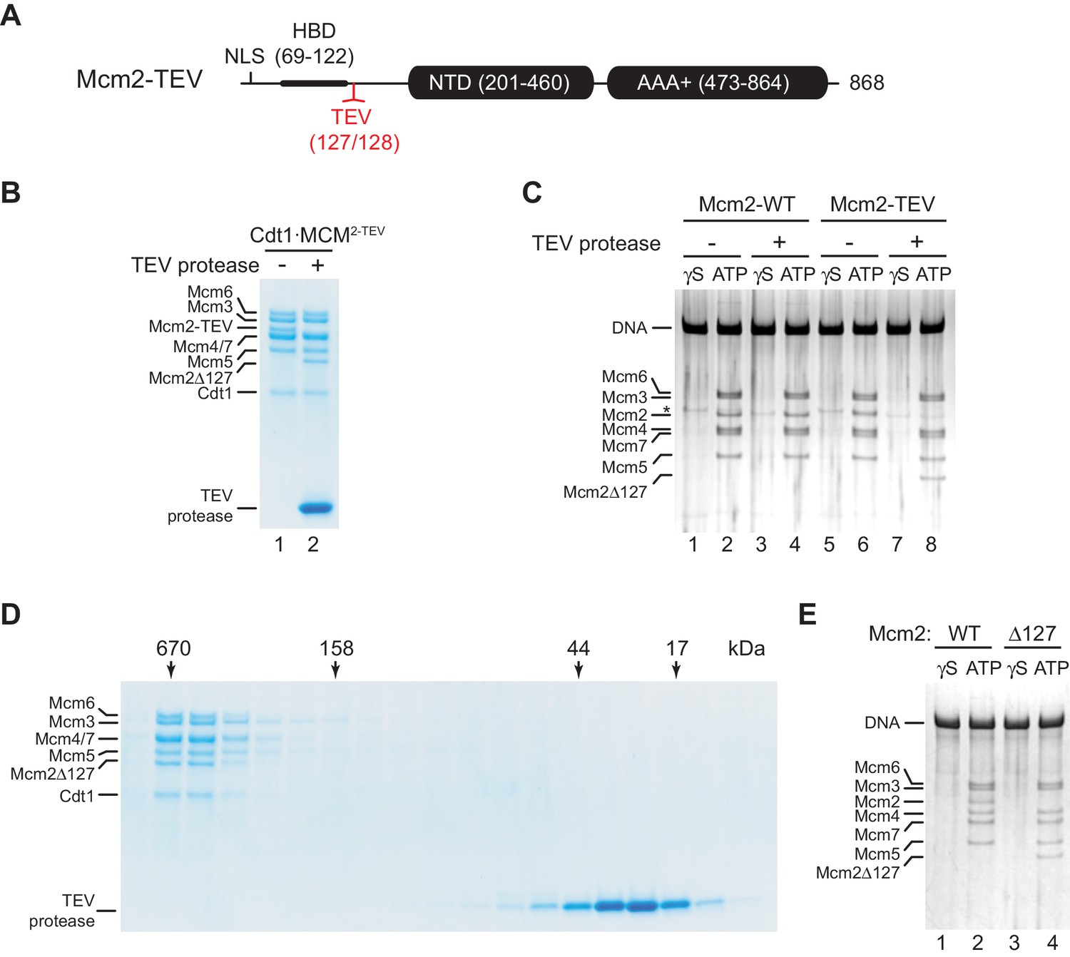
Residues 1–127 of the Mcm2 NTE are dispensable for MCM DH stability.
(A) Schematic of Mcm2 domain structure. Numbers indicate amino acid positions. The position of the TEV cleavage site is highlighted in red. NLS: Nuclear localization sequence; HBD: Histone binding domain; NTD: N-terminal domain; AAA+: ATPase domain. (B) Cdt1·MCM2-TEV was mock-treated or digested with TEV protease for 1 hr at 30°C, as indicated. Reactions were fractionated on SDS-PAGE and stained with Coomassie blue. (C) MCM loading reactions were performed on 3 kbp ARS305-containing DNA in the presence of ATPγS (γS) or ATP as indicated. DNA-bound material was washed with high-salt buffer, mock-treated or digested with TEV protease as indicated, washed again with high-salt buffer, and analyzed by SDS-PAGE and silver staining. * denotes Orc1 protein. (D) Gel-filtration analysis of purified Cdt1·MCM2-TEV following digestion with TEV protease. The digestion reaction was fractionated on a Superdex 200 column and fractions analyzed by SDS-PAGE and Coomassie stain. (E) Mcm2-7 loading reactions with either wildtype Cdt1·MCM (lanes 1+2) or Cdt1·MCM2-Δ127 (lanes 3+4). Reactions were performed either in the presence of ATPγS or ATP as indicated and DNA-beads subsequently washed with high-salt buffer. DNA-bound fractions were analyzed by SDS-PAGE and silver stain.
-
Figure 1—source data 1
- https://cdn.elifesciences.org/articles/58571/elife-58571-fig1-data1-v2.pdf
-
Figure 1—source data 2
- https://cdn.elifesciences.org/articles/58571/elife-58571-fig1-data2-v2.pdf
-
Figure 1—source data 3
- https://cdn.elifesciences.org/articles/58571/elife-58571-fig1-data3-v2.pdf
Figure 1—figure supplement 1
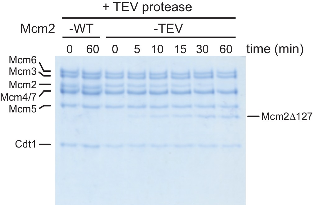
Time course analysis of Cdt1·MCM2-TEV and Cdt1·MCM2-WT cleavage by TEV protease.
Fractions of the reactions were analyzed by SDS-PAGE and Coomassie stain.
-
Figure 1—figure supplement 1—source data 1
- https://cdn.elifesciences.org/articles/58571/elife-58571-fig1-figsupp1-data1-v2.pdf
Figure 2 with 2 supplements
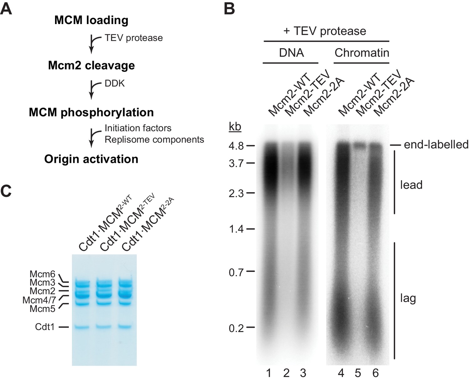
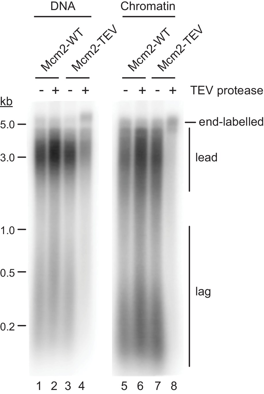
The Mcm2 NTE is important for DNA replication.
(A) Experimental outline. (B) In vitro DNA replication reactions were performed on naked (lanes 1–3) or chromatinized (lanes 4–6) circular plasmid DNA (p1017, 4.8 kbp). TEV protease was added to each reaction following MCM loading for 1 hr at 30°C, before addition of DDK and standard initiation/replisome factors. Chromatin replication reactions additionally contained FACT and Nhp6. Products were analyzed by 0.8% denaturing agarose gel-electrophoresis and autoradiography. Lead: Leading strand product; lag: Lagging strand product. (C) Purified Cdt1·MCM complexes containing either wildtype Mcm2 (Cdt1·MCM2-WT), Mcm2-TEV (Cdt1·MCM2-TEV), or Mcm2-2A (Cdt1·MCM2-2A).
Figure 2—figure supplement 1
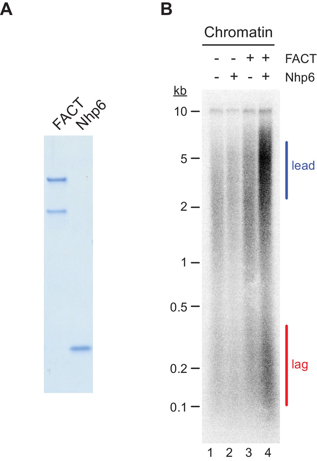
FACT/Nhp6-dependent chromatin replication.
(A) Purified FACT and Nhp6. Samples were analyzed by SDS-PAGE and Coomassie stain. (B) In vitro DNA replication reaction was performed on chromatinized p470 (10 kbp) in the absence or presence of FACT and Nhp6 as indicated. Reaction products were analyzed by denaturing agarose gel-electrophoresis and autoradiography.
-
Figure 2—figure supplement 1—source data 1
- https://cdn.elifesciences.org/articles/58571/elife-58571-fig2-figsupp1-data1-v2.pdf
-
Figure 2—figure supplement 1—source data 2
- https://cdn.elifesciences.org/articles/58571/elife-58571-fig2-figsupp1-data2-v2.pdf
Figure 2—figure supplement 2
Attenuation of DNA synthesis in the presence of Mcm2-TEV is dependent on TEV protease cleavage.
Standard DNA replication reactions using naked or chromatinized p1017 (4.8 kbp) as template were performed with Cdt1·MCM2-TEV or Cdt1·MCM2-WT as indicated. TEV protease or mock buffer was added to the reaction following MCM loading and preceding origin activation as indicated. Reaction products were analyzed by denaturing agarose gel-electrophoresis and autoradiography.
-
Figure 2—figure supplement 2—source data 1
- https://cdn.elifesciences.org/articles/58571/elife-58571-fig2-figsupp2-data1-v2.pdf
Figure 3
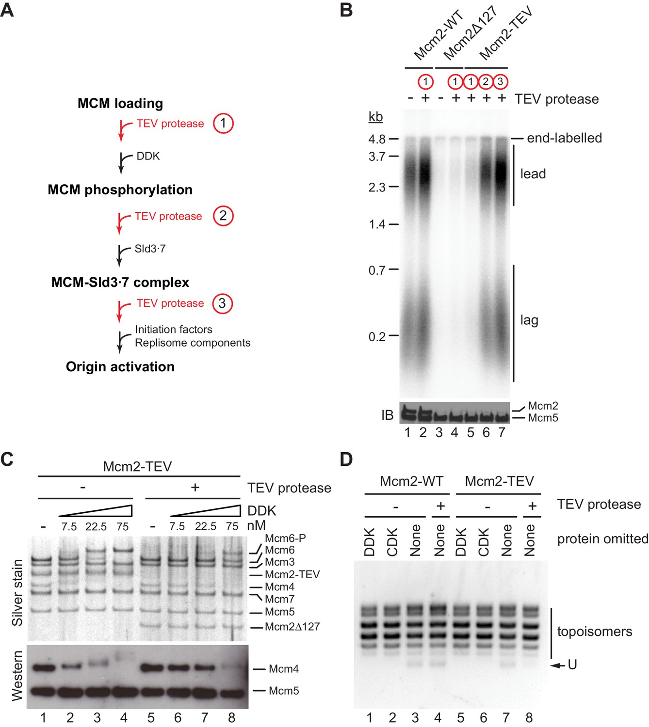
The Mcm2 NTE promotes DDK function during origin activation.
(A) Experimental outline for experiment in B. Variable addition points for TEV protease are highlighted in red. (B) Standard in vitro DNA replication reactions were performed using p1017 (4.8 kb) as a template. TEV protease or mock buffer was added for 1 hr at 30°C as indicated. Reaction products were analyzed by denaturing agarose gel-electrophoresis and autoradiography (top). A fraction of each reaction was analyzed by SDS-PAGE and western blot using antibodies against Mcm2 and Mcm5 (bottom); note that the N-terminal epitope recognized by the Mcm2 antibody is lost after TEV protease cleavage. (C) MCM DHs assembled with Cdt1·MCM2-TEV were either mock-treated (lanes 1–4) or digested with TEV protease (lanes 5–8). DDK was subsequently added to the reactions at the indicated concentrations and reactions analyzed by SDS-PAGE and silver stain or western blot using antibodies against Mcm4 and Mcm5. (D) Plasmid unwinding assay. CMGs were assembled with Cdt1·MCM2-WT (lanes 1–4) or Cdt1·MCM2-TEV (lanes 5–8) using p79 (3 kbp) as substrate. TEV protease was added to the reactions after the MCM loading step, prior to the addition of DDK, CDK, Sld2, Sld3⋅7, Dpb11, GINS, Cdc45, Pol ε, RPA, and Mcm10 as indicated. DNA was repurified from the reaction and analyzed by native agarose gel-electrophoresis and EtBr stain. U: U-form DNA.
-
Figure 3—source data 1
Figure 3B, autoradiograph.
- https://cdn.elifesciences.org/articles/58571/elife-58571-fig3-data1-v2.pdf
-
Figure 3—source data 2
Figure 3B, immunoblot: Mcm2, Mcm5.
- https://cdn.elifesciences.org/articles/58571/elife-58571-fig3-data2-v2.pdf
-
Figure 3—source data 3
Figure 3C, silver stain.
- https://cdn.elifesciences.org/articles/58571/elife-58571-fig3-data3-v2.pdf
-
Figure 3—source data 4
Figure 3C, immunoblot: Mcm4, Mcm5.
- https://cdn.elifesciences.org/articles/58571/elife-58571-fig3-data4-v2.pdf
-
Figure 3—source data 5
- https://cdn.elifesciences.org/articles/58571/elife-58571-fig3-data5-v2.pdf
Figure 4

The Mcm2 NTE promotes binding of DDK to MCM DHs.
(A) MCM DHs were assembled on bead-immobilized DNA, washed with high-salt buffer, and subsequently incubated with ATPγS and DDK at the indicated concentrations. As a control, Cdt1·MCM was omitted from the MCM loading reaction in lane 6. After incubation with DDK, DNA-bound material was isolated and analyzed by SDS-PAGE and silver stain (top) or western blot using antibodies against Mcm7, Dbf4, or Cdc7 (bottom). (B) MCM loading reactions were carried out either in the presence of ATP (lane 1) or ATPγS (lanes 2–8). DNA beads were subsequently washed with low-salt buffer and incubated with DDK in the presence of ATPγS. DNA-bound material was analyzed as in A. (C) MCM DHs were assembled from Cdt1·MCM2-TEV, mock-treated or digested with TEV protease as indicated and incubated with purified DDK at the indicated concentrations. DNA-bound material was analyzed as in A. (D) MCM DHs were assembled from Cdt1·MCM2-TEV and mock-treated or digested with TEV protease as indicated. In lane 3, DDK was added after TEV protease, in lane 4 DDK was added before TEV protease. DDK was included at 150 nM. DNA-bound material was analyzed as in A. (E) DNA-bound DDK-MCM DH complexes were washed with buffer containing the indicated concentration of KOAc, and where indicated followed by a wash with buffer containing 500 mM NaCl. (F) MCM DHs were assembled on bead-immobilized DNA, washed to remove free ATP, and subsequently incubated with DDK in the presence of ATP or ATP analogues, as indicated. DNA-bound material was analyzed as in A.
-
Figure 4—source data 1
Figure 4A, silver stain.
- https://cdn.elifesciences.org/articles/58571/elife-58571-fig4-data1-v2.pdf
-
Figure 4—source data 2
Figure 4 A+E, immunoblot: Mcm7, Cdc7.
- https://cdn.elifesciences.org/articles/58571/elife-58571-fig4-data2-v2.pdf
-
Figure 4—source data 3
Figure 4 A+E, immunoblot: Dbf4.
- https://cdn.elifesciences.org/articles/58571/elife-58571-fig4-data3-v2.pdf
-
Figure 4—source data 4
Figure 4B, silver stain.
- https://cdn.elifesciences.org/articles/58571/elife-58571-fig4-data4-v2.pdf
-
Figure 4—source data 5
Figure 4B, immunoblot: Cdc7.
- https://cdn.elifesciences.org/articles/58571/elife-58571-fig4-data5-v2.pdf
-
Figure 4—source data 6
Figure 4B, immunoblot: Dbf4.
- https://cdn.elifesciences.org/articles/58571/elife-58571-fig4-data6-v2.pdf
-
Figure 4—source data 7
Figure 4C, silver stain.
- https://cdn.elifesciences.org/articles/58571/elife-58571-fig4-data7-v2.pdf
-
Figure 4—source data 8
Figure 4C, immunoblot: Mcm7, Cdc7.
- https://cdn.elifesciences.org/articles/58571/elife-58571-fig4-data8-v2.pdf
-
Figure 4—source data 9
Figure 4C, immunoblot: Dbf4.
- https://cdn.elifesciences.org/articles/58571/elife-58571-fig4-data9-v2.pdf
-
Figure 4—source data 10
Figure 4D, silver stain.
- https://cdn.elifesciences.org/articles/58571/elife-58571-fig4-data10-v2.pdf
-
Figure 4—source data 11
Figure 4D, immunoblot: Mcm7.
- https://cdn.elifesciences.org/articles/58571/elife-58571-fig4-data11-v2.pdf
-
Figure 4—source data 12
Figure 4D, immunoblot: Dbf4.
- https://cdn.elifesciences.org/articles/58571/elife-58571-fig4-data12-v2.pdf
-
Figure 4—source data 13
Figure 4D, immunoblot: Cdc7.
- https://cdn.elifesciences.org/articles/58571/elife-58571-fig4-data13-v2.pdf
-
Figure 4—source data 14
Figure 4E, silver stain.
- https://cdn.elifesciences.org/articles/58571/elife-58571-fig4-data14-v2.pdf
-
Figure 4—source data 15
Figure 4F, silver stain.
- https://cdn.elifesciences.org/articles/58571/elife-58571-fig4-data15-v2.pdf
-
Figure 4—source data 16
Figure 4F, immunoblot: Cdc7.
- https://cdn.elifesciences.org/articles/58571/elife-58571-fig4-data16-v2.pdf
-
Figure 4—source data 17
Figure 4F, immunoblot: Mcm7.
- https://cdn.elifesciences.org/articles/58571/elife-58571-fig4-data17-v2.pdf
-
Figure 4—source data 18
Figure 4F, immunoblot: Dbf4.
- https://cdn.elifesciences.org/articles/58571/elife-58571-fig4-data18-v2.pdf
Figure 5 with 1 supplement
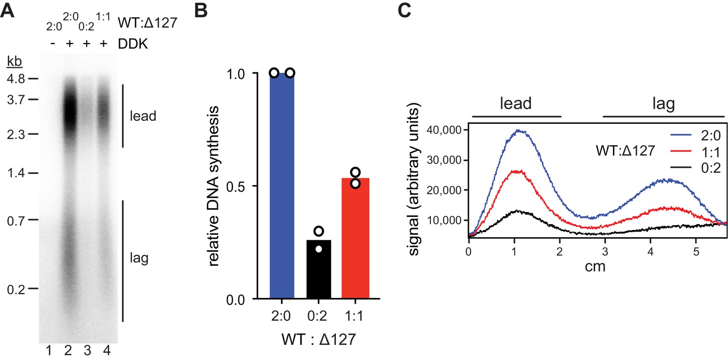
Mcm2-WT does not rescue the Mcm2Δ127 replication defect.
(A) Standard DNA replication reaction using p1017 (4.8 kb) as template. Cdt1·MCM2-Δ127 and Cdt1·MCM2-WT were included at the MCM loading step at the indicated ratios; the total concentration of Cdt1·MCM was 80 nM in the Mcm2-7 loading reaction. (B) Quantification of total relative DNA synthesis in reactions of experiment in C. Bars represent the average of two independent experiments. (C) Lane traces of experiment in C.
-
Figure 5—source data 1
Figure 5A, autoradiograph.
- https://cdn.elifesciences.org/articles/58571/elife-58571-fig5-data1-v2.pdf
Figure 5—figure supplement 1
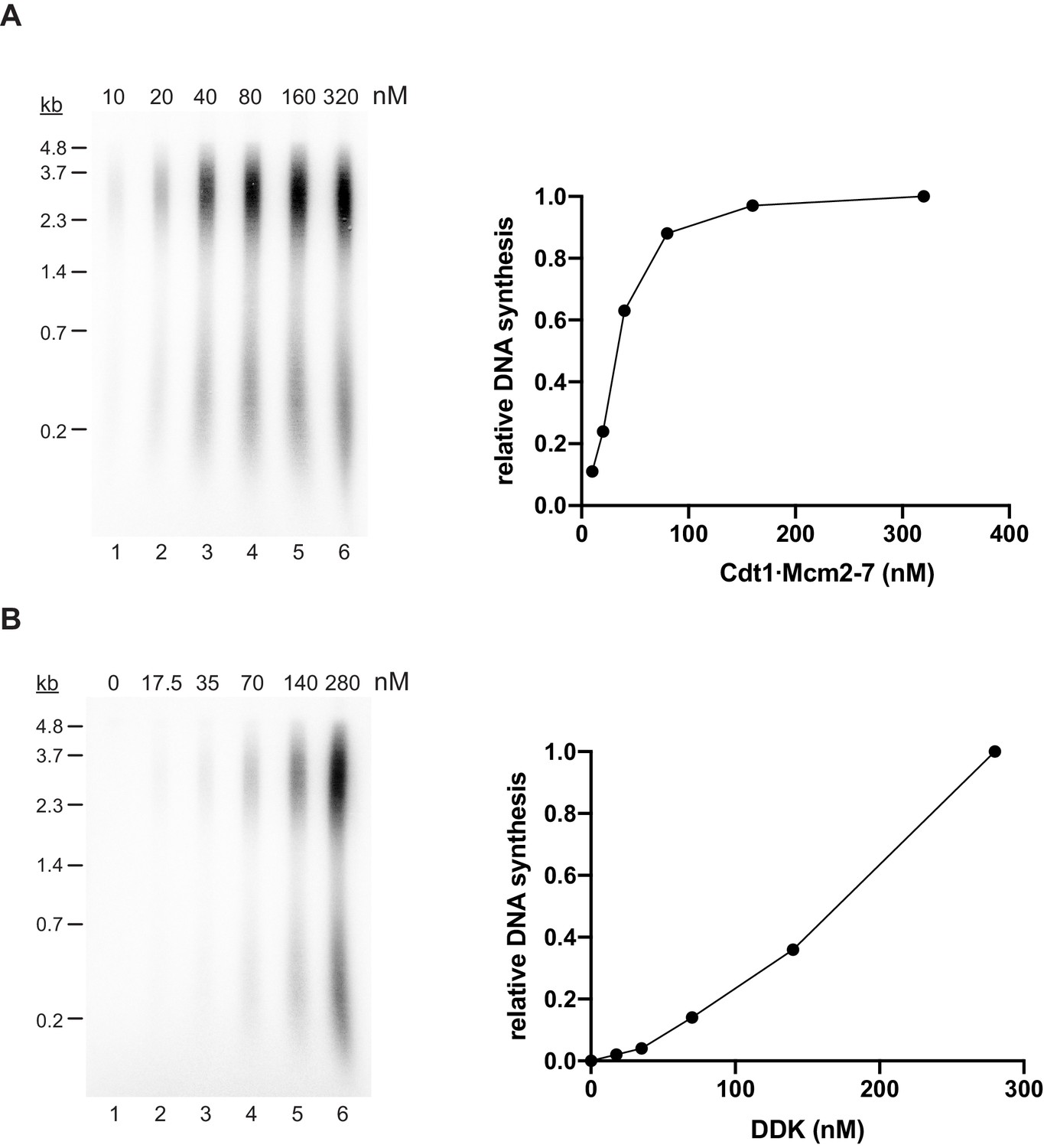
The effect of Cdt1·MCM and DDK concentrations on DNA replication in vitro.
(A) Cdt1·MCM titration experiment using standard DNA replication conditions. Template: p1017 (4.8 kbp). Left: Reaction products were analyzed by denaturing agarose gel-electrophoresis and autoradiography. Right: Plot of total normalized DNA synthesis. (B) DDK titration experiment using standard DNA replication conditions, but Cdt1·MCM2-Δ127 in place of Cdt1·MCM2-WT. Template: p1017 (4.8 kbp). Left: Reaction products were analyzed by denaturing agarose gel-electrophoresis and autoradiography. Right: Plot of total normalized DNA synthesis.
-
Figure 5—figure supplement 1—source data 1
- https://cdn.elifesciences.org/articles/58571/elife-58571-fig5-figsupp1-data1-v2.pdf
-
Figure 5—figure supplement 1—source data 2
- https://cdn.elifesciences.org/articles/58571/elife-58571-fig5-figsupp1-data2-v2.pdf
Figure 6

Steric inhibition of DDK binding to MCM DHs by Rad53.
(A) DDK binding to purified MCM DHs was monitored in the presence of ATP and in the absence or presence of Rad53-WT or Rad53-kd, as indicated. In lanes 5+6 DDK and Rad53 were co-incubated in the presence of ATP prior to addition to DNA-bound MCM-7 DHs; in lanes 7+8 DDK was incubated with purified MCM DHs before addition of Rad53. DNA-bound material was analyzed SDS-PAGE and silver stain or western blot as indicated. (B) Standard DNA replication reaction using p1017 (4.8 kb) as template. Rad53 and DDK were either co-incubated prior to simultaneous addition after the MCM loading step (lanes 3+4) or Rad53 was added after DDK prior to the addition of activation factors (lanes 5+6). Replication products were analyzed by denaturing agarose gel-electrophoresis and autoradiography. The results of two experiment repeats are plotted in the graph on the right. (C) DDK binding to DNA-bound MCM DHs was monitored in the presence of AMP-PNP. DDK and Rad53 were either co-incubated in the presence of AMP-PNP prior to addition to purified DNA-bound MCM DHs (lane 4), or added sequentially to MCM DHs (lane 5) as indicated.
-
Figure 6—source data 1
Figure 6A, silver stain.
- https://cdn.elifesciences.org/articles/58571/elife-58571-fig6-data1-v2.pdf
-
Figure 6—source data 2
Figure 6A, immunoblot: Mcm7.
- https://cdn.elifesciences.org/articles/58571/elife-58571-fig6-data2-v2.pdf
-
Figure 6—source data 3
Figure 6A, immunoblot: Dbf4.
- https://cdn.elifesciences.org/articles/58571/elife-58571-fig6-data3-v2.pdf
-
Figure 6—source data 4
Figure 6A, immunoblot: Cdc7.
- https://cdn.elifesciences.org/articles/58571/elife-58571-fig6-data4-v2.pdf
-
Figure 6—source data 5
Figure 6B, autoradiograph.
- https://cdn.elifesciences.org/articles/58571/elife-58571-fig6-data5-v2.pdf
-
Figure 6—source data 6
Figure 6C, silver stain.
- https://cdn.elifesciences.org/articles/58571/elife-58571-fig6-data6-v2.pdf
-
Figure 6—source data 7
Figure 6C, immunoblot: Mcm7, Dbf4.
- https://cdn.elifesciences.org/articles/58571/elife-58571-fig6-data7-v2.pdf
-
Figure 6—source data 8
Figure 6C, immunoblot: Cdc7.
- https://cdn.elifesciences.org/articles/58571/elife-58571-fig6-data8-v2.pdf
Figure 7

Rad53-WT, but not Rad53-kd, can form a stable complex with DDK.
(A) Gel-filtration analysis of purified Rad53-WT (top) or Rad53-kd (bottom), as indicated. Samples were analyzed by SDS-PAGE and Coomassie stain. (B) Gel-filtration analysis of Rad53-WT + DDK (top), DDK alone (center), or Rad53-kd + DDK (bottom). Samples were analyzed by SDS-PAGE and Coomassie stain or western blot, as indicated. (C) Model illustrating the inhibition of DDK-MCM DH complex formation by competitive binding of activated Rad53 to DDK.
-
Figure 7—source data 1
Figure 7A, Rad53-WT.
- https://cdn.elifesciences.org/articles/58571/elife-58571-fig7-data1-v2.pdf
-
Figure 7—source data 2
Figure 7A, Rad53-kd.
- https://cdn.elifesciences.org/articles/58571/elife-58571-fig7-data2-v2.pdf
-
Figure 7—source data 3
Figure 7B, Rad53-WT + DDK.
- https://cdn.elifesciences.org/articles/58571/elife-58571-fig7-data3-v2.pdf
-
Figure 7—source data 4
Figure 7B, Rad53-WT + DDK, immunoblot: Dbf4.
- https://cdn.elifesciences.org/articles/58571/elife-58571-fig7-data4-v2.pdf
-
Figure 7—source data 5
Figure 7B, Rad53-WT + DDK, immunoblot: Cdc7.
- https://cdn.elifesciences.org/articles/58571/elife-58571-fig7-data5-v2.pdf
-
Figure 7—source data 6
Figure 7B, DDK.
- https://cdn.elifesciences.org/articles/58571/elife-58571-fig7-data6-v2.pdf
-
Figure 7—source data 7
Figure 7B, DDK, immunoblot: Dbf4, Cdc7.
- https://cdn.elifesciences.org/articles/58571/elife-58571-fig7-data7-v2.pdf
-
Figure 7—source data 8
Figure 7B, Rad53-kd + DDK.
- https://cdn.elifesciences.org/articles/58571/elife-58571-fig7-data8-v2.pdf
-
Figure 7—source data 9
Figure 7B, Rad53-kd + DDK, immunoblot: Dbf4, Cdc7.
- https://cdn.elifesciences.org/articles/58571/elife-58571-fig7-data9-v2.pdf
Tables
Key resources table
| Reagent type (species) or resource | Designation | Source or reference | Identifiers | Additional information |
|---|---|---|---|---|
| Strain, strain background (Saccharomyces cerevisiae) | YDR125 | This paper | Overexpression and purification of FACT (see Table 1) | |
| Strain, strain background (Saccharomyces cerevisiae) | YJF38 | PMID:23474987 | Overexpression and purification of Cdt1·Mcm2-7WT | |
| Strain, strain background (Saccharomyces cerevisiae) | YMC5 | PMID:24566988 | Overexpression and purification of DDK | |
| Strain, strain background (Saccharomyces cerevisiae) | YSA11 | This paper | Overexpression and purification of Cdt1·Mcm2-72-TEV (see Table 1) | |
| Strain, strain background (Saccharomyces cerevisiae) | YSA27 | This paper | Overexpression and purification of Cdt1·Mcm2-72-2A (see Table 1) | |
| Strain, strain background (Saccharomyces cerevisiae) | YSA35 | This paper | Overexpression and purification of DDK (see Table 1) | |
| Antibody | Anti-Cdc7 (yN-18) (goat polyclonal) | Santa Cruz Biotechnology | Cat. #: sc-11964 RRID:AB_638349 | (1:2000) |
| Antibody | Anti-Dbf4 (yA-16) (goat polyclonal) | Santa Cruz Biotechnology | Cat. #: sc-5706 RRID:AB_637654 | (1:2000) |
| Antibody | Anti-Mcm2 (yN-19) (goat polyclonal) | Santa Cruz Biotechnology | Cat. #: sc-6680 RRID:AB_648843 | (1:2000) |
| Antibody | Anti-Mcm4 (yC-19) (goat polyclonal) | Santa Cruz Biotechnology | Cat. #: sc-6685 RRID:AB_648862 | (1:2000) |
| Antibody | Anti-Mcm5 (yN-19) (goat polyclonal) | Santa Cruz Biotechnology | Cat. #: sc-6687 RRID:AB_648872 | (1:2000) |
| Antibody | Anti-Mcm7 (yN-19) (goat polyclonal) | Santa Cruz Biotechnology | Cat. #: sc-6688 RRID:AB_647936 | (1:2000) |
| Antibody | Anti-goat IgG-HRP (mouse monoclonal) | Santa Cruz Biotechnology | Cat. #: sc-6688 RRID:AB_628490 | (1:5000) |
| Recombinant DNA reagent | p79 (pARS1.4.1) | PMID:3281162 | Plasmid unwinding assay | |
| Recombinant DNA reagent | p470 (pARS305) | PMID:27989437 | Template for MCM loading, phosphorylation, DDK binding, and replication assays | |
| Recombinant DNA reagent | p779 (pRS306G-MCM2/FLAG-MCM3) | PMID:23474987 | Yeast overexpression of Mcm2 and FLAG-Mcm3, template for Mcm2 modifications | |
| Recombinant DNA reagent | p993 (pRS305G-CBP-POB3++) | This paper | Yeast overexpression of CBP-Pob3 | |
| Recombinant DNA reagent | p1000 (pRS306G-SPT16++) | This paper | Yeast overexpression of Spt16 | |
| Recombinant DNA reagent | p1017 (pARS1) | PMID:27989437 | Template for replication assay | |
| Recombinant DNA reagent | p1034 (pRS306G-MCM2-TEV/FLAG-MCM3) | This paper | Yeast overexpression of Mcm2-TEV and FLAG-Mcm3 | |
| Recombinant DNA reagent | p1035 (pet15b-NHP6) | This paper | Bacterial overexpression of His-Nhp6 | |
| Recombinant DNA reagent | p1162 (pRS306G-MCM2-2A/FLAG-MCM3) | This paper | Yeast overexpression of Mcm2-2A and FLAG-Mcm3 | |
| Recombinant DNA reagent | p1220 (pRS305G-CDC7-myc/DBF4-ybbR-FLAG) | This paper | Yeast overexpression of DDK with Cdc7-myc and Dbf4-ybbR-FLAG | |
| Sequence-based reagent | DR772 | IDT | PCR primer | /5PCBio/CCATTATCGAAGGCA |
| Sequence-based reagent | DR2417 | BioSynthesis | PCR primer | TACTGAAATGGTATAC[5-Fluoro-2'-dC]GGTAGATGCATAACGAATTCGCTGCGTAGCATTTGGAG |
| Peptide, recombinant protein | Nap1 (6xHis-Nap1) | PMID:27989437 | ||
| Peptide, recombinant protein | ISW1a (Isw1-3xFLAG) | PMID:27989437 | ||
| Peptide, recombinant protein | ORC (CBP-Orc1) | PMID:23474987 | ||
| Peptide, recombinant protein | Cdc6 | PMID:24566988 | ||
| Peptide, recombinant protein | Cdt1·Mcm2-7WT(3xFLAG-Mcm3) | PMID:23474987 | ||
| Peptide, recombinant protein | Cdt1·Mcm2-72-TEV(3xFLAG-Mcm3) | This paper | Purified from Saccharomyces cerevisiae cells | |
| Peptide, recombinant protein | Cdt1·Mcm2-72-2A(3xFLAG-Mcm3) | This paper | Purified from Saccharomyces cerevisiae cells | |
| Peptide, recombinant protein | DDK (Cdc7-myc) | PMID:24566988 | ||
| Peptide, recombinant protein | DDK (Dbf4-ybbR-3xFLAG/Cdc7 myc) | This paper | Purified from Saccharomyces cerevisiae cells (see Materials and methods) | |
| Peptide, recombinant protein | Sld3·7 (10xHis-Smt3-Sld3) | PMID:27989437 | ||
| Peptide, recombinant protein | Cdc45 (Cdc45-3xFLAGint) | PMID:27989437 | ||
| Peptide, recombinant protein | CDK (Clb5-CBP) | PMID:27989437 | ||
| Peptide, recombinant protein | GINS (Psf1-CBP) | PMID:27989437 | ||
| Peptide, recombinant protein | Pol ε (CBP-Pol2) | PMID:27989437 | ||
| Peptide, recombinant protein | Dpb11 (Dpb11-CBP) | PMID:32341532 | ||
| Peptide, recombinant protein | Sld2 (Sld2-3xFLAG) | PMID:32341532 | ||
| Peptide, recombinant protein | RPA | PMID:27989437 | ||
| Peptide, recombinant protein | Pol α (CBP-Pri1) | PMID:27989437 | ||
| Peptide, recombinant protein | Ctf4 (6xHis-Ctf4) | PMID:27989437 | ||
| Peptide, recombinant protein | RFC (Rfc1-FLAG-HAT) | PMID:27989437 | ||
| Peptide, recombinant protein | PCNA (6xHis-PCNA) | PMID:27989437 | ||
| Peptide, recombinant protein | Pol δ (GST-Pol3) | PMID:27989437 | ||
| Peptide, recombinant protein | Csm3·Tof1 (CBP-Csm3) | PMID:32341532 | ||
| Peptide, recombinant protein | Mrc1 (Mrc1-3xFLAG) | PMID:32341532 | ||
| Peptide, recombinant protein | Mcm10 (6xHis-Mcm10) | PMID:24566988 | ||
| Peptide, recombinant protein | Top1 (Top1-CBP) | PMID:27989437 | ||
| Peptide, recombinant protein | Top2 (CBP-Top2) | PMID:27989437 | ||
| Peptide, recombinant protein | Nhp6 (6xHis-Nhp6) | This paper | Purified from E. coli BL21-CodonPlus (DE3)-RIL cells (see Materials and methods) | |
| Peptide, recombinant protein | FACT (CBP-Pob3) | This paper | Purified from Saccharomyces cerevisiae cells (see Materials and methods) | |
| Peptide, recombinant protein | Rad53 (6xHis-Rad53) | PMID:32341532 | ||
| Peptide, recombinant protein | Rad53D339A(6xHis-Rad53D339A) | PMID:32341532 | ||
| Peptide, recombinant protein | HpaII methyltransferase | NEB | Cat. #: M0214S | |
| Commercial assay or kit | SilverQuest Silver Staining Kit | Invitrogen (ThermoFisher) | Cat. #: LC6070 | |
| Chemical compound, drug | ATP | Thermo Scientific (Thermo Fisher) | Cat. #: R1441 | |
| Chemical compound, drug | ATPγS | Roche (MilliporeSigma) | Cat. #: 11162306001 | |
| Chemical compound, drug | AMP-PNP | Roche (MilliporeSigma) | Cat. #: 10102547001 | |
| Software, algorithm | ImageJ software | ImageJ (http://imagej.nih.gov/ij/) | RRID:SCR_003070 | |
| Software, algorithm | GraphPad Prism software | GraphPad Prism (https://graphpad.com) | RRID:SCR_015807 |
Table 1
Yeast strains.
| Strain name | Genotype | Purpose |
|---|---|---|
| YDR125 | W303-1a MATa ade2-1 trp1-1 can1-100 pep4::kanMX bar::hphNAT1 (hygromycinB) his3-11,15::P/Gal 1,10-GAL4 (HIS3) leu2-3,112::P/Gal 1,10-CBP-POB3++ (LEU2) ura3-1::P/Gal 1,10-SPT16++ (URA3) | Overexpression and purification of FACT |
| YSA11 | W303-1a MATa ade2-1 can1-100 pep4::kanMX bar1::hphNAT1 (hygromycinB) his3-11,15::GAL4-P/Gal1,10-CDT1 (HIS3) trp1-1::MCM5-P/Gal1,10-MCM4 (TRP1) leu2-3,112::MCM7-P/Gal1,10-MCM6 (LEU2) ura3-1::MCM2-TEV-P/Gal1,10-FLAG-MCM3 (URA3) | Overexpression and purification of Cdt1·Mcm2-72-TEV |
| YSA27 | W303-1a MATa ade2-1 can1-100 pep4::kanMX bar1::hphNAT1 (hygromycinB) his3-11,15::GAL4-P/Gal1,10-CDT1 (HIS3) trp1-1::MCM5-P/Gal1,10-MCM4 (TRP1) leu2-3,112::MCM7-P/Gal1,10-MCM6 (LEU2) ura3-1::MCM2-2A-P/Gal1,10-FLAG-MCM3 (URA3) | Overexpression and purification of Cdt1·Mcm2-72-2A |
| YSA35 | W303-1a MATa ade2-1 ura3-1 trp1-1 can1-100 pep4::kanMX bar::hphNAT1 (hygromycinB) his3-11,15::P/Gal 1,10-GAL4 (HIS3) leu2-3,112::CDC7-myc-P/Gal 1,10-DBF4-ybbR-FLAG (LEU2) | Overexpression and purification of DDK |
Additional files
Download links
A two-part list of links to download the article, or parts of the article, in various formats.
Downloads (link to download the article as PDF)
Open citations (links to open the citations from this article in various online reference manager services)
Cite this article (links to download the citations from this article in formats compatible with various reference manager tools)
Antagonistic control of DDK binding to licensed replication origins by Mcm2 and Rad53
eLife 9:e58571.
https://doi.org/10.7554/eLife.58571


