microRNA-mediated regulation of microRNA machinery controls cell fate decisions
- Department of Biochemistry and Molecular Biology, Mayo Clinic, United States
Figures
Figure 1 with 2 supplements
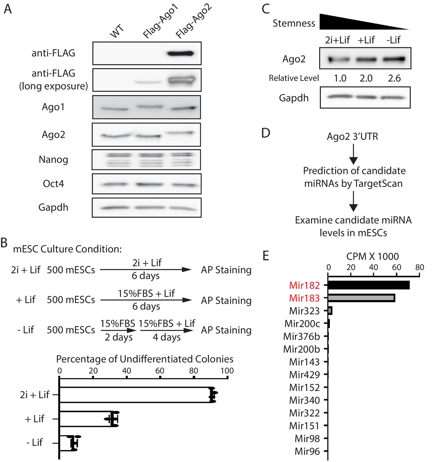
Ago2 is the major developmentally regulated Argonaute protein in mouse embryonic stem cells (mESCs).
(A) Western blotting in the wild-type (WT), Flag-Ago1, and Flag-Ago2 mESCs. (B) Colony formation assay for the mESCs. The WT mESCs were cultured under the indicated conditions, and the resultant colonies were fixed and stained for AP (alkaline phosphatase activity). The results represent the means (± SD) of four independent experiments. (C) Western blotting in the WT mESCs cultured under the indicated conditions. (D) Outline of identifying miRNAs that can potentially regulate Ago2. (E) Expression levels of the identified miRNAs from (D) in mESCs. CPM: counts per million reads.
-
Figure 1—source data 1
Tiff files of raw gel images for Figure 1A and C; Figure 1—figure supplement 1C.
Excel files of numbers for Figure 1B and E; Figure 1—figure supplement 1A, D, Figure 1—figure supplement 2.
- https://cdn.elifesciences.org/articles/72289/elife-72289-fig1-data1-v2.zip
Figure 1—figure supplement 1
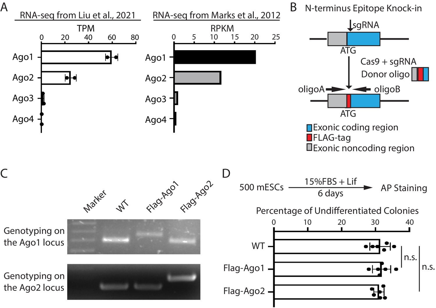
Expression of Argonaute proteins in mouse embryonic stem cells (mESCs).
Figure 1—figure supplement 2
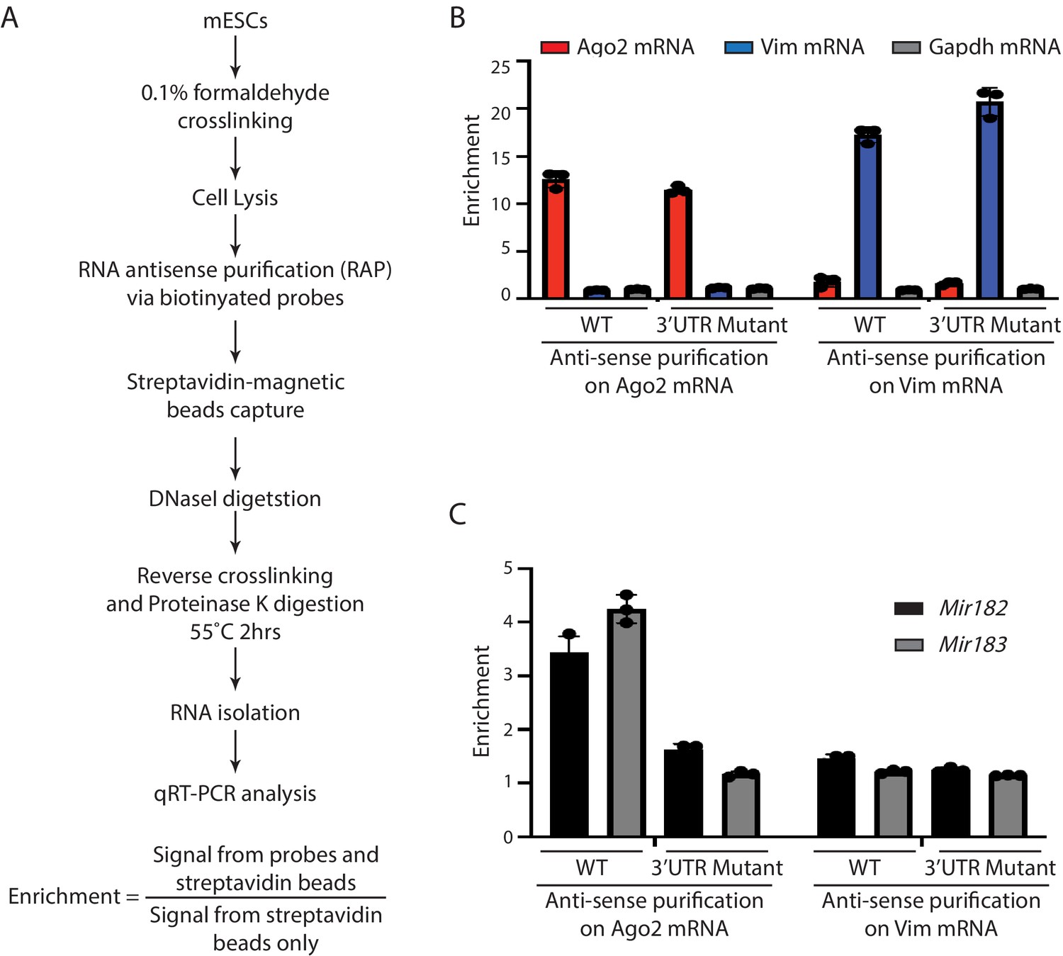
Mir182 and Mir183 are associated with Ago2 mRNA in mouse embryonic stem cells (mESCs).
Figure 2 with 1 supplement
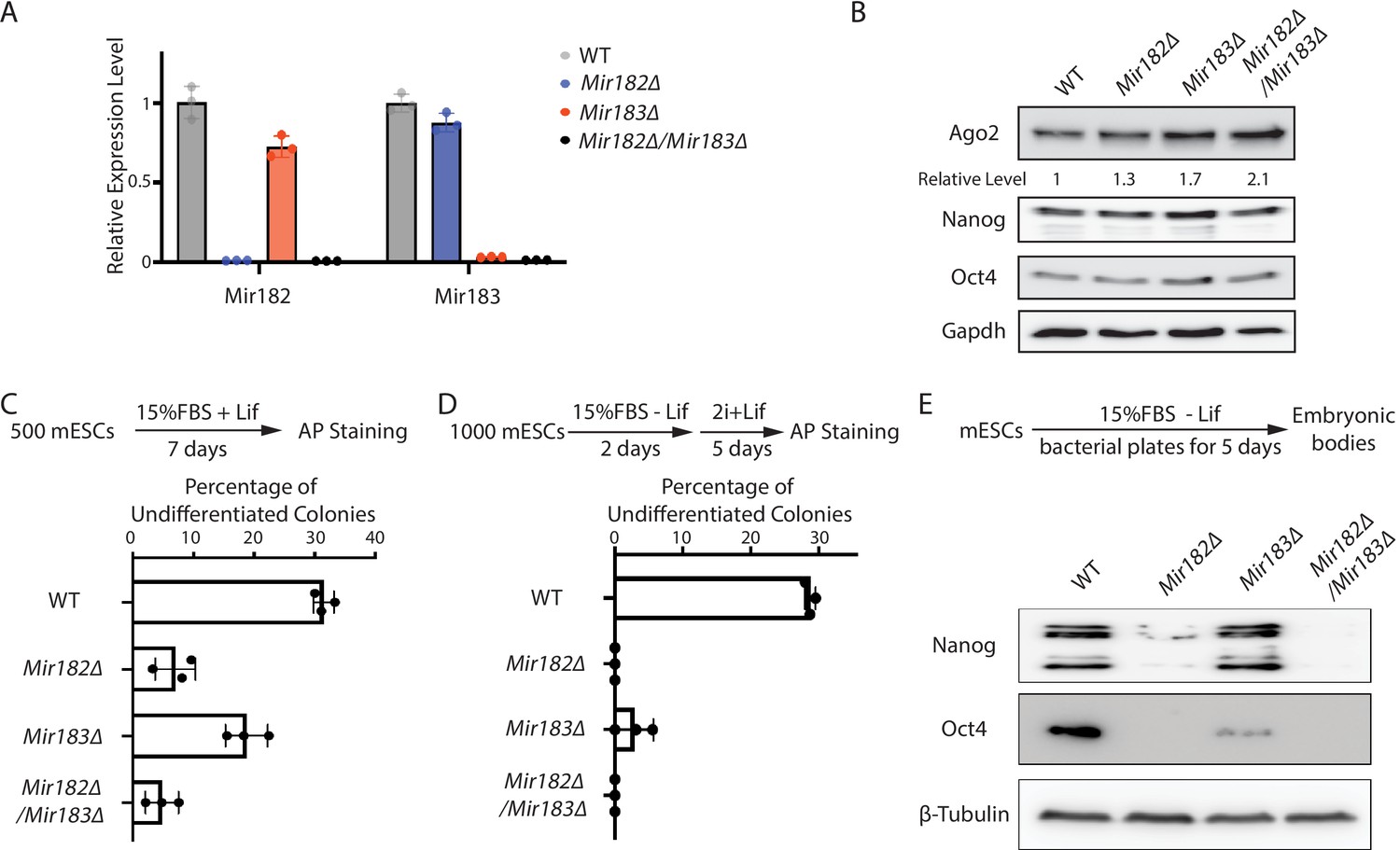
Mir182/Mir183 regulate Ago2 and maintain stemness in mouse embryonic stem cells (mESCs).
(A) qRT-PCR on Mir182 and Mir183. For each miRNA, the expression level in wild-type (WT) cells was set as one for relative comparison. U6 RNA was used for normalization. The results represent the means (± SD) of three independent replicates. (B) Western blotting in the WT, Mir182Δ, Mir183Δ, and Mir182Δ/Mir183Δ mESCs. GAPDH was used for normalization in calculating the relative expression levels. (C) Colony formation assay for mESCs. The mESCs were cultured in 15% FBS+ Lif for 7 days, and the resultant colonies were fixed and stained for alkaline phosphatase (AP). (D) Exit pluripotency assay for mESCs. The mESCs were induced to exit pluripotency in medium without Lif for 2 days and then switched to 2i + Lif medium for 5 days. The resultant colonies were fixed and stained for AP. In (C and D), the colony morphology and AP intensity were evaluated through microscopy; 100–200 colonies were examined each time to determine the percentage of undifferentiated colonies. The results represent the means (± SD) of three independent experiments. (E) Western blotting of pluripotency factors during embryoid body (EB) formation.
-
Figure 2—source data 1
Tiff files of raw gel images for Figure 2B and E; Figure 2—figure supplement 1C.
Excel files of numbers for Figure 2A, C and D; Figure 2—figure supplement 1B.
- https://cdn.elifesciences.org/articles/72289/elife-72289-fig2-data1-v2.zip
Figure 2—figure supplement 1

Ago2 mRNA is a target of Mir182 and Mir183 in mouse embryonic stem cells (mESCs).
Figure 3 with 1 supplement
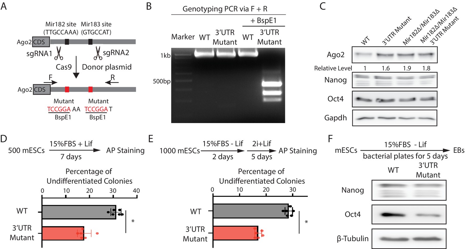
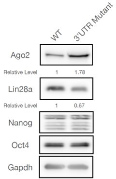
Mir182/Mir183-mediated repression of Ago2 is required for maintaining pluripotency.
(A) Mutating Mir182- and Mir183-binding sites in Ago2 mRNA’s 3’UTR via genome editing. (B) Genotyping of the Ago2 3’UTR mutant. The PCR was performed using the oligos (F and R) indicated in (A). (C) Western blotting in the wild-type (WT), Ago2 3’UTR mutant, Mir182Δ/ Mir183Δ, and Mir182Δ/ Mir183Δ/Ago2 3’UTR mutant. (D) Colony formation assay for mouse embryonic stem cells (mESCs). (E) Exit pluripotency assay for mESCs. In (D and E), the colony morphology and alkaline phosphatase (AP) intensity were evaluated through microscopy. The results represent the means (± SD) of four independent experiments. *p < 0.05 by the Student’s t-test. Western blotting of pluripotency factors in day 5 embryoid bodies (EBs).
-
Figure 3—source data 1
Tiff files of raw gel images for Figure 3C and F; Figure 3—figure supplement 1B.
Excel files of numbers for Figure 3D and E; Figure 3—figure supplement 1A, C.
- https://cdn.elifesciences.org/articles/72289/elife-72289-fig3-data1-v2.zip
Figure 3—figure supplement 1
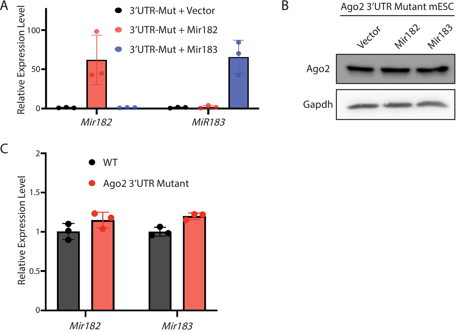
Inhibition of Mir182/Mir183-mediated regulation of Ago2 in mouse embryonic stem cells (mESCs).
Figure 4
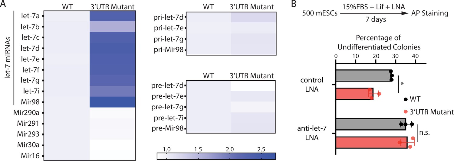
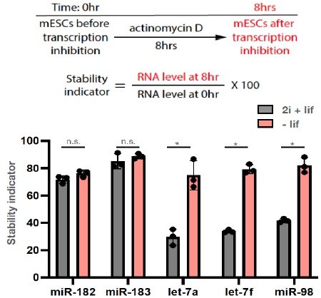
The stemness defects in the 3’UTR mutant mouse embryonic stem cells (mESCs) are caused by elevated let-7 microRNAs (miRNAs).
(A) Relative levels of miRNAs, let-7 pri-miRNAs, and let-7 pre-miRNAs in the wild-type (WT) and the Ago2 3’UTR mutant mESCs. For each miRNA, pri-miRNA, and pre-miRNA, the expression level in WT cells was set as one for relative comparison. U6 RNA was used for normalization in miRNA and pre-miRNA quantification; 18 S rRNA was used for normalization in pri-miRNA quantification. The heatmap was generated from the means of three independent replicates. (B) Colony formation assay for WT and the Ago2 3’UTR mutant mESCs cultured in the presence of 500 nM anti-let-7 locked nucleic acid (LNA) or a control LNA. The results represent three independent experiments. *p < 0.05, and n.s. not significant (p > 0.05) by the Student’s t-test.
-
Figure 4—source data 1
Excel files of numbers for Figure 4A and B.
- https://cdn.elifesciences.org/articles/72289/elife-72289-fig4-data1-v2.zip
Figure 5 with 1 supplement
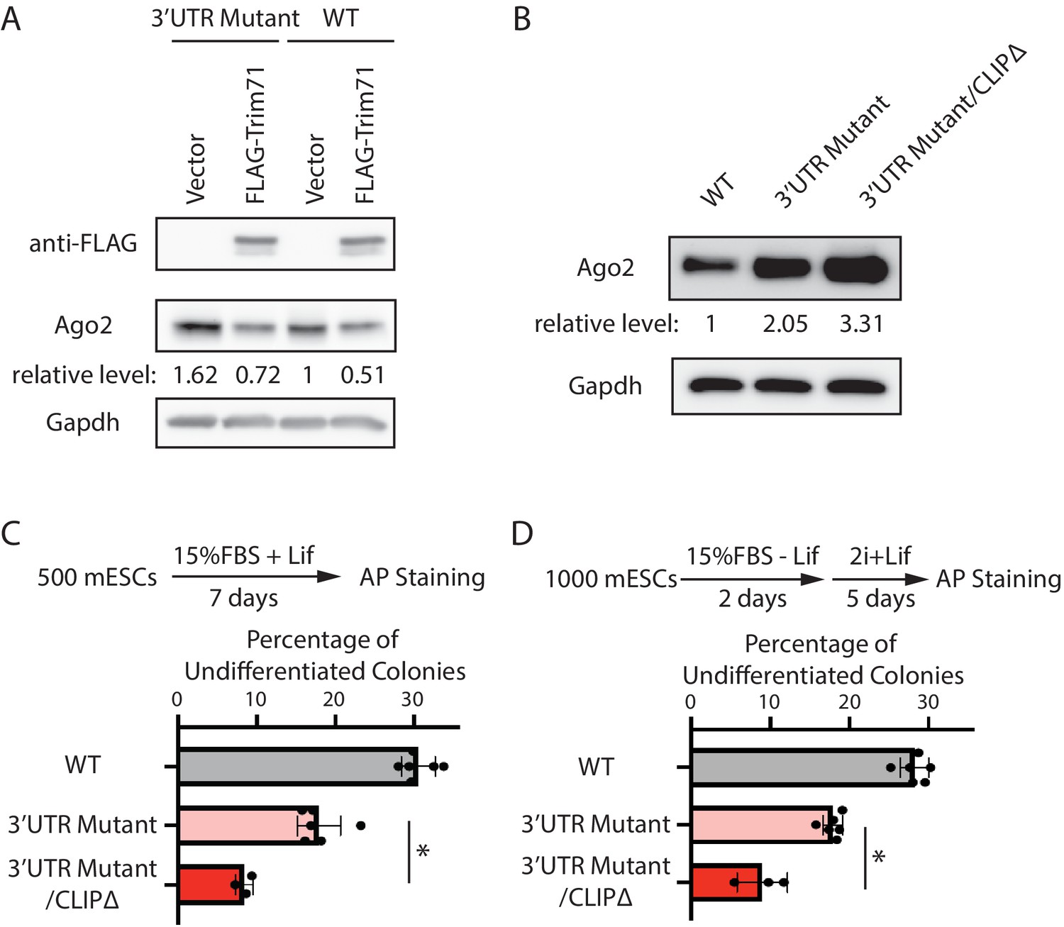
Mir182/Mir183 and Trim71 function in parallel to repress Ago2 mRNA in mouse embryonic stem cells (mESCs).
(A) Western blotting in the wild-type (WT) mESCs expressing either a vector or FLAG-Trim71 and in the 3’UTR mutant mESCs expressing either a vector or FLAG-Trim71. (B) Western blotting in the WT, 3’UTR mutant, and 3’UTR mutant/CLIPΔ mESCs. In (A and B), GAPDH was used for normalization in calculating the relative expression levels. (C) Colony formation assay for mESCs. (D) Exit pluripotency assay for mESCs.
-
Figure 5—source data 1
Tiff files of raw gel images for Figure 5A and B; Figure 5—figure supplement 1.
An Excel file of numbers for Figure 5C and D.
- https://cdn.elifesciences.org/articles/72289/elife-72289-fig5-data1-v2.zip
Figure 5—figure supplement 1

Generation of the CLIPΔ in the 3’UTR mutant mouse embryonic stem cells (mESCs).
Author response image 1
Author response image 2
Tables
Key resources table
| Reagent type (species) or resource | Designation | Source or reference | Identifiers | Additional information |
|---|---|---|---|---|
| Antibody | (Mouse monoclonal) anti-FLAG M2 | Sigma-Aldrich | Cat# F1804 | WB (1:5000) |
| Antibody | (Mouse monoclonal) anti-GAPDH (6 C5) | Santa Cruz Biotechnology | Cat# sc-32233 | WB (1:5000) |
| Antibody | (Rabbit monoclonal) anti-beta-Tubulin | Selleckchem | Cat# A5032 | WB (1:5000) |
| Antibody | (Rabbit monoclonal) anti-Ago1 (D84G10) | Cell Signaling Technology | Cat# 5053 | WB (1:1000) |
| Antibody | (Rabbit monoclonal) anti-Ago2 | Bimake | Cat# A5701 | WB (1:3000) |
| Antibody | (Mouse monoclonal) anti-Oct-4 | BD Transduction Laboratories | Cat# 611202 | WB (1:5000) |
| Antibody | (Rabbit monoclonal) anti-Nanog (D2A3) | Cell Signaling Technology | Cat# 8822 | WB (1:3000) |
| Antibody | Goat Anti-Rabbit IgG (H L)-HRP Conjugate | Bio-Rad | Cat# 170-6515 | WB (1:5000) |
| Antibody | Goat Anti-Mouse IgG (H L)-HRP Conjugate | Bio-Rad | Cat# 170-6516 | WB (1:5000) |
| Chemical compound, drug | DMEM/F-12 | Gibco | Cat# 12500096 | |
| Chemical compound, drug | FBS | Millipore | Cat# ES-009-B | |
| Chemical compound, drug | mLIF | Millipore | Cat# ESG1107 | |
| Chemical compound, drug | PD0325901 | APExBio | Cat# A3013 | |
| Chemical compound, drug | CHIR99021 | APExBio | Cat# A3011 | |
| Chemical compound, drug | N2 | Millipore | Cat# SCM012 | |
| Chemical compound, drug | N27 | Millipore | Cat# SCM013 | |
| Chemical compound, drug | MEM NEAA | Gibco | Cat# 11140–50 | |
| Chemical compound, drug | Penicillin-Streptomycin | Gibco | Cat# 11548876 | |
| Chemical compound, drug | L-Glutamin | Sigma-Aldrich | Cat# G7513 | |
| Chemical compound, drug | β-Mercaptoethanol | Sigma-Aldrich | Cat# M3148 | |
| Chemical compound, drug | Accutase | Millipore | Cat# SF006 | |
| Chemical compound, drug | Fugene6 | Promega | Cat# E2691 | |
| Chemical compound, drug | Puromycin | Sigma-Aldrich | Cat# P9620 | |
| Chemical compound, drug | Doxycycline | Sigma-Aldrich | Cat# D9891 | |
| Chemical compound, drug | Protease inhibitors | Bimake | Cat# B14001 | |
| Chemical compound, drug | Gelatin | Sigma-Aldrich | Cat# G1890 | |
| Chemical compound, drug | One Step-RNA Reagent | Bio Basic | Cat# BS410A | |
| Chemical compound, drug | DNaseI | NEB | Cat# M0303L | |
| Chemical compound, drug | SuperScript II Reverse Transcriptase | Invitrogen | Cat# 18064014 | |
| Chemical compound, drug | SsoAdvanced Universal SYBR Green Supermix | Bio-Rad | Cat# 1725270 | |
| Chemical compound, drug | Q5 High-Fidelity DNA Polymerase | NEB | Cat# M0491L | |
| Chemical compound, drug | Control LNA | Qiagen | Cat# 339137 | |
| Chemical compound, drug | anti-let-7 LNA | Qiagen | Cat# YFI0450006 | |
| Commercial assay or kit | Alkaline Phosphatase Assay Kit | System Biosciences | Cat# AP100R-1 | |
| Commercial assay or kit | Gibson Assembly Master Mix | NEB | Cat# E2611L | |
| Commercial assay or kit | Pierce BCA Protein Assay Kit | Thermo Fisher Scientific | Cat# 23225 | |
| Commercial assay or kit | Mir-X miRNA First Strand Synthesis Kit | Takara | Cat# 638313 | |
| Cell line (Mus musculus) | ES-E14TG2a mESC | ATCC | CRL-1821 | |
| Cell line (Mus musculus) | FLAG-Ago1 mESC | This paper | ||
| Cell line (Mus musculus) | FLAG-Ago2 mESC | PMID:33599613 | ||
| Cell line (Mus musculus) | Mir182∆ mESC | This paper | ||
| Cell line (Mus musculus) | Mir183∆ mESC | This paper | ||
| Cell line (Mus musculus) | Mir182∆/Mir183∆ mESC | This paper | ||
| Cell line (Mus musculus) | 3'UTR Mutant mESC | This paper | ||
| Cell line (Mus musculus) | Mir182∆/Mir183∆/3'UTR Mutant mESC | This paper | ||
| Recombinant DNA reagent | PiggyBac-based dox-inducible expression vector | PMID:33599613 | pWH406 | |
| Recombinant DNA reagent | Inducible GFP expressing vector | PMID:33599613 | pWH1055 | |
| Recombinant DNA reagent | Inducible mouse Mir182 expressing vector | This paper | pWH1039 | |
| Recombinant DNA reagent | Inducible mouse Mir183 expressing vector | This paper | pWH1040 | |
| Recombinant DNA reagent | sgRNA and Cas9 expressing vector (pX458) pWH464 | Addgene | Cat# 48138 | |
| Recombinant DNA reagent | Super PiggyBac Transposase expressing vector (pWH252) | System Biosciences | Cat# PB210PA-1 |
Additional files
-
Supplementary file 1
Antibodies, plasmids, and oligonucleotides used in this study.
- https://cdn.elifesciences.org/articles/72289/elife-72289-supp1-v2.xlsx
-
Transparent reporting form
- https://cdn.elifesciences.org/articles/72289/elife-72289-transrepform1-v2.docx
Download links
A two-part list of links to download the article, or parts of the article, in various formats.
Downloads (link to download the article as PDF)
Open citations (links to open the citations from this article in various online reference manager services)
Cite this article (links to download the citations from this article in formats compatible with various reference manager tools)
microRNA-mediated regulation of microRNA machinery controls cell fate decisions
eLife 10:e72289.
https://doi.org/10.7554/eLife.72289


