Altered regulation of Ia afferent input during voluntary contraction in humans with spinal cord injury
- Shirley Ryan AbilityLab, Northwestern University, and Edward Hines Jr., VA Medical Center, United States
Figures
Figure 1
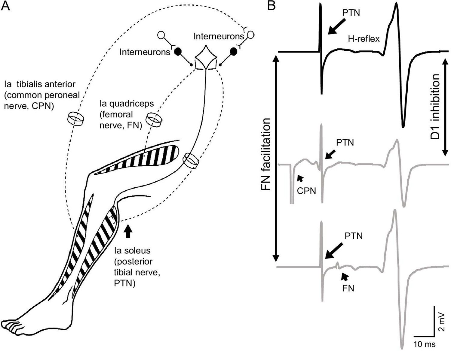
Experimental setup.
(A) Schematic representation of afferent fibers and motor neurons stimulated during our procedures. The soleus reflex was evoked by electrical stimulation of Ia afferents on the posterior tibial nerve (PTN). We assessed Ia afferent input to motor neurons by measuring the depression of the soleus H-reflex evoked by stimulating Ia afferents on the common peroneal nerve (CPN; referred as ‘D1 inhibition’) and the monosynaptic Ia facilitation of the soleus H-reflex evoked by stimulating Ia afferents on femoral nerve (referred as ‘FN facilitation’) at rest, and during tonic voluntary contraction. (B) Representative traces showing the soleus H-reflex evoked by PNT stimulation, the D1 inhibition evoked by stimulation of the CPN preceding the PTN at a conditioning-test interval of 15 ms, and the FN facilitation evoked by stimulation of the FN after the PTN at a conditioning-test interval of –8 ms (negative value of the interval indicates that the stimuli to the PTN precedes the FN stimuli).
Figure 2
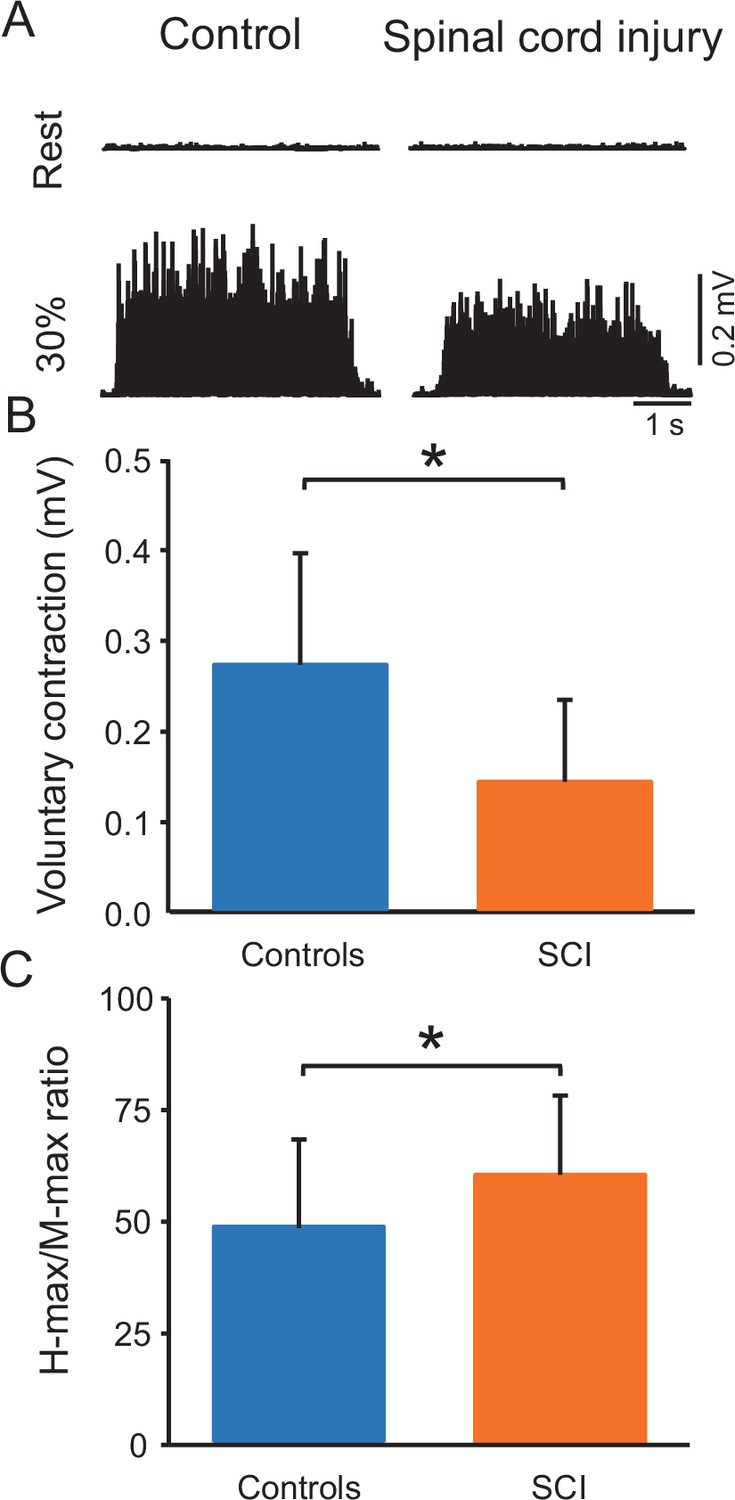
Voluntary contraction and maximal H-reflex and maximal motor response (M-max) ratio (H-max/M-max ratio).
(A) Electromyographic (EMG) traces tested at rest and during 30% of maximal voluntary contraction (MVC) with the soleus muscle in a control and in a spinal cord injury (SCI) participant. (B) Bar graph shows the MVC group data. The abscissa shows the groups tested (controls = blue bar, SCI = orange bar) and the ordinate shows the MVC (in millivolts). (C) Bar graph shows the H-max/M-max ratio group data. The abscissa shows the groups tested (controls = blue bar, SCI = orange bar) and the ordinate shows the H-max/M-max ratio. *p<0.05, one-way ANOVA with Holm-Sidak post-hoc analysis. n = 20 per group, error bars show standard diviation (SD).
Figure 3
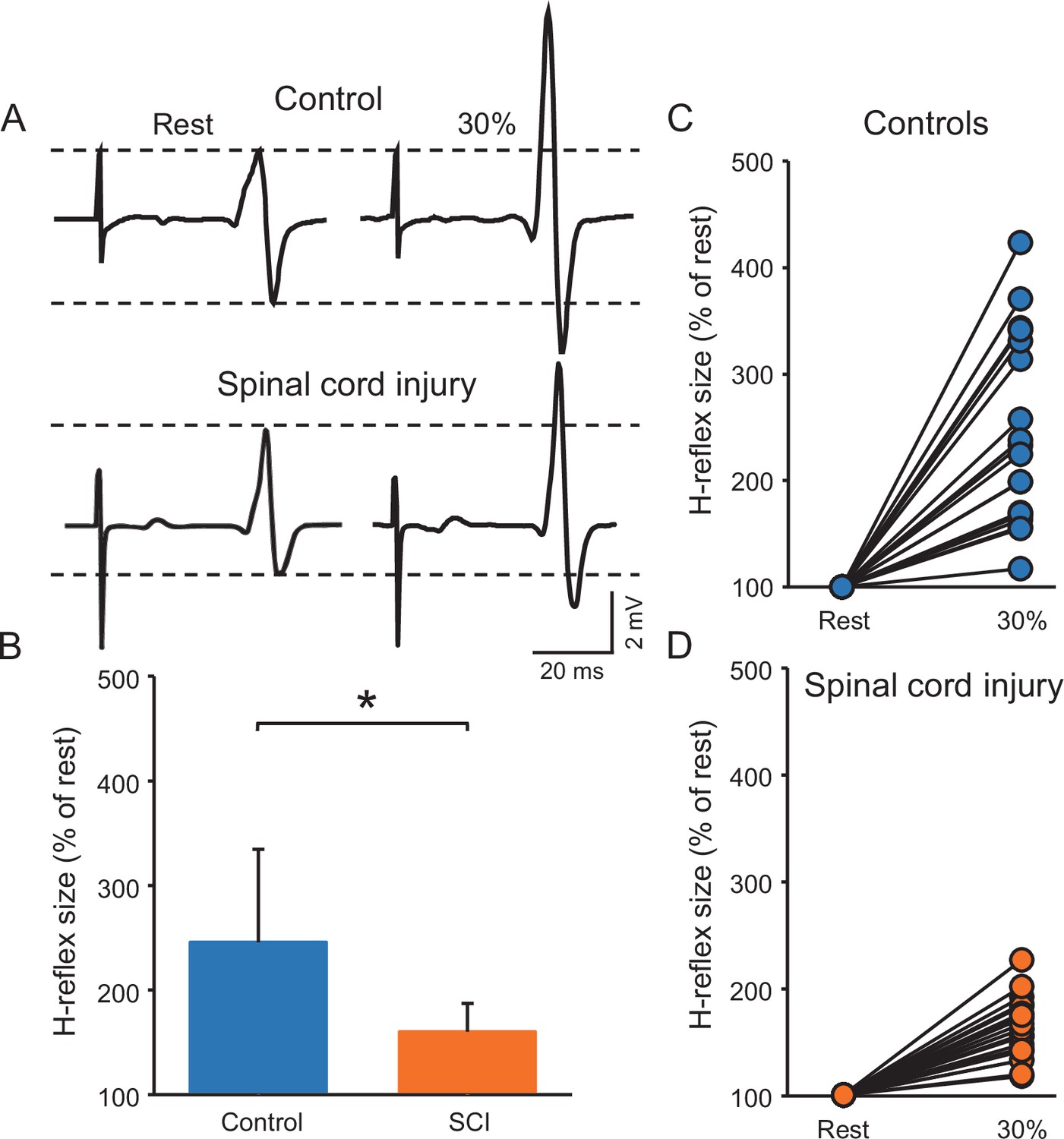
Soleus H-reflex.
(A) Representative EMG traces showing the soleus H-reflex tested at rest and during 30% of MVC in a control and in a SCI participant. (B) Graph shows the group H-reflex data. The abscissa shows the groups tested (controls = blue bar, SCI = orange bar) and the ordinate shows the H-reflex size during 30% of MVC expressed as a % of the H-reflex size at rest. Graphs show individual H-reflex data in controls (C) and SCI (D) participants. The abscissa shows the conditions tested (rest, 30% of MVC) and the ordinate shows the H-reflex size during 30% of MVC expressed as a % of the H-reflex size at rest. *p<0.05, repeated measures ANOVA with Holm-Sidak post-hoc analysis. n = 20 per group, error bars show SD.
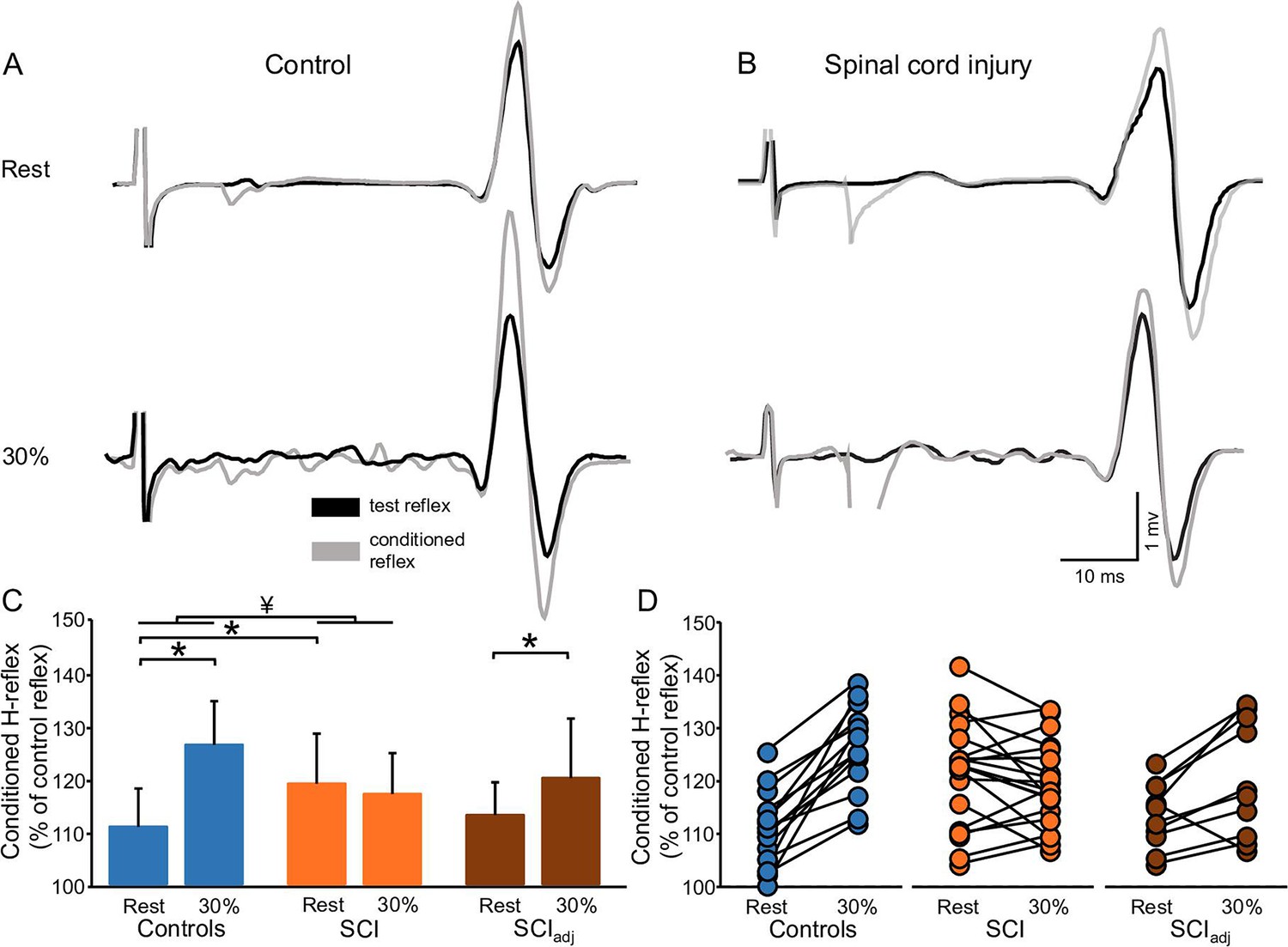
Figure 4
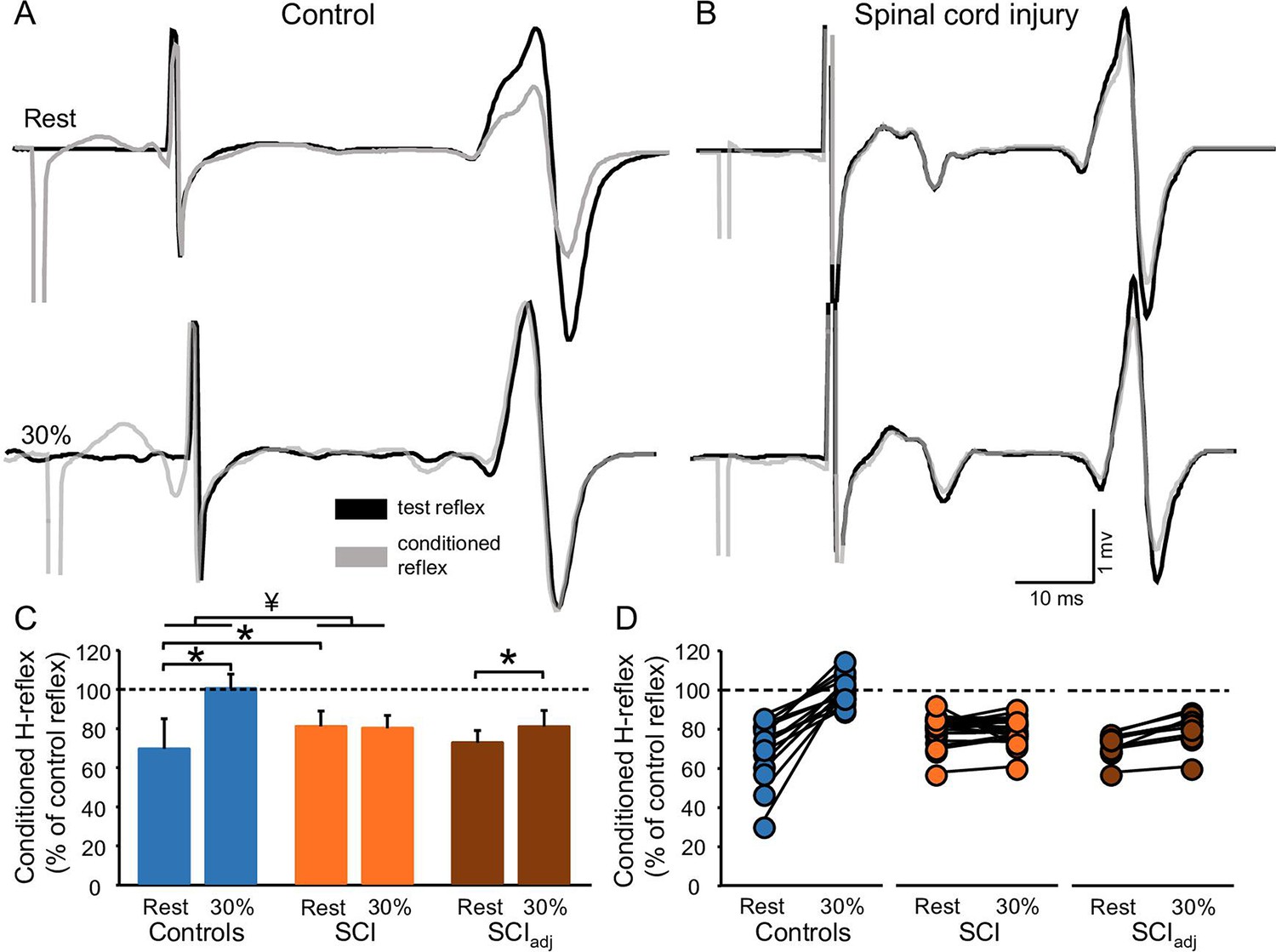
D1 inhibition.
Representative traces showing the H-reflex (control reflex, in black) and the H-reflex conditioned by common peroneal nerve (CPN) stimulation (conditioned H-reflex, in gray) tested at rest and during 30% of MVC in a control (A) and in a SCI (B) participant. Note that during 30% of MVC we show the test H-reflex adjusted. The bar graph shows the conditioned H-reflex normalized to the control H-reflex in both groups (C). The abscissa shows the groups tested at rest and during 30% of MVC (controls = blue bars, SCI = orange bars, SCIadj = brown bars). Note that here the SCIadj condition refers to testing of the D1 inhibitionadj. The ordinate shows the size of conditioned H-reflex expressed as a % of the control H-reflex (use to assess the D1 inhibition). Data from individual subjects (D) showing the conditioned H-reflex normalized to the control H-reflex in all groups tested (controls = blue circles, SCI = orange circles, SCIadj = brown circles). *p<0.05, repeated measures ANOVA with Holm-Sidak post-hoc analysis. Controls n = 14, SCI n = 20, SCIadj n = 10, error bars show SD.
Figure 5
FN facilitation.
Representative traces showing the H-reflex (control reflex, in black) and the H-reflex conditioned by FN stimulation (conditioned H-reflex, in gray) tested at rest and during 30% of MVC in a control (A) and in a SCI (B) participant. Note that during 30% of MVC we show the test H-reflex adjusted. The bar graph shows the conditioned H-reflex normalized to control H-reflex in both groups (C). The abscissa shows the groups tested at rest and during 30% of MVC (controls = blue bars, SCI = orange bars, SCIadj = brown bars). Note that here the SCIadj condition refers to the testing of the FN facilitationadj. The ordinate shows the size of the conditioned H-reflex expressed as a % of the control H-reflex (used to assess the FN facilitation). Data from individual subjects (D) showing the conditioned H-reflex normalized to the control H-reflex in all groups tested (controls = blue circles, SCI = orange circles, SCIadj = brown circles). *p<0.05, repeated measures ANOVA with Holm-Sidak post-hoc analysis. Controls n = 14, SCI n = 20, SCIadj n = 10, error bars show SD.
Figure 6

Correlation.
Graphs show individual data from controls (blue circles) and SCI (orange circles) participants. The abscissa shows the size of the H-reflex during 30% of MVC expressed as a % of the H-reflex tested at rest (A and B) and the ordinate shows the D1 inhibition during 30% of MVC expressed as a % of the D1 inhibition tested at rest (A), the FN facilitation during 30% of MVC expressed as a % of the FN facilitation tested at rest (B). Data from individual subjects (C) showing the correlation between the FN facilitation during 30% of MVC expressed as a % of the FN facilitation tested at rest (C, abscissa) and the D1 inhibition during 30% of MVC expressed as a % of the D1 inhibition tested at rest (C, ordinate). Dashed lines represent the regression line of all the data points included in the plot. *p<0.05, Controls n = 14, SCI n = 20.
Tables
Table 1
Spinal cord injury (SCI) participants.
| ID | Age (years) | Gender | AIS | MAS | Level | Time post injury (years) | Medication |
|---|---|---|---|---|---|---|---|
| 1 | 61 | F | D | 1 | T8 | 19 | NO |
| 2 | 48 | M | D | 3 | C4 | 12 | BAC |
| 3 | 56 | M | D | 2 | C4 | 9 | GAB |
| 4 | 41 | M | D | 2 | C3 | 2 | BAC,GAB |
| 5 | 46 | M | D | 3 | C5 | 8 | BAC,GAB |
| 6 | 30 | M | C | 1 | C2 | 8 | NO |
| 7 | 37 | M | D | 4 | C7 | 10 | NO |
| 8 | 71 | M | D | 3 | T10 | 6 | NO |
| 9 | 73 | M | C | 1 | C5 | 8 | NO |
| 10 | 38 | M | D | 2 | C5 | 22 | NO |
| 11 | 33 | M | D | 2 | C5 | 8 | NO |
| 12 | 64 | M | C | 2 | C3 | 40 | BAC,GAB |
| 13 | 42 | M | D | 1 | C5 | 11 | NO |
| 14 | 63 | M | C | 1 | C5 | 6 | TIZ |
| 15 | 59 | M | D | 1 | C7 | 11 | BAC |
| 16 | 28 | M | D | 4 | C4 | 1 | BAC |
| 17 | 19 | M | D | 2 | C5 | 2 | NO |
| 18 | 80 | M | D | 1 | C4 | 9 | NO |
| 19 | 58 | F | C | 1 | C3 | 5 | GAB |
| 20 | 72 | M | D | 0 | C3 | 12 | NO |
-
M = male; F = female; AIS = American Spinal Injury Association impairment scale; MAS = Modified Ashworth Scale; BAC = baclofen; GAB = gabapentin; DIA = diazepam; TIZ = tizanidine.
Additional files
Download links
A two-part list of links to download the article, or parts of the article, in various formats.
Downloads (link to download the article as PDF)
Open citations (links to open the citations from this article in various online reference manager services)
Cite this article (links to download the citations from this article in formats compatible with various reference manager tools)
Altered regulation of Ia afferent input during voluntary contraction in humans with spinal cord injury
eLife 11:e80089.
https://doi.org/10.7554/eLife.80089


