SIRT-1 is required for release of enveloped enteroviruses
- Department of Microbiology and Immunology and Center for Pathogen Research, University of Maryland, Baltimore, United States
Figures
Figure 1 with 3 supplements

Enterovirus D68 (EV-D68) infection changes SIRT-1’s subcellular localization.
H1HeLa cells were transfected with either scramble control or SIRT-1 siRNAs for 48 hr. The cells were then infected with EV-D68 (multiplicity of infection [MOI = 0.1]) for 5 hr. The intracellular (A) and extracellular (B) particles were collected for plaque assay. (C) Cells were transfected with the indicated plasmids for 24 hr before being infected with EV-D68 (MOI = 0.1) for 5 hr. The extracellular particles were collected and analyzed by a plaque assay. (D) H1HeLa cells were infected for the indicated time points for immunofluorescence analysis (IFA) against SIRT-1. (E) H1HeLa cells were infected (MOI = 30) for 4 hr. Lysates were collected for western blot against SIRT-1. (F) Densitometry quantitation of E. Error bars denote the mean ± standard error of the mean (SEM) of three independent repeats. Unpaired Student’s t-test was used for the statistical analyses (**p < 0.01; *p ≤ 0.05; ns = not significant). Scale bar = 6.2 µm.
Figure 1—figure supplement 1

SIRT-1 in LC3 puncta formation and virus release.
H1HeLa cells were pretreated with 50 µM EX527 for 24 hr, followed by 0.1 MOI enterovirus D68 (EV-D68) infection. The intracellular (A) and extracellular (B) viral titers were determined by a plaque assay. (C) H1HeLa cells were transfected with scramble control or SIRT-1 siRNA for 24 hr, after which the cells were reseeded and transfected with the DNA constructs for 24 hr. The cells were then infected with EV-D68 (MOI = 0.1) for 5 hr. A plaque assay was used to determine the viral titer. (D) Cells were treated as in C for a western blot (E) H1HeLa cells were transfected with either scramble or SIRT-1 siRNA for 48 hr. GFP-LC3 transfection was initiated at 24 hr post-siRNA transfection for 24 hr. The cells were then fixed, and images were acquired with a revolve microscope (**p < 0.01; *p ≤ 0.05; ns = not significant). n = 3 independent experiments. Scale bar = 6 µm.
Figure 1—figure supplement 2

Leptomycin B (LMB) inhibits Transcription Factor EB (TFEB) nuclear export but not SIRT-1 translocation during enterovirus D68 (EV-D68) infection.
Scale bar = 6 µm. (A) Cells were left untreated, starved, starved, and then refeed with complete media for 1 hr, or starved and refeed with complete media containing 10 nM leptomycin B for 1 hr. The cells were fixed, and immunofluorescence analysis (IFA) was performed against endogenous TFEB. (B) Cells were pretreated with 50 nM of LMB for 2 hr. The cells were then infected (MOI = 30) for 30 min (adsorption) and incubated with and without LMB until the end of the infection (4 hr) for IFA against SIRT-1. Starvation for 4 hr was included as a control.
Figure 1—figure supplement 3
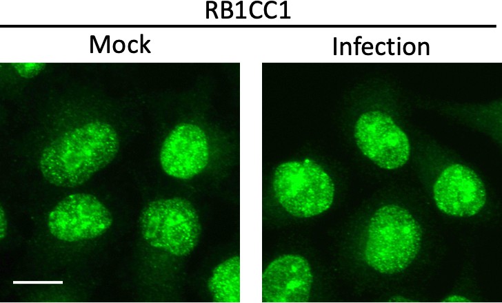
Enterovirus D68 (EV-D68) infection does not alter RB1CC1 subcellular localization.
H1HeLa cells were mock-infected or infected with EV-D68 at an MOI of 30 for 4 hr, before subjecting the cells to immunofluorescence analysis (IFA) against RB1CC1. Scale bar = 10 µm.
Figure 2 with 1 supplement
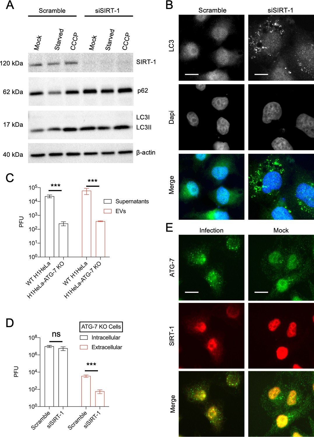
SIRT-1 promotes autophagy but decreases enterovirus D68 (EV-D68) extracellular titers in autophagy-deficient ATG-7 KO cells.
(A) H1HeLa cells were transfected with scramble or SIRT-1 siRNA for 48 hr. The cells were subsequently starved or treated with CCCP (10 µM) for 4 hr. Lysates were harvested and analyzed by western blot. (B) Cells were transfected with the indicated siRNAs for 48 hr before being fixed and subjected to immunofluorescence analysis (IFA) against endogenous LC3. (C) H1HeLa and ATG-7 KO cells were infected with 0.1 MOI of EV-D68 for 5 hr. The extracellular vesicles were isolated as described in the materials and methods and viral titers were determined by a plaque assay. (D) ATG-7 KO cells were transfected with scramble or SIRT-1 siRNAs for 48 hr and infected as in C, followed by plaque assay-based viral titer determination. (E) H1Hela cells were infected with EV-D68 (MOI = 30) for 4 hr. The cells were fixed and immuno-stained with antibodies against SIRT-1 and ATG-7. Error bars denote the mean ± standard error of the mean (SEM) of three independent repeats. Unpaired Student’s t-test was used for the statistical analyses (***p < 0.001; ns = not significant). Scale bar = 6 µm.
Figure 2—figure supplement 1

SIRT-1 does not colocalize with p62 during starvation.
(A) H1HeLa cells were starved for the indicated periods, fixed, and subjected to immunofluorescence analysis (IFA) against SIRT-1. Scale bar = 6 µm. (B) H1HeLa cells were treated with and without the Axe starvation media for 2 hr. The cells were then fixed and immunostained using antibodies against SIRT-1 and p62. Scale bar = 10 µm.
Figure 3
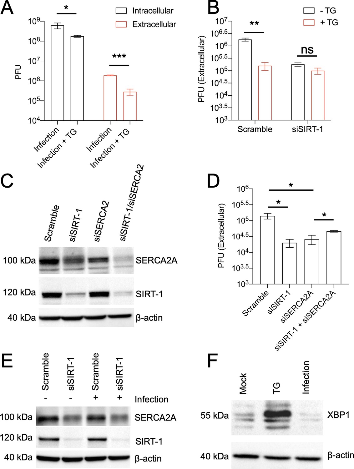
The proviral activity of SIRT-1 is mediated through endoplasmic reticulum stress (ER stress).
(A) H1HeLa cells were infected with enterovirus D68 (EV-D68) (MOI = 0.1) for 30 min. The cells were washed and replenished with complete media with or without 2 µM thapsigargin (TG) for 5 hr. Viral titers were determined by plaque assay. (B) Cells were transfected with either scramble control siRNA or SIRT-1 siRNA for 48 hr. The cells were infected and treated as in A, and viral extracellular titers were similarly measured by plaque assay. Cells were transfected with the indicated siRNAs for 48 hr for western blot (C) or viral titer determination (D) following EV-D68 infection (MOI = 0.1) for 5 hr. (E) H1HeLa cells were transfected for 48 hr with the indicated siRNAs, and either mock-infected or infected (with EV-D68 MOI = 30) for 4 hr. Lysates were collected, and a western blot was performed against the indicated proteins. (F) H1HeLa cells were mock-infected, treated with TG for 4 hr, or infected with EV-D68 (MOI = 30) for 4 hr for western blot against XBP1. Error bars indicate mean ± standard error of the mean (SEM) of at least three independent experiments. Unpaired Student’s t-test was used for the statistical analysis (***p < 0.001; **p < 0.01; *p ≤ 0.05; ns = not significant).
Figure 4

Induction of endoplasmic reticulum stress (ER stress) impairs enterovirus D68 (EV-D68) release in hSABCi-NS1.1 cells.
(A) hSABCi-NS1.1 cells were infected with 0.1 MOI of EV-D68 for 30 min. The cells were washed and replenished with basal media or media containing 2 µM thapsigargin (TG) for 6 hr. Viral titers were determined by plaque assay. (B) hSABCi-NS1.1 cells were mock-infected, infected with EV-D68, and treated with or without TG for 6 hr, or treated with TG alone for 6 hr. The lysates were harvested and a western blot was performed against the indicated proteins. n = 2 independent experiments. (C) hSABCi-NS1.1 were pretreated with 50 µM EX527 for 48 hr. The cells were then infected with EV-D68 (MOI = 0.1) for 6 hr for a plaque assay. (D) hSABCi-NS1.1 cells were mock-infected or infected with EV-D68 (MOI = 30) for 6 hr. The cells were fixed for immunofluorescence analysis (IFA) against endogenous SIRT-1. Error bars represent mean ± standard error of the mean (SEM) (**p < 0.01; *p ≤ 0.05; ns = not significant). Scale bar = 7.5 µm.
Figure 5 with 1 supplement

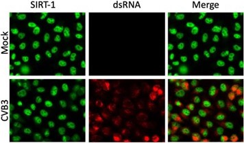
SIRT-1 is critical for poliovirus (PV), but not coxsackievirus B3 (CVB3), release from cells.
(A) H1HeLa cells were mock-infected or infected with PV or CVB3 for 4 hr. The cells were fixed, and immunofluorescence analysis (IFA) was done against SIRT-1 MOI = 30. (B, C) Cells were transfected with scramble or SIRT-1 siRNAs for 48 hr before being infected with PV (MOI = 0.1) for 5 hr. Viral titers were determined by a plaque assay. (D, E) Cells were transfected and infected with CVB3 as in B. n = 3 independent experiments, and error bars represent mean ± standard error of the mean (SEM) (***p < 0.001; *p ≤ 0.05; ns = not significant). Scale bar = 7.5 µm.
Figure 5—figure supplement 1

EX527 pretreatment reduced poliovirus (PV), but not coxsackievirus B3 (CVB3) titers.
H1HeLa cells pretreated with EX527 (50 µM) for 24 hr were infected with PV and CVB3 (MOI = 0.1) for 5 hr. The intracellular and extracellular titers for PV (A, B) and for CVB3 (C, D) were determined by a plaque assay (**p < 0.01; ns = not significant.). n = 3 independent repeats.
Figure 6

SARS-CoV-2 infection requires SIRT-1 and induces SIRT-1 translocation to the cytosol.
(A) A549-ACE2 cells were infected with SAR-CoV-2 (MOI = 0.5) for 24 hr. The cells were fixed and subjected to immunofluorescence analysis (IFA) against SIRT-1 and SARS-CoV-2 nucleoprotein. (B) A549-ACE2 cells were transfected with SIRT-1 siRNA for 48 hr. Lysates were analyzed by western blot. (C) Cells were transfected as in B and infected (MOI 0.01) for 48 hr before being subjected to a plaque assay for viral titer measurement (**p < 0.01). Scale bar = 30 µm. n = 3 independent experiments.
Figure 7 with 1 supplement

SIRT-1 KD reduces extracellular vesicle-mediated release of infectious enterovirus D68 (EV-D68) viral particles.
H1HeLa cells were transfected with SIRT-1 and Scramble siRNAs for 48 hr. (A) Cells were infected with EV-D68 (MOI = 0.1) for 5 hr, and EVs were isolated for viral titer measurement by plaque assay. (B) Cells were transfected as in A, and the whole-cell lysates (WCL) were collected and prepared for western blot against CD63. (C) H1HeLa cells were transfected with the indicated siRNAs for 48 hr. The cells were either left uninfected or infected with MOI 30 of EV-D68 for 4 hr. The EVs and WCL were prepared for western blot against the indicated proteins. (D) Cells were plated on cover slides and transfected with scramble or SIRT-1 siRNA for 48 hr. The cells were then fixed and subjected to immunofluorescence analysis against CD63. Arrows indicate large CD63 puncta. Error bars indicate mean ± standard error of the mean (SEM) of three independent experiments. Unpaired Student’s t-test was used for the statistical analysis (**p < 0.01). Scale bar = 6.5 µm.
Figure 7—figure supplement 1
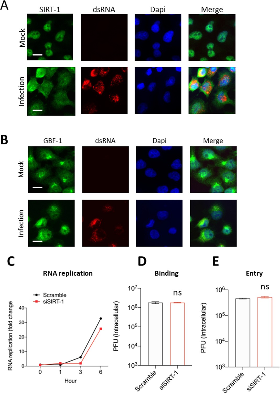
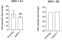
SIRT-1 is not required for enterovirus D68 (EV-D68) entry and replication.
(A) Cells were infected for the indicated time point, fixed, and processed for immunofluorescence analysis (IFA) against SIRT-1 and dsRNA. (B) Cells were infected as in A for IFA against GBF-1, and dsRNA. (C) H1HeLa cells were transfected with scramble or SIRT-1 siRNA for 48 hr. The cells were then infected (MOI = 0.1) for the indicated time point for the quantitative polymerase chain reaction (qPCR) assay. (D, E) Cells were transfected as in B, and virus binding and entry assays were performed as described in the materials and methods (ns = nonsignificant). Scale bar = 6.2 µm.
Author response image 1
Author response image 2
Author response image 3
Additional files
-
MDAR checklist
- https://cdn.elifesciences.org/articles/87993/elife-87993-mdarchecklist1-v1.docx
-
Source data 1
Western blot source data.
- https://cdn.elifesciences.org/articles/87993/elife-87993-data1-v1.zip
Download links
A two-part list of links to download the article, or parts of the article, in various formats.
Downloads (link to download the article as PDF)
Open citations (links to open the citations from this article in various online reference manager services)
Cite this article (links to download the citations from this article in formats compatible with various reference manager tools)
SIRT-1 is required for release of enveloped enteroviruses
eLife 12:RP87993.
https://doi.org/10.7554/eLife.87993.3


