Optogenetic stimulation of the locus coeruleus enhances appetitive extinction in rats
- Department of Psychology, University of Toronto, Canada
- Cell and Systems Biology, University of Toronto, Canada
Figures
Figure 1
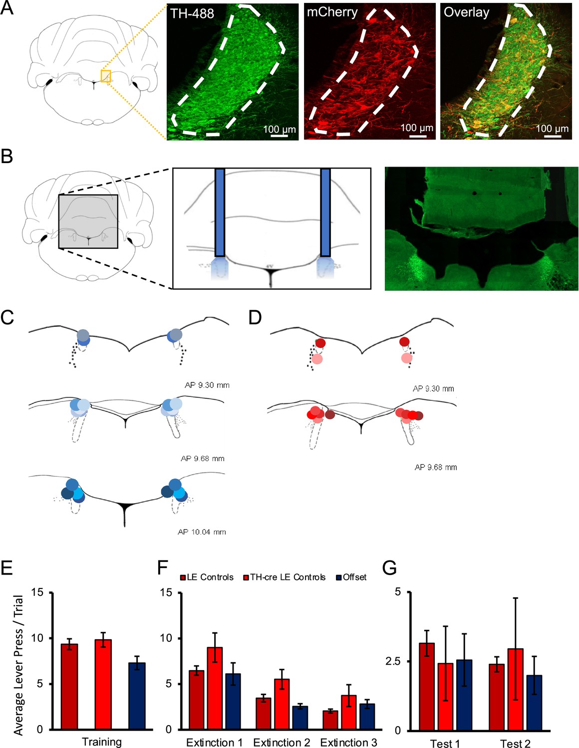
Histological verification of optogenetic targeting of noradrenergic locus coeruleus (LC) neurons.
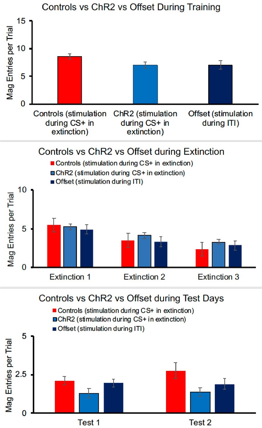
(A) To verify viral expression, the LC was stained with tyrosine hydroxylase (TH) primary antibody along with secondary antibody with Alexa Fluor 488 to identify TH-positive neurons in the region of the LC. The same tissue was counterstained with mCherry primary antibody and enhanced with Alexa Fluor 568 to determine the overlap of virally infected TH-positive cells. Cell counts and calculation of infection rates were conducted on sections of the LC positioned between –9.68 and –10.04 mm in the anterior–posterior plane (Paxinos and Watson, 2007). Each section provided ~116 ± 25 TH-positive LC neurons as determined by DAPI and TH/Alexa Fluor 488 staining. Viral expression was identified by colocalized TH and mCherry expression. Animals that did not have viral expression in the LC were not included in the experimental groups. TH-Cre Long–Evans (LE) rats showed clear viral expression in the LC with approximately 73 ± 2.8% of all TH+ cells in the LC expressing AAV5-EF1a-DIO-ChR2(H134R)-mCherry, with an example provided in the overlay, and 97 ± 2.5% of infected cells staining positive for TH indicating robust and selective targeting of noradrenergic LC neurons (A). Males and females showed very similar infection rates (males, 74%; females, 72%). (B) The approximate probe tip locations in each animal are displayed confirming successful targeting of the LC, with shades of blue representing animals expressing ChR2 (C), and shades of red representing control animals infected with AAV5-EF1a-DIO-mCherry (D). When the behaviour of multiple control groups was compared, TH-Cre and LE rats expressing control virus and receiving light delivery during stimulus presentation in extinction, as well as offset controls expressing ChR2 virus and receiving light delivery during the inter-trial intervals (ITI) of extinction sessions showed no significant differences in response rates on the final day of training (E), during extinction sessions with light delivery (F), or during tests of spontaneous recovery conducted without stimulation the next day (test 1) and again 1 wk later (test 2; G; p>0.05). These multiple control groups are therefore collapsed in subsequent analyses. Error bars represent the standard error of the mean. Please see Results for full reporting of statistical results.
Figure 2
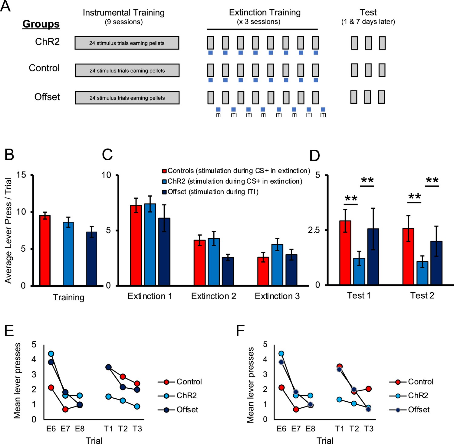
Optogenetic stimulation of the locus coeruleus (LC) enhances long-term expression of extinction.
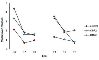
(A) Schematic representation of the study design. All groups received identical discriminated operant training where lever-pressing during 20 s presentations of a discriminative stimulus was reinforced with a food pellet. Responding in the absence of the discriminative stimuli was not reinforced. The groups differed in the period of time in which optical manipulation of the LC took place during extinction sessions with the timing of light delivery indicated by the blue bars; for control and ChR2 groups, light was delivered during stimulus presentations (grey bars) when reward was expected based on previous training, but not delivered; the offset group received similar LC stimulation but offset from stimulus presentations (i.e. during the inter-trial intervals [ITI]). The three groups (control, ChR2, and offset groups) showed similar response rates in training (B; p>0.05). Responding in all groups extinguished when reward was omitted (p<0.05) and no differences were observed between groups (C; p>0.05). When tested the next day for spontaneous recovery, the ChR2 group displayed significantly fewer lever presses than the control or offset groups, suggesting that LC stimulation strengthened extinction learning (D, test 1; p<0.05). A similar effect was observed 1 wk later, indicating a persistent effect of LC stimulation on later retention of extinction (D, test 2; p<0.05). (E, F) To directly test for spontaneous recovery, we compared responding during the last trial of extinction to the first test trial for each group. In test 1, the control group increased responding from the end of extinction to the beginning of the test the following day (E, p<0.001), thus demonstrating spontaneous recovery. A similar pattern was observed in the offset group, although this effect was marginal (p=0.055). Of note, there was no change in responding across time in the ChR2 group (p=0.890) and responding remained low in this group, indicating suppressed spontaneous recovery. To test whether this was a lasting effect, we tested the same rats again 1 wk later where we observed a similar effect of group [test 2; D; F(2,40) = 3.732, p=0.033], resulting again from reduced responding in the ChR2 group compared to the control [t(35) = 2.663, p=0.006] or offset groups [t(19) = 2.117, p=0.024], which did not differ [t(26) = 0.506, p=0.309]. To demonstrate spontaneous recovery, trial data were again considered. The control group increased responding from the end of extinction to the beginning of the test conducted 1 wk later (F; p=0.002), thus demonstrating spontaneous recovery. A similar pattern was observed in the offset group (p=0.049). Of note, there was no change in responding across time in the ChR2 group (p=0.256), suggesting that LC stimulation can enhance extinction learning, increasing its resilience against spontaneous recovery. Error bars represent the standard error of the mean. Please see Results for full reporting of statistical results.
Figure 3
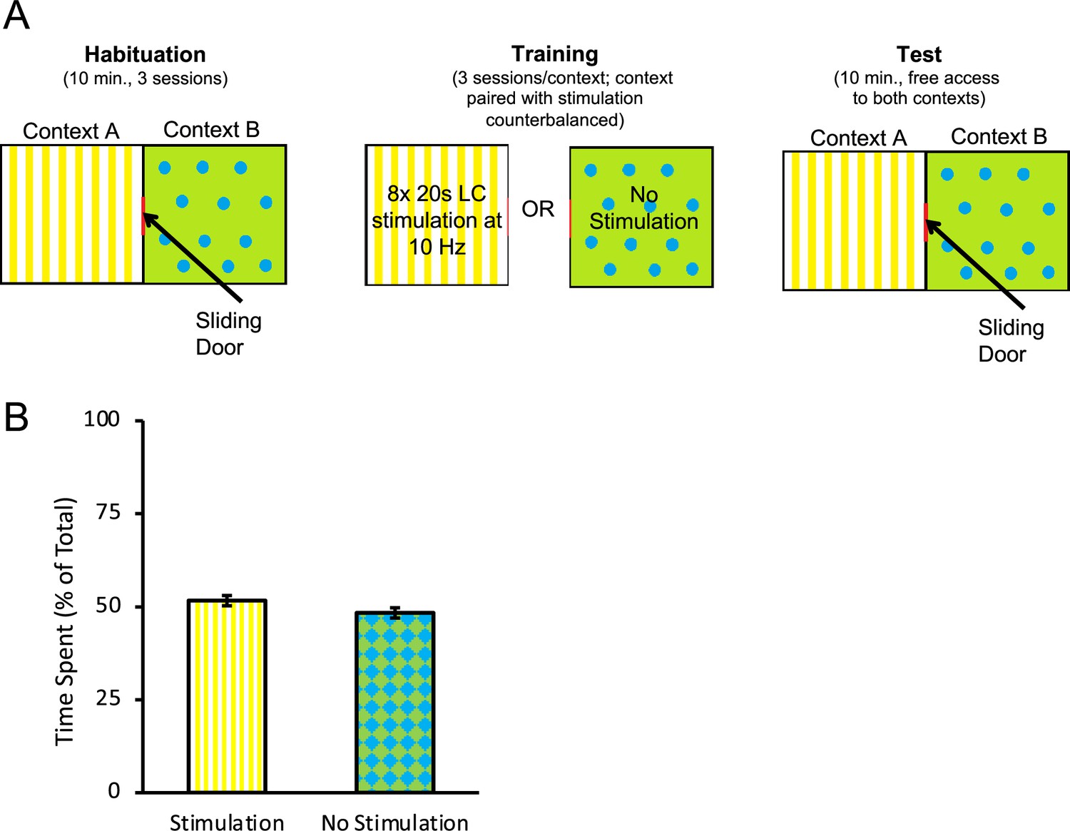
Optogenetic stimulation of the locus coeruleus (LC) does not produce conditioned aversion.
A conditioned place preference/aversion test was performed to determine whether LC stimulation produced aversive conditioning. Rats expressing ChR2 were placed in an apparatus with two chambers separated by a sliding door, depicted in (A). One chamber, labelled context A, had a yellow background with vertical stripes. A second chamber, labelled context B, had a green background with blue circles. All rats were given 3 d to freely explore both chambers for 10 min per day. Afterwards, the door was closed, and rats were confined to one context (A or B, counterbalanced) where they received eight 20 s bouts of LC stimulation at 10 Hz (160 s total stimulation time) within the 10 min session. On alternating days, rats were confined to the other context (B or A, counterbalanced) without stimulation. On the test day, the sliding door opened and rats were given 10 min to freely explore. The total time spent in each context was recorded. A rat was considered to be in one chamber when both hindfeet were inside the chamber. No preference for either context was observed as rats spent a near-equal amount of time in both chambers (B), suggesting no aversive associations with the context formed as a result of optogenetic stimulation of the LC with the parameters used here. Error bars represent the standard error of the mean. Please see Results for full reporting of statistical results.
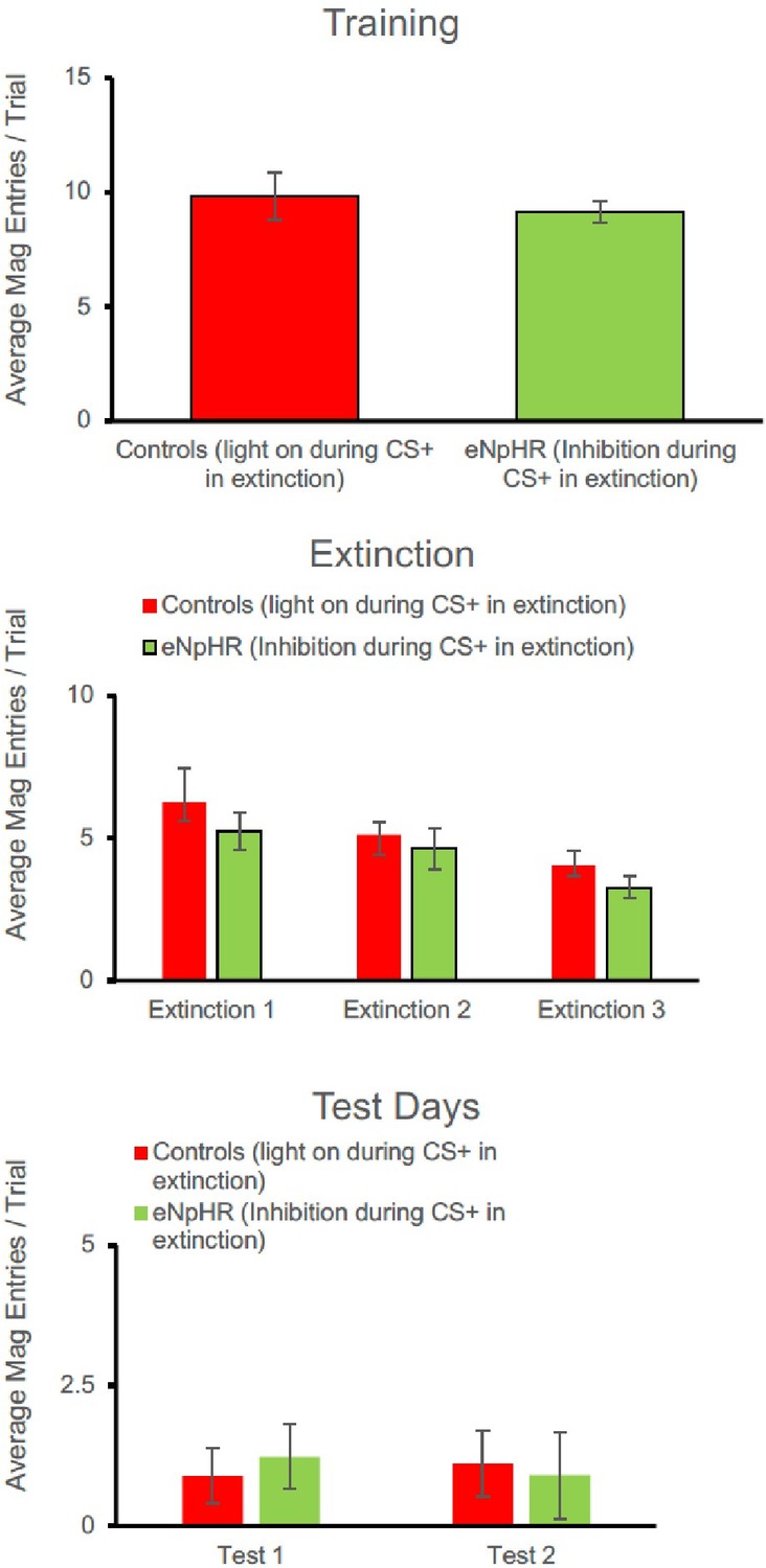
Figure 4
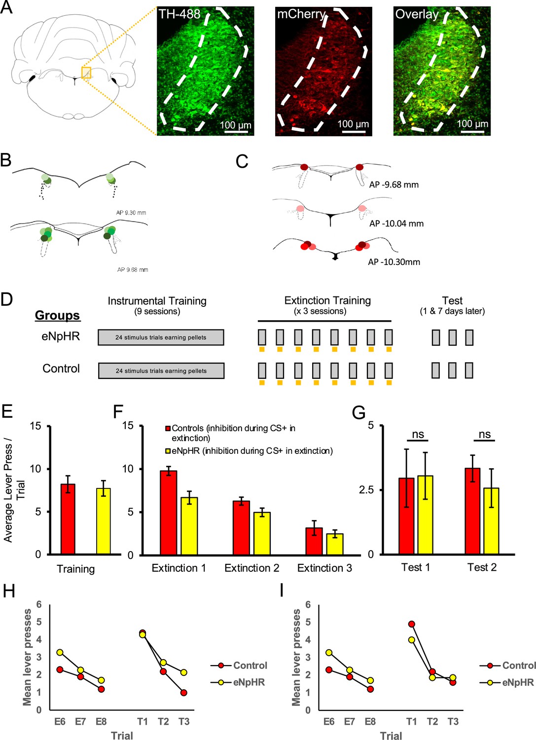
Optogenetic inhibition of the locus coeruleus (LC) did not impair the long-term expression of extinction.
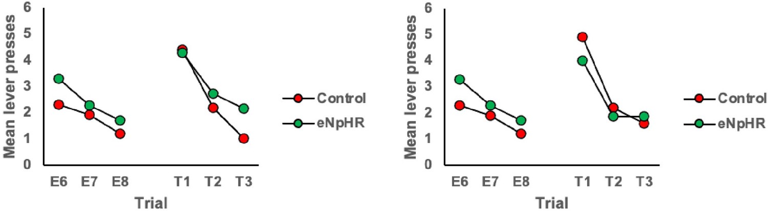
LC neurons were successfully targeted with halorhodopsin-expressing virus (AAV5-EF1a-DIO-eNpHR3.0-mCherry) or a control virus (AAV5-EF1a-DIO-mCherry) as shown in (A). Green represents tyrosine hydroxylase (TH)-positive neurons, red represents virally infected neurons expressing mCherry, and infected TH-positive cells are shown in the overlay (55 ± 8.7% infection rate). The locations of probe tips are shown with shades of green representing animals expressing eNpHR (B) and shades of red representing control animals (C). (D) Schematic representation of training and testing. Grey bars represent stimulus presentations, and yellow bars represent light delivery. Both groups underwent identical discriminated operant training and showed comparable response rates at the end of acquisition (E; p>0.05). Control and eNpHR groups were then given LC light delivery during stimulus trials over three extinction sessions. Photoinhibition of the LC during extinction training reduced mean response rates compared to control rats (F; F(1,13) = 6.899, p=0.021) but this difference did not persist to tests conducted 1 (test 1) and 7 (test 2) d later where no group differences were observed in spontaneous recovery (G; p>0.05). (H, I) To directly test for spontaneous recovery, we compared responding during the last trial of extinction to the first test trial for each test. There was a significant effect of trial with responding increasing from the end of extinction to the beginning of the test the following day (H; p=0.020). There was no effect of group and no trial × group interaction (p=0.780), suggesting that spontaneous recovery was similar in the two groups. Similar results were found for the test conducted 1 wk later where responding increased from extinction to testing, thus demonstrating spontaneous recovery (I; p=0.011). But again there was no effect of group or trial × group interaction. Together these results suggest that spontaneous recovery was observed and retention of extinction was equivalent between groups. Error bars represent the standard error of the mean. Please see Results for full reporting of statistical results.
Author response image 1
Author response image 2

Author response image 3

Author response image 4

Author response image 5

Author response image 6

Author response image 7
Author response image 8
Author response image 9
Author response image 10

Author response image 11
Additional files
Download links
A two-part list of links to download the article, or parts of the article, in various formats.
Downloads (link to download the article as PDF)
Open citations (links to open the citations from this article in various online reference manager services)
Cite this article (links to download the citations from this article in formats compatible with various reference manager tools)
Optogenetic stimulation of the locus coeruleus enhances appetitive extinction in rats
eLife 12:RP89267.
https://doi.org/10.7554/eLife.89267.3


