Co-evolution: Immune to defeat
- University of Texas Southwestern Medical Center, United States
Organisms that grow in close contact with one another often co-evolve. This is particularly evident in pathogens and their hosts, with each always needing to stay one small step ahead of the other in order to survive. This evolutionary competition has given rise to the systems of immune molecules that hosts use to recognize and neutralize invading pathogens, and also to the effector molecules that pathogens use to survive attack by the immune system. A prime example of this can be seen in the interactions between a class of enzymes called the immune-related GTPases (IRG proteins) and the pathogens that they recognize and destroy (Kim et al., 2012). It is known that IRG proteins work by assembling on the surface of the vacuoles within which the pathogens replicate, but the details of this mechanism are poorly understood.
Now, in eLife, Jonathan Howard of the University of Cologne and colleagues present striking evidence that the co-evolution of IRG proteins with Toxoplasma gondii—the parasite that causes toxoplasmosis—has been an important driving force in the functional evolution of the IRG system (Lilue et al., 2013). Howard and colleagues—including Jingtao Lilue as first author, Urs Muller and Tobias Steinfeldt—found that mice derived from wild mice were resistant to strains of the parasite that were lethal to laboratory mice. Remarkably, they demonstrated that this resistance was due to genetic variation in one particular form of IRG protein, named Irgb2-b1: they found that certain alleles of Irgb2-b1 appeared to inhibit the activity of the effector molecules secreted by the parasite, which themselves have evolved to inactivate the IRG system.
As it enters a new host cell, Toxoplasma secretes an active kinase called ROP18 and a diverse family of pseudokinases, called ROP5. The ROP5 pseudokinases appear to sequester the IRG proteins and then present them for phosphorylation by ROP18 (Fleckenstein et al., 2012). The process of phosphorylation inactivates the IRG proteins, which prevents them from assembling on the surface of the vacuole. This, in turn, allows the parasites to replicate (Figure 1; Steinfeldt et al., 2010; Fentress et al., 2010). There are considerable differences in the virulence (to mice) of the major strains of Toxoplasma, and genetic variations in ROP5 and ROP18 can explain almost all of these differences (Saeij et al., 2006; Taylor et al., 2006; Behnke et al., 2011; Reese et al., 2011). This highlights the importance of the IRG system in the control of Toxoplasma infection in mice, and the pressure that competition with this system has placed on the parasite.
Figure 1
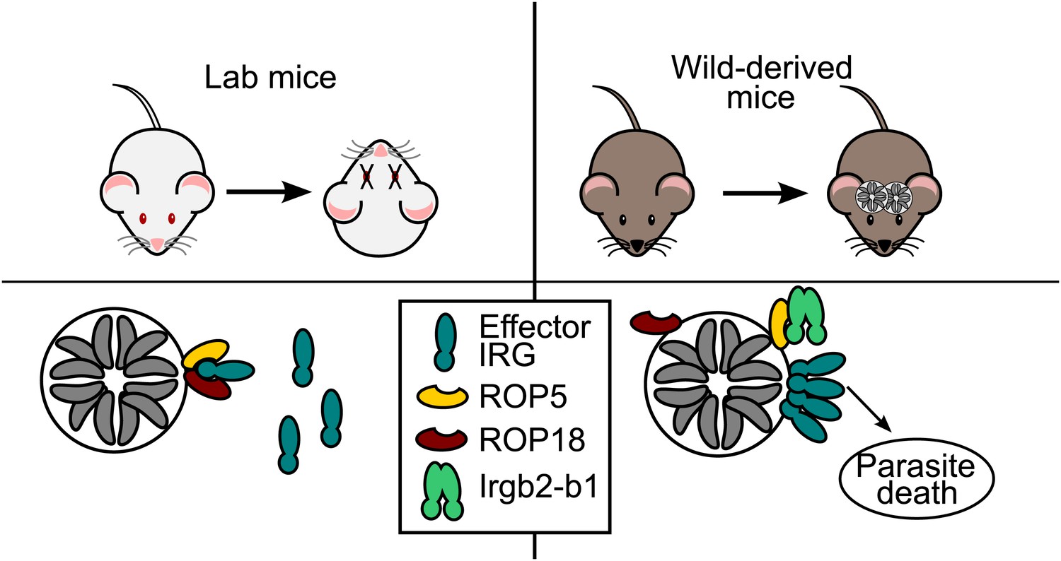
Hosts vs parasites.
Toxoplasma parasites can kill laboratory mice (left) because they secrete effector molecules (ROP5 & ROP18) that prevent the effector IRG proteins (dark green) in the immune system of the mice from doing their job, which is to prevent the Toxoplasma parasites (grey) replicating themselves inside a vacuole. Lilue et al. show that in mice derived from wild mice (right), a particular isoform of an IRG protein (Irgb2-b1) binds to the ROP5 molecules, which means that the effector IRG proteins can assemble on the surface of the vacuole. This allows wild-derived mice to control an otherwise lethal infection in a way that, seemingly paradoxically, enables the survival of both the host and the parasite (which survives inside cysts in the brain and muscle tissue of the host).
As is typical of genes under strong selective pressure, the IRG genes differ both in copy number and in coding sequence in closely related species and even, as described by Lilue et al., among individuals within the same species. Previously the different forms of an IRG protein have been grouped into two distinct functional classes: effector IRG proteins that destroy the vacuoles inside which the pathogens replicate, and regulatory IRGs that are thought to inhibit premature activation of the system (Hunn et al., 2011). However, Irgb2-b1—the form of the protein identified by Lilue et al.—appears to belong to a third class of IRG proteins that recognize a particular pathogen effector rather than the pathogen itself. They found that Irgb2-b1 only attached itself to Toxoplasma vacuoles that were already coated with ROP5. Moreover, Irgb2-b1 loading appeared to block the activity of the ROP5 and ROP18 proteins. This left the effector IRG proteins free to assemble on the surface of the vacuole, thus leading to the destruction of the parasite (Figure 1). Strikingly, mice that carried the protective allele of Irgb2-b1 were able to survive acute infection with a highly virulent strain of the parasite, whereas infection by just one parasite of the same strain would kill a laboratory mouse.
Toxoplasma is also a survivor—as the acute infection ends, the parasite switches to a slow-growing form that becomes enclosed within cysts in the brain and muscle of the host, as was the case with the wild mice studied by Lilue et al. When a host is killed and eaten by a carnivore, the parasite switches back to a fast growing stage to infect its new host. Because strains of the parasite that secrete the virulent versions of both ROP5 and ROP18 are able to completely defeat the IRG system in laboratory mice, it was thought that these strains could not have co-evolved with mice, because the death of the host can be an evolutionary dead-end for a pathogen. The fact that Irgb2-b1 derived from wild mice represents a specific countermeasure to the parasite effectors provides a strong argument for co-evolution. The work of Lilue, Muller, Steinfeldt and Howard thus reminds us to look beyond lab-adapted organisms and examine biology in the wild to discover its full diversity.
References
-
Virulence differences in Toxoplasma mediated by amplification of a family of polymorphic pseudokinasesProc Natl Acad Sci USA 108:9631–9636.https://doi.org/10.1073/pnas.1015338108
-
IFN-inducible GTPases in host cell defenseCell Host Microbe 12:432–444.https://doi.org/10.1016/j.chom.2012.09.007
-
Polymorphic family of injected pseudokinases is paramount in Toxoplasma virulenceProc Natl Acad Sci USA 108:9625–9630.https://doi.org/10.1073/pnas.1015980108
Article and author information
Author details
Publication history
- Version of Record published: October 29, 2013 (version 1)
Copyright
© 2013, Reese
This article is distributed under the terms of the Creative Commons Attribution License, which permits unrestricted use and redistribution provided that the original author and source are credited.
Metrics
-
- 561
- views
-
- 50
- downloads
-
- 0
- citations
Views, downloads and citations are aggregated across all versions of this paper published by eLife.
Download links
A two-part list of links to download the article, or parts of the article, in various formats.
Downloads (link to download the article as PDF)
Open citations (links to open the citations from this article in various online reference manager services)
Cite this article (links to download the citations from this article in formats compatible with various reference manager tools)
Co-evolution: Immune to defeat
eLife 2:e01599.
https://doi.org/10.7554/eLife.01599
Further reading
-
- Microbiology and Infectious Disease
- Structural Biology and Molecular Biophysics
African trypanosomes replicate within infected mammals where they are exposed to the complement system. This system centres around complement C3, which is present in a soluble form in serum but becomes covalently deposited onto the surfaces of pathogens after proteolytic cleavage to C3b. Membrane-associated C3b triggers different complement-mediated effectors which promote pathogen clearance. To counter complement-mediated clearance, African trypanosomes have a cell surface receptor, ISG65, which binds to C3b and which decreases the rate of trypanosome clearance in an infection model. However, the mechanism by which ISG65 reduces C3b function has not been determined. We reveal through cryogenic electron microscopy that ISG65 has two distinct binding sites for C3b, only one of which is available in C3 and C3d. We show that ISG65 does not block the formation of C3b or the function of the C3 convertase which catalyses the surface deposition of C3b. However, we show that ISG65 forms a specific conjugate with C3b, perhaps acting as a decoy. ISG65 also occludes the binding sites for complement receptors 2 and 3, which may disrupt recruitment of immune cells, including B cells, phagocytes, and granulocytes. This suggests that ISG65 protects trypanosomes by combining multiple approaches to dampen the complement cascade.
-
- Medicine
- Microbiology and Infectious Disease
Background:
End-stage renal disease (ESRD) patients experience immune compromise characterized by complex alterations of both innate and adaptive immunity, and results in higher susceptibility to infection and lower response to vaccination. This immune compromise, coupled with greater risk of exposure to infectious disease at hemodialysis (HD) centers, underscores the need for examination of the immune response to the COVID-19 mRNA-based vaccines.
Methods:
The immune response to the COVID-19 BNT162b2 mRNA vaccine was assessed in 20 HD patients and cohort-matched controls. RNA sequencing of peripheral blood mononuclear cells was performed longitudinally before and after each vaccination dose for a total of six time points per subject. Anti-spike antibody levels were quantified prior to the first vaccination dose (V1D0) and 7 d after the second dose (V2D7) using anti-spike IgG titers and antibody neutralization assays. Anti-spike IgG titers were additionally quantified 6 mo after initial vaccination. Clinical history and lab values in HD patients were obtained to identify predictors of vaccination response.
Results:
Transcriptomic analyses demonstrated differing time courses of immune responses, with prolonged myeloid cell activity in HD at 1 wk after the first vaccination dose. HD also demonstrated decreased metabolic activity and decreased antigen presentation compared to controls after the second vaccination dose. Anti-spike IgG titers and neutralizing function were substantially elevated in both controls and HD at V2D7, with a small but significant reduction in titers in HD groups (p<0.05). Anti-spike IgG remained elevated above baseline at 6 mo in both subject groups. Anti-spike IgG titers at V2D7 were highly predictive of 6-month titer levels. Transcriptomic biomarkers after the second vaccination dose and clinical biomarkers including ferritin levels were found to be predictive of antibody development.
Conclusions:
Overall, we demonstrate differing time courses of immune responses to the BTN162b2 mRNA COVID-19 vaccination in maintenance HD subjects comparable to healthy controls and identify transcriptomic and clinical predictors of anti-spike IgG titers in HD. Analyzing vaccination as an in vivo perturbation, our results warrant further characterization of the immune dysregulation of ESRD.
Funding:
F30HD102093, F30HL151182, T32HL144909, R01HL138628. This research has been funded by the University of Illinois at Chicago Center for Clinical and Translational Science (CCTS) award UL1TR002003.


