
Peer review process
Revised: This Reviewed Preprint has been revised by the authors in response to the previous round of peer review; the eLife assessment and the public reviews have been updated where necessary by the editors and peer reviewers.
Read more about eLife’s peer review process.Editors
- Reviewing EditorSoumen BasakNational Institute of Immunology, New Delhi, India
- Senior EditorSatyajit RathNational Institute of Immunology, New Delhi, India
Reviewer #1 (Public review):
Summary:
Drp1 promotes mitochondrial fission, whereas viral sensing can trigger mitochondrial fusion, promoting MAVS aggregation and the type I interferon response. In this manuscript, Fang et al. describe an unexpected role of caspases induced upon Rift Valley fever virus (RVFV) infection in inactivating Drp1. They show that multiple caspases cleave Drp1 in virus-infected cells and propose that this cleavage boosts mitochondrial elongation and type I interferon signaling. Finally, the authors establish the generalisability of the proposed mechanism in the context of cellular infections with H1N1, SeV, and HSV-1. Caspase-dependent and caspase-independent cell death are important components of host defense against viruses. This work suggests an additional antiviral role for caspases through the mitochondrial type I interferon axis. Although the authors have addressed most of the reviewers' concerns in a rigorously revised manuscript, one aspect of the proposed mechanism requires further validation.
The data in Figures 6E and 6F remain difficult to reconcile with the study's central thesis. The authors propose that caspase-mediated cleavage of full-length Drp1 enhances the type I interferon response in virus-infected cells. To test this model, they overexpressed the caspase-resistant mutant Drp1-3DE. Yet RVFV infection reduced Drp1-WT and Drp1-3DE levels to a similar extent (Fig. 6E), while progeny virus yield was higher in cells overexpressing Drp1-3DE (Fig. 6F). This observation contradicts the notion that the abundance of Drp1 in virus-infected cells is primarily regulated by Caspases and also weakens the potential link between Caspases and viral propagation through Drp1. The authors' rebuttal that the transcription-inhibitory function of NSs may have contributed to the reduction of both Drp1-WT and Drp1-3DE is neither persuasive nor helpful in resolving the mechanistic discrepancy. Because this is a very significant point, clarity on this matter will be critical.
Reviewer #2 (Public review):
In the present study, the authors report the role of virus-induced apoptosis in positively regulating innate immune response. Upon infection, host cell apoptosis is triggered as a defense mechanism against virus replication. Culmination of infected-cell death impairs replicative potential for viruses hence attenuating virus propagation. Reports exist denoting inhibitory effect of apoptosis upon innate immune signaling. Contrary to that, findings of this manuscript underscore possible role of apoptosis in enhancing innate immune signaling and effector response. Infection-induced activation of caspases (3, 6, 7 and 8) has been demonstrated to cleave DRP1 protein. DRP1, acting as positive regulator for mitochondrial fission, upon degradation leads to altered mitochondrial morphology (elongation).
Mitochondria being a hub for innate immune signaling (via operation of RLR-MAVS-downstream effector molecule-axis), upon elongation as a result of DRP1 depletion results in greater innate immune signal flux and interferon induction. Increased interferon induction thus acts to inhibit virus propagation, as demonstrated by the authors using cell-culture models.
Strengths:
(1) The findings presented by the authors have been validated by employing elaborate biochemical experimental approaches. The study entails extensive biochemical characterization of DRP1 residues targeted by activated caspases, in vitro assays validating caspase-mediated DRP1 cleavage & caspase-DRP1 interaction.
(2) This study possesses broad implications since authors demonstrate role of caspase-mediated DRP1 cleavage in promoting innate immunity, in context of infection by diverse viruses (both RNA and DNA viruses).
Weakness:
Although the authors undertook a thorough experimental approach attempting to validate their findings, all the experiments were performed using either cell-culture models for infection or in vitro biochemical assays (cleavage and protein-protein interaction). Additional experimentation using animal models (in vivo) will further help strengthen biological significance of their findings under more physiological settings.
Comments on revised version.
I am satisfied with the revised version of the manuscript.
Author response:
The following is the authors’ response to the original reviews.
Public Reviews:
Reviewer #1 (Public Review):
Drp1 supports mitochondrial fission (doi: 10.1038/s41586-019-1296-y). Viral sensing triggers mitochondrial fusion, leading to MAVS aggregation and improved type-1 IFN response. It was suggested that impairment of Drp1 upon phosphorylation by Tbk1 enhances mitochondrial fusion in virus-infected cells (doi.org/10.1016/j.molcel.2020.10.018). In this manuscript, Fang et al. describe an unexpected role of caspases activated upon Rift Valley fever virus (RVFV) infection in inactivating Drp1. They show that Drp1 is targeted by multiple caspases, including caspase-3, -6, -7 and -8. Indeed, cleavage of Drp1 leads to mitochondrial elongation, boosting the type-1 IFN response of infected cells. Finally, the authors establish the general disability of the proposed mechanism in the context of cellular infections with H1N1, SeV, and HSV-1. Caspase-dependent and independent cell death processes provide important host defence mechanisms against obligatorily intracellular viral pathogens. This work suggests that caspases reinforce antiviral response involving also the mitochondria-type 1 IFN axis. As such, the manuscript is well written, and the proposal pertaining to caspase-mediated targeting of Drp1 may have implications beyond host-virus interaction studies. However, several loose ends remain, and these concerns need to be addressed to substantiate the mechanistic model.
Reviewer #2 (Public Review):
In the present study, authors report the role of virus-induced apoptosis in positively regulating the innate immune response. Upon infection, host cell apoptosis is triggered as a defence mechanism against virus replication. Culmination of infected-cell death impairs replicative potential for viruses, hence attenuating virus propagation. Reports exist denoting the inhibitory effect of apoptosis upon innate immune signalling. Contrary to that, the findings of this manuscript underscore the possible role of apoptosis in enhancing innate immune signalling and effector response. Infection-induced activation of caspases (3, 6, 7, and 8) has been demonstrated to cleave DRP1 protein. DRP1, a positive regulator for mitochondrial fission, degradation leads to altered mitochondrial morphology (elongation).
Mitochondria, being a hub for innate immune signalling (via operation of RLR-MAVS-downstream effector molecule-axis), upon elongation as a result of DRP1 depletion, results in greater innate immune signal flux and interferon induction. Increased interferon induction thus acts to inhibit virus propagation, as demonstrated by the authors using cell-culture models.
Strengths:
(1) The findings presented by the authors have been validated by employing elaborate biochemical experimental approaches. The study entails extensive biochemical characterization of DRP1 residues targeted by activated caspases, in vitro assays validating caspase-mediated DRP1 cleavage & caspase-DRP1 interaction.
(2) This study possesses broad implications since the authors demonstrate the role of caspase-mediated DRP1 cleavage in promoting innate immunity in the context of infection by diverse viruses (both RNA and DNA viruses).
Weaknesses:
Although the authors undertook a thorough experimental approach attempting to validate their findings, all the experiments were performed using either cell-culture models for infection or in vitro biochemical assays (cleavage and protein-protein interaction). Additional experimentation using animal models (in vivo) will further help strengthen the biological significance of their findings under more physiological settings.
Reviewer #3 (Public Review):
Summary:
The authors demonstrated that the NSs protein of RVFV triggers the activation of apoptotic caspases, which cleave the mitochondrial fission factor DRP1 resulting in mitochondrial elongation.
Strengths:
The manuscript provides an insightful investigation into a novel mechanism through which apoptotic caspases promote anti-viral immunity by regulating mitochondrial morphodynamics.
We are very grateful for this constructive review. Our specific responses are enclosed below.
Recommendations for the authors:
Reviewing Editor Comments:
It is advised that the deficiencies and weaknesses pointed out by all three reviewers are meticulously addressed. This will be important to mechanistically substantiate the proposed model. While the absence of animal data represents a deficiency, the authors may graphically/textually articulate future courses of in vivo experiments that may shed further light on the physiological significance of the proposed mechanism.
Reviewer #1 (Recommendations for the Authors):
(1) Figure 1: While Figure 1 is convincing, could it be possible to add some quantification for mitochondrial morphodynamics? For example, the average number, size, perimeter, interconnectivity, and elongation of mitochondria from uninfected and infected cells may be compared in a violin plot. This approach should then be followed throughout the manuscript.
We appreciate the reviewer’s suggestion. We agree that quantitative analysis of mitochondrial morphology would be valuable to the study. However, it seems that a widely accepted quantification standard for the mitochondrial elongation phenotype is currently not available. We also tried to use the MiNA (Mitochondrial Network Analysis) plugin (ImageJ; 2017 Acta Histochem, doi:10.1016/j.acthis.2017.03.001) to analyze mitochondrial morphology. Unfortunately, this plugin could not be properly executed due to unknown reasons. For this reason, we propose not to include the quantitative data in the main manuscript, also because the immunofluorescence analysis showed a rather distinctive phenotype. If the reviewer can suggest a method for this kind of quantification, we could try with the suggested method.
(2) Figure 2: NSs of RVFV interact with the general transcription factor TF-IIH and primarily reside in the nucleus. Then, what could be the mode of caspase activation by NSs? Is the interaction between NSs and TF-IIH important for caspase activation? In general, the link between NSs and caspases may be discussed more elaborately.
Regarding the mode of caspase activation, our recent publication (together with other’s previous work) showed that the NSs-mediated TFIIH degradation leads to transcriptional inhibition resulting in rapid downregulation of the short-lived pro-survival protein MCL-1, contributing to BAK activation (2024 Cell, doi: 10.1016/j.cell.2024.09.008; 2024 PLoS Pathog, DOI: 10.1371/journal.ppat.1012387). Activation of BAK may trigger mitochondrial apoptosis (2022 Nat Commun. doi: 10.1038/s41467-021-27851-y).
(3) Figure 3: Some data quantification will be important for immunoblot analyses. This is relevant for other figure panels as well.
We have now included semi-quantitative analyses for the key immunoblot data in Figure 3.
(4) Figure 3: Previous studies implicated MITOL-mediated pathway in Drp1 degradation (doi: 10.1242/jcs.257808). Importantly, the ubiquitin-proteasome system was shown to degrade PKR in RVFV-infected cells (doi.org/10.1371/journal.ppat.1005437). It could be important to determine the PKR level in immunoblot analyses performed in Figure 3A to ensure that MG-132-mediated inhibition was not inadequate in those experiments.
We have now included detection of PKR in the revised Figure 3A and updated the corresponding text in the manuscript. The results showed that MG132 treatment effectively rescued the RVFV-induced degradation of PKR. In contrast, MG132 treatment did not inhibit RVFV-induced DRP1 downregulation.
These results indicate that while proteasomal degradation of DRP1 may occur in other contexts, it is not the primary mechanism for DRP1 downregulation in the context of RVFV infection.
(5) Figure S2F and Figure S2G assure the necessity and sufficiency of NSs in caspase activation and should be placed in the main text. These figures MUST also include an additional panel comparing the level of full-length and cleaved Drp1.
We have reorganized the figures in the revised manuscript accordingly. The changes are as following:
(1) The original Figure 3G (showing caspase activation during RVFV infection) has been moved to the Supplementary Information as new Figure S2F to streamline the main figures.
(2) The data showing that NSs expression alone is sufficient to trigger caspase activation and PARP-1 cleavage remains as Figure S2G, with DRP1 downregulation shown separately in Figure 2E from the same samples. This arrangement was maintained for narrative flow in the main text, but we have updated the text to explicitly guide the reader between these figures.
(3) We have put the original Figure S2F to the main text as new Figure 3G. This figure now includes the DRP1 cleavage data from the same experiment. And we compared the level of full-length and cleaved Drp1.
(6) Figure 4: Figure 4A needs a positive control to show that caspase 1, -4, and -9 were active in the in vitro assay condition.
As suggested, the corresponding positive controls of caspase-1, -4, and -9 have now been included in the revised Supplementary Figure S3.
The enzymatic activities were confirmed as follows:
Caspase-1 and Caspase-4: We tested cGAS as a reported substrate (2017 Immunity, doi:10.1016/j.immuni.2017.02.011). While the cleavage fragments for cGAS were not observed potentially due to the epitope affinity of the antibody, a clear reduction in full-length cGAS level was observed upon incubation with both caspase-1 and caspase-4 (Figure S3A), indicating substrate degradation by these caspases.
Caspase-9: Its activity was demonstrated by its ability to process a catalytically inactive mutant of procaspase-3 (Figure S3C).
(7) There are some anomalies related to the size of the cleaved Drp1, and this needs serious attention. Figure 3E suggests the size of the cleaved Drp1 produced in RVFV-infected cells to be ~ 55 kDa. However, Figure 4A and Figure 4D indicate that the size of the cleavage products is around 15 kDa [Drp1(1)] and 35 kDa [Drp1(2)]. This must be clarified. Did the Drp1(D556E) mutant still produce the 55 kDa band upon virus infection?
Below, we provide a point-by-point response.
(1) Clarification on the apparent size discrepancy of cleaved DRP1 fragments:
The apparent size difference arises from the use of different antibodies targeting distinct regions of the DRP1 protein.
In Figure 3E, we detected endogenous DRP1 using an anti-DRP1 monoclonal antibody (CST, #5391) by Western blot. Under these conditions, this monoclonal antibody recognize a single linear epitope. The detection of the ~55 kDa fragment provides evidence that the epitope for this monoclonal antibody is located N-terminal to the D500/D503 cleavage site. Therefore, the ~55 kDa band represents the large N-terminal fragment generated after caspase cleavage.
In contrast, Figures 4A and 4D analyze exogenously expressed, C-terminally strep-tagged DRP1 (DRP1-strep) using an anti-strep antibody. This setup visualizes the C-terminal cleavage fragments (~15 kDa or ~35 kDa) that contain the strep tag.
These experiments are designed to detect different portions of the cleaved DRP1 protein. We have also revised the respective figure legends to state the protein source and detection method to prevent confusion.
Specific Cleavage Pattern of the DRP1(D556E) Mutant:
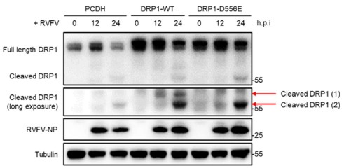
The full-length DRP1-strep protein is 83.2 kDa. Cleavage at D556 produces an estimated ~61.9 kDa N-terminal fragment and an estimated ~21.3 kDa C-terminal fragment (containing the strep-tag). Cleavage at D503 produces an estimated ~56.3 kDa N-terminal fragment and an estimated ~26.9 kDa C-terminal fragment (containing the strep-tag). Therefore, the DRP1(D556E) mutant would not produce the N-terminal ~61.9 kDa fragment (cleaved DRP1 (1), Author response image 1) and the C-terminal ~21.3 kDa fragment. However, since the D503 site remains intact, this mutant can still be cleaved at D503, yielding the ~56.3 kDa N-terminal fragment (cleaved DRP1 (2), shown in Author response image 1) and the ~26.9 kDa C-terminal fragment. When detected with an anti-DRP1 antibody, the ~56.3 kDa N-terminal fragment from D503 cleavage is observed. This fragment migrates at approximately 55 kDa on immunoblots. The result (see Author response image 1) are consistent with this interpretation and reflect the situation during virus infection.
Author response image 1.
(8) Figure 5: It will be meaningful to add a panel on mitochondrial morphodynamics upon Drp1 knockdown in this Figure. This may provide a correlative argument linking mitochondria to type 1 IFN response through Drp1.
According to this suggestion, we performed this experiment and included a new panel showing mitochondrial morphology in DRP1 knockout cells. The new Figure 5D demonstrates that DRP1 knockout induces significant mitochondrial elongation compared to control cells.
(9) Figure 6 and Figure 7: One may consider combining these in one consolidated panel.
We have now combined the original Figure 6 and Figure 7 into a single consolidated panel (new Figure 6 in the revised manuscript).
(10) Figure 7: Figure 7B needs particular attention. It seems that RVFV infection causes a similar downmodulation of both Drp1-wt and Drp1-3DE. However, Drp1-3DE did not produce the 15 kDa cleavage product and did not support enhanced type 1 IFN response as much as its WT counterpart did. Could the proteasomal pathway have a role in degrading caspase-resistant Drp1? Could the cleavage product have a role in enhancing anti-viral pathways? This must be meticulously addressed involving additional experimental designs, keeping in mind the results obtained using Drp1 knockdown cells (Figure 5).
We thank the reviewer for this comment. (1) Regarding the similar downmodulation of DRP1-WT and DRP1-3DE, the RVFV NSs has a potent function to inhibit host transcription. The exogenous DRP1-WT and DRP1-3DE are expressed from a CMV promoter via lentiviral transduction. This high-level exogenous expression system is sensitive to the transcriptional inhibition caused by RVFV-NSs, potentially explaining the observed similar reduction in protein levels. (2) Regarding the role of proteasomal pathway in degrading caspase-resistant Drp1, our data indicate that the proteasomal pathway is not the primary mechanism for the downregulation of the caspase-resistant DRP1-3DE. This is supported by the observation that the downregulation of endogenous DRP1 was largely insensitive to MG132 treatment (Figure 3A). (3) Whether the cleavage product plays a role in enhancing anti-viral pathways is an interesting question, which we propose to address in future study considering the scope of the current study.
(11) Figure 7: A panel may be added to compare the effect of overexpression of WT and mutant Drp1 on mitochondrial morphodynamics.
According to this suggestion, we have performed the experiment, and the data are included as a new panel in the revised Figure 6H. The results indicate that upon RVFV infection, control cells (PCDH) exhibited significant mitochondrial elongation. In contrast, cells overexpressing either DRP1-WT or DRP1-3DE did not show significant mitochondrial elongation morphology.
(12) Figure 8: Considering the known role of Drp1 in MAVS aggregation (doi.org/10.1016/j.molcel.2020.10.018), this figure in its present form could appear less informative. One may argue that a comparison of WT and mutant Drp1 for controlling virus-induced MAVS aggregation could be more meaningful.
We thank the reviewer for this comment. The major finding of our work is revealing a novel, caspase-dependent mechanism of DRP1 down-regulation. Figure 8 (revised Figure 7) showed that depletion of Drp1 promoted MAVS aggregation resulting in a strong antiviral effect. Regarding the suggestion to compare DRP1-WT and DRP1-3DE for MAVS aggregation during virus infection, we have focused our analysis on the DRP1 knockout system. This approach mimics the natural outcome of infection, where virus-induced caspase activation leads to the depletion of endogenous DRP1. Overexpression of WT and mutant Drp1 could hamper virus-induced depletion of Drp1 due to it’s high abundance, and may therefore affect the MAVS aggregation analysis.
(13) Figure 9: This figure establishes that Drp1 is cleaved by caspases in response to cellular infection by multiple viruses. However, the significance of caspase-mediated Drp1 cleavage per se in the progeny virus titre remains unclear.
We agree with this comment. The purpose of the original Figure 9 (revised Figure 8) is to show that the caspase-dependent Drp1 cleavage may occur in response to different virus infection. Whether and how this may affect progeny virus production, would be an interesting topic for future investigation. According to this comment, we reworded our description in the corresponding Results section.
(14) Would caspase inhibition impact mitochondrial morphodynamics and progeny virus titres? This question needs to be experimentally addressed.
Our data in Figures 3C, 3D, and 3F showed that the pan-caspase inhibitor Z-VAD-FMK effectively blocks virus-induced DRP1 cleavage in a dose-dependent manner. However, this upstream inhibition did not result in a significantly variation of nucleoprotein (NP) expression under RVFV infection. It is possible that the broad-spectrum inhibition of caspases by Z-VAD-FMK may simultaneously affect multiple aspects of the host cell and affect virus-host interactions.
For this reason, we employed the caspase-resistant DRP1-3DE mutant (revised Figure 6), which allowed us to specifically analyze the functional impact of Drp1 cleavage without introducing the effects of global caspase inhibition and caspase-dependent processes. The result that DRP1-3DE enhances viral replication provides evidence for the role of DRP1 cleavage in restricting virus production.
(15) One weakness is a lack of in vivo data despite the availability of Drp1-floxed mice. What are the results that one may anticipate based on the current study? This should be indicated in the Discussion section.
We thank the reviewer for this suggestion. According to this suggestion, we have now added discussion of the anticipated outcomes of in vivo studies.
Reviewer #2 (Recommendations for the Authors):
(1) Based on the DRP1-residues that caspase-3 targets for cleavage (500/503 and 556), 3 fragments having molecular weights of 55, 5.5 and 22 kDa, approximately, should be generated. In Figure 3E, the authors show the presence of DRP1 cleavage product having the size of approx. 50 Kda. Since the antibody (anti-DRP1) used by the authors recognizes the C-terminal region of DRP1, immunoblot for DRP1 under RVFV infection context should also demonstrate the presence of ~20 Kda DRP1 fragment. The authors should provide immunoblots showing the same.
We agree with the reviewer that, based on the identified cleavage sites at D500/D503 and D556, a C-terminal fragment of approximately 20 kDa should be detectable.
In our experiments using whole-cell lysates, we consistently observed the ~55 kDa cleaved DRP1 fragment but were unable to detect the smaller ~20 kDa fragment with the anti-DRP1 monoclonal antibody (CST, #5391). According to the manufacturer’s information, this antibody was raised against the carboxy-terminal region of human DRP1; however, the exact epitope has not been disclosed (we tried to contact the company, but did not get an answer). Since this is a monoclonal antibody, we infer that this single epitope likely resides upstream of the caspase-3 cleavage sites (D500/D503 and D556). Consequently, the C-terminal cleavage fragment may lack this epitope and is not recognized by the antibody.
To verify the generation of this C-terminal fragment, we performed in vitro cleavage assays using C-terminally strep-tagged DRP1 (DRP1-strep), where an anti-strep antibody clearly detects the ~20 kDa band (as shown in Figures 4A and 4D–4F). We have included this information in the manuscript for clarity.
(2) In Figure 4A, although the band for active caspase3 (from in vitro cleavage assay) is missing, DRP1 shows cleavage. Similarly, in Figure 4E, the band for active caspase3 is also missing. The authors should provide explanations for these data.
The reason is that under our standardized in vitro cleavage assay conditions, the anti-His antibody did not efficiently detect these particular commercial recombinant proteins, likely due to variations in epitope accessibility, protein conformation, or the detection sensitivity of the Western blot. The enzymatic activity of caspases used in our assays was verified using established positive control substrates. For caspase-3, activity was confirmed by cleavage of the well-characterized substrate PARP1 See Figure S3B.
(3) In Figure 4F, WT-DRP1 full length-band exhibits reduction in intensity upon treatment with caspase3, which is expected given the ability of caspase3 to cleave DRP1. However, although mutating 'D500/556E & D500/503/556E' abolishes DRP1 cleavage by caspase3, total DRP1 band intensity exhibits marked reduction in both cases. The authors should provide an explanation for this anomaly.
The recombinant protein used in this assay is C-terminally strep-tagged DRP1 (DRP1-strep), detected with an anti-strep antibody. The mutation of sites D500/503/556E prevented cleavage at these positions. However, the observed decrease in full-length protein indicates that caspase-3 cleavage may occur at other, alternative sites within DRP1, such as D221 (Figure 4C). The detection of cleavage fragment is dependent on the anti-strep antibody recognizing the C-terminal tag. Cleavage at alternative sites located between the mutated sites and the C-terminus is likely to produce shorter C-terminal fragments that may be difficult to be detected. Therefore, it is possible that although caspase-3 cleavage proceeds at these alternative sites in the mutant proteins, the fragments remain undetectable with our detection method.
(4) In Figures 7B and 7D, infection by RVFV of cells overexpressing DRP1-3DE (caspase-mediated cleavage-resistant) also leads to a reduction in full-length DRP1, although no cleavage product was seen. Does this indicate a possible role for other DRP1 residues being targeted by caspases upon virus infection? Treatment of DRP1-3DE-overexpressing RVFV-infected cells with a pan-caspase inhibitor will further provide insight into whether other DRP1 sites can also be targeted by caspase activity. Moreover, the authors should treat RVFV-infected cells with pan-caspase inhibitor and verify whether WT-DRP1 cleavage is restored concomitant with the abrogation of interferon induction and signaling.
We thank the reviewer for this comment. (1) For the reduction of full-length DRP1-3DE in RVFV-infected cells, similar with the response to the above comment, it is possible that the transcription inhibition effect of RVFV lead to reduction of Drp1-3DE expression level, an effect which may be more pronounced in the exogenous overexpression system. Additionally, the existence of other potential caspase cleavage sites, such as D221 (Figure 4C), may also in part account for the observed reduction. (2) Regarding the suggestion to use pan-caspase inhibition to analyze interferon signaling, we feel that blocking multiple caspase activities with the pan-caspase inhibitor Z-VAD-FMK will likely affect the global caspase activity in the cell affecting the scope beyond the interferon signaling pathways and is likely to complicate the interpretation of the results. Therefore, we employed the DRP1-3DE mutant for the analysis (revised Figure 6). This way we would not introduce the unspecific effects of global inhibition of multiple caspase-dependent processes. The result that DRP1-3DE enhances viral replication (revised Figures 6E-6G) provides evidence for the role of DRP1 cleavage in antiviral immunity.
(5) MAVS aggregation shown by SDD-AGE in Figures 8B, 8C & 8D does not display a typical appearance characterizing MAVS aggregation. The pattern shown in the above-mentioned immunoblots is very atypical for aggregates and indicates an increased MAVS abundance in RVFV-infected (8B)/DPR1-KO (8C, 8D) lanes. The aggregation of MAVS when demonstrated in SDD-PAGE gels exhibits a completely different pattern. The authors should include positions for molecular weight markers indicating their corresponding molecular sizes in the immunoblot. That will help in differentiating MAVS monomer from the aggregates and will help assess the extent of MAVS aggregation upon infection/DPR1-KO.
We thank the reviewer for this comment.
In the meantime, we checked literatures detecting the pattern of MAVS aggregation using the same technique (e.g., 2011 Cell, DOI: 10.1016/j.cell.2011.06.041; 2017 Nature Communications, DOI: 10.1038/ncomms15138), where, if we understood correctly, MAVS aggregation showed a similar high-molecular-weight smear. It is common in SDD-AGE not to use protein markers, as this method separates by molecular size under semi-denaturing conditions and is designed to resolve high-molecular-weight aggregates rather than provide precise molecular weights.
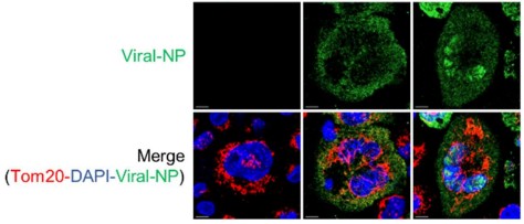
(6) In Figure 9D, the authors should also immunostain cells with virus-specific antibodies. This will help delineate that only virus-infected, but not uninfected cells exhibit elongation of mitochondria (as indicated by the appearance of TOM20 staining). In addition to that, the authors should provide an immunoblot for SeV antigen in Figure 9F. Instead of upregulating the H1N1 antigen, caspase inhibition by zVAD-FMK displayed a reduction in viral protein concomitant with DRP1 cleavage inhibition. Whereas, HSV-1 antigen abundance was seen to be enhanced upon the inhibition of apoptosis, which is consistent with the activity of axis entailing apoptosis-DRP1 cleavage-mitochondrial elongation-enhanced innate immunity- reduced virus propagation. The authors should explain this discrepancy.
Regarding Figure 9D (revised Figure 8D) and virus-specific staining, we now provided the viral antigen (NP) staining for RVFV and H1N1 (See Author response image 2). However, highly specific antibodies for SeV and HSV-1 immunofluorescence (IFA) are not available for us at the moment. We therefore propose to keep the original layout of the figure.
Author response image 2.
Regarding the immunoblot for SeV antigen in Figure 9F, we now provided the immunoblot for SeV antigen. See Figures 8F.
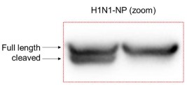
Regarding the H1N1-NP level upon Z-VAD-FMK treatment in Figure 9E, the reduction in NP signal with Z-VAD-FMK can be explained by the fact that the H1N1-NP protein itself is a substrate for caspase cleavage, as previously reported (DOI: 10.1007/s00705-007-0003-8). A zoom-in view of the blot in Figure 9E reveals a smaller migrating band corresponding to the cleaved NP. Z-VAD-FMK treatment inhibited the generation of this cleavage band, concurrently with the inhibition of DRP1 cleavage. When the ratio of internal control tubulin was considered, the NP level in the Z-VAD-FMK treatment group would not indicate a reduction of signal.
Figures 8E in the revised manuscript and Author response image 3.
Author response image 3.
(7) Previous report (Wu et al, 2022, Nat cell death & disease, DOI: 10.1038/s41419-022-05156-2) demonstrates the inhibitory role of apoptosis in interferon-induction upon HSV/SeV in macrophage cells. However, in this study, the authors suggest apoptosis positively regulates interferon response (upon SeV/HSV-1 infection of macrophage cells). The authors should provide an explanation for possible reasons accounting for this difference in the discussions section.
We propose that the functional consequence of caspases activation, which process different substrates, will likely pose two-sided effects on immune signaling as a two-sided “sword”. While the established caspase-dependent cleavage of molecules such as cGAS, MAVS, or NF-κB may pose inhibitory effect, the caspase-dependent cleavage of other substrates (such as Drp1 and yet-to-be identified other substrates) may pose the opposite effect. The overall effect would need to be determined in a context-dependent manner. We have included this notion in the revised Discussion section.
Reviewer #3 (Recommendations for the Authors):
(1) Please add the full name in the Abstract.
We have now added the full names for all relevant abbreviations upon their first appearance in both the Abstract and the Introduction sections.
(2) There is no scale bar in Figure 1A, 2A, 2B, 9D upper panel, Figure 8A.
We have now added scale bars to the revised figures: upper panels of Figure 1A, 2A, 2B, and 8D (original 9D), and Figure 7A (original 8A), as suggested.
(3) Can the author explain more in the text why DRP1, but not other mitochondria proteins?
Our initial investigation simultaneously examined the key regulators of both mitochondrial fusion (MFN1, MFN2, OPA1) and fission (DRP1) using the same set of cell samples. This comparison showed that DRP1 was specifically down-regulated upon RVFV infection (Figure 2C).
(4) Can the author explain more in the text why further study H1N1, SeV, HSV-1, but not other viruses?
H1N1, SeV and HSV-1 are model RNA or DNA viruses that are commonly used and were selected as model viruses in our system. We propose that other viruses could also be investigated in further studies.


