Peer review process
Revised: This Reviewed Preprint has been revised by the authors in response to the previous round of peer review; the eLife assessment and the public reviews have been updated where necessary by the editors and peer reviewers.
Read more about eLife’s peer review process.Editors
- Reviewing EditorMaría ZambranoCorpoGen, Bogotá, Colombia
- Senior EditorRichard WhiteUniversity of Oxford, Oxford, United Kingdom
Reviewer #1 (Public review):
[Editors' note: this version has been assessed by the Reviewing Editor without further input from the original reviewers. The authors have addressed most of the comments raised in the previous round of review.]
Summary:
This study addresses the emerging role of fungal pathogens in colorectal cancer and provides mechanistic insights into how Candida albicans may influence tumor-promoting pathways. While the work is potentially impactful and the experiments are carefully executed, the strength of evidence is limited by reliance on in vitro models, small patient sample size, and the absence of in vivo validation, which reduces the translational significance of the findings.
Strengths:
(1) Comprehensive mechanistic dissection of intracellular signaling pathways.
(2) Broad use of pharmacological inhibitors and cell line models.
(3) Inclusion of patient-derived organoids, which increases relevance to human disease.
(4) Focus on an emerging and underexplored aspect of the tumor microenvironment, namely fungal pathogens.
Reviewer #2 (Public review):
The authors in this manuscript studied the role of Candida albicans in Colorectal cancer progression. The authors have undertaken a thorough investigation and used several methods to investigate the role of Candida albicans in Colorectal cancer progression. The topic is highly relevant, given the increasing burden of colon cancer globally and the urgent need for innovative treatment options.
Strengths:
Authors have undertaken a thorough investigation and used several methods to investigate the role of Candida albicans in Colorectal cancer progression.
Author response:
The following is the authors’ response to the original reviews.
Public Reviews:
Reviewer #1 (Public review):
Summary:
This study addresses the emerging role of fungal pathogens in colorectal cancer and provides mechanistic insights into how Candida albicans may influence tumor-promoting pathways. While the work is potentially impactful and the experiments are carefully executed, the strength of evidence is limited by reliance on in vitro models, small patient sample size, and the absence of in vivo validation, which reduces the translational significance of the findings.
Strengths:
(1) Comprehensive mechanistic dissection of intracellular signaling pathways.
(2) Broad use of pharmacological inhibitors and cell line models.
(3) Inclusion of patient-derived organoids, which increases relevance to human disease.
(4) Focus on an emerging and underexplored aspect of the tumor microenvironment, namely fungal pathogens.
Weaknesses:
(1) Clinical association data are inconsistent and based on very small sample numbers.
We thank the reviewer for this important comment. We have investigated 4 colorectal tumors (2 in early stage and 2 in late stage), and we observed Candida albicans in the 2 late-stage samples while not in the early-stage ones. This result is consistent with TCGA data (which is large-scale) that Candida albicans mainly detectable in the late-stage colorectal tumors (Fig. 1c) and suggests that Candida albicans contributed to colorectal cancer progression, which is the main research direction of this study.
(2) No in vivo validation, which limits the translational significance.
We appreciate the reviewer’s concern regarding the lack of in vivo validation. While we recognize the value of in vivo models, our current institutional biosafety protocols and animal facility designations do not support the handling of pathogenic microorganisms like Candida albicans in live infection models. Consequently, these experiments were beyond the immediate technical scope of this study. To validate the findings using cell lines, we have performed Candida albicans infection experiments using organoids collected from colorectal cancer patients instead (Fig. 7). We have revised the Discussion section to acknowledge this limitation and clarify that the current work serves as a mechanistic study based on in vitro and ex vivo systems. We have also incorporated references to recent studies demonstrating the in vivo effects of C. albicans in tumor models, which support the biological relevance of our findings.
(3) Species- and cell type-specificity claims are not well supported by the presented controls.
We thank the reviewer for this insightful comment. We agree that our current dataset does not warrant definitive conclusions regarding species- or cell type-specificity. Accordingly, we have tempered our claims throughout the manuscript, describing the observed effects as context-dependent across different epithelial models. Specifically, we observed differential responses among the cell lines and epithelial systems evaluated, suggesting variability rather than strict specificity. Furthermore, the Discussion has been expanded to address potential underlying factors, such as variations in EGFR expression levels and other signaling determinants. We have also added a dedicated section to acknowledge this limitation and emphasize the need for future systematic investigations using a more diverse array of fungal species and cell models.
(4) Reliance on colorectal cancer cell lines alone makes it difficult to judge whether findings are specific or general epithelial responses.
We appreciate the reviewer’s thoughtful concern. Although most of our mechanistic experiments were performed in colorectal cancer cell lines, we also evaluated our finding across a broader range of epithelial models, including normal human colon-derived organoids and the breast epithelial cancer line MCF7 (Fig. 8). Neither model exhibited HIF-1α activation upon C. albicans exposure, supporting that the hypoxia response we observed might not be universal. Interestingly, the observed response in non-colorectal epithelial cancer lines (e.g., HCC1937, NUGC-3) suggests that this mechanism is not strictly confined to CRC. Based on these observations, we propose that the specificity is likely related to EGFR levels but may involve additional epithelial determinants, which we aim to investigate in future work.
Reviewer #2 (Public review):
The authors in this manuscript studied the role of Candida albicans in Colorectal cancer progression. The authors have undertaken a thorough investigation and used several methods to investigate the role of Candida albicans in Colorectal cancer progression. The topic is highly relevant, given the increasing burden of colon cancer globally and the urgent need for innovative treatment options. However, there are some inconsistencies in the figures and some missing details in the figures, including:
(1) The authors should clearly explain in the results section which patient samples are shown in Figure 1B.
We thank the reviewer for pointing out this omission. We apologize for the lack of clarity in the initial submission. The patient samples shown in Figure 1B are from the CRC patients with Stage III. We have revised the manuscript to explicitly state this information in the legend for Figure 1B to ensure better clarity for the reader.
(2) What do a, ab, b, b written above the bars in Figure 1F represent? Maybe authors should consider removing them, because they create confusion. Also, there is no explanation for those letters in the figure legend.
We thank the reviewer for this helpful comment. The letters above the bars represent statistical groupings from post-hoc multiple-comparison tests (a standard convention used after ANOVA or similar analyses): bars sharing the same letter are not significantly different, whereas different letters indicate statistically distinct groups. We chose this letter-based system over asterisks to avoid the visual clutter and potential confusion that often arise from numerous pairwise comparisons; therefore, we will retain the letter-based grouping. In the revised manuscript, we have explicitly defined this notation in the figure legend to be ease of interpretation for the reader.
(3) The authors should submit all the raw images of Western blot with appropriate labels to indicate the bands of protein of interest along with molecular weight markers.
We appreciate the reviewer’s request for raw data. We have now included the raw images of the Western blots in the supplementary materials, with clear annotations of the bands corresponding to the proteins of interest as well as molecular weight markers.
(4) The authors should do the quantification of data in Figure 2d and include it in the figure.
We thank the reviewer for this valuable suggestion. In the revised manuscript, we have quantified the subcellular localization of HIF-1α in PBS-treated versus C. albicans–infected cells shown in Figure 2d. The quantification results are shown in the following figure and provided in Supplementary Figure 3c.
(5) In Figure 2h, the authors should indicate if the quantification represents VEGF expression after 6h or 12h of C. albicans co-culture with cells.
We thank the reviewer for pointing this out. We have updated Figure 2h to specify that the quantification represents VEGF expression after 12 hours of co-culture with Candida albicans.
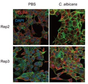
(6) In Figure 2i, quantification of VEGF should be done and data from three independent experiments should be submitted. The authors should also mention the time point.
We thank the reviewer for this helpful comment. In the revised manuscript, we have quantified VEGFA fluorescence intensity based on three independent experiments (the other 2 replicates were shown in Author response image 1). The corresponding time point (12 hours of co-culture) has been clearly indicated in the figure legend.
Author response image 1.
Recommendations for the authors:
Reviewing Editor Comments:
(1) Some of the statements regarding Candida albicans and CRC progression in Figure 1 may be overstated (since the association with stage/survival may be cross-confounded). That is, analyses of survival ought to be stage-adjusted.
We thank the editor for this important comment. We agree that the association between C. albicans and patient survival may be influenced by tumor stage as a confounding factor. In the revised manuscript, we have moderated our statements regarding the clinical associations and clarified the limitations of these analyses, now presenting these findings as correlative observations rather than causal relationships. We have also noted in the Discussion that stage-adjusted analyses would be required to more rigorously assess the independent contribution of C. albicans to patient outcomes.
(2) Fan et al. (citation 26) is incorrectly referenced. The paper states that Bacteroides fragilis does not affect Candida albicans colonization. Instead, Bacteroides thetaiotamicron was shown to reduce C. albicans colonization, but B. fragilis was used in the current study as a control.
We thank the editor for pointing out this error, and we have corrected the citation accordingly. As noted, the referenced study indicates that Bacteroides thetaiotaomicron, rather than Bacteroides fragilis, reduces C. albicans colonization. We selected B. fragilis as a control in this study because it is a prevalent gut commensal and has been previously implicated in colorectal cancer progression. Although prior reports suggest that B. fragilis does not significantly affect C. albicans growth, we observed that co-culture with B. fragilis led to a noticeable inhibition of C. albicans growth under our experimental conditions. This discrepancy may reflect differences in experimental settings. We believe these findings provide additional context for the complex interactions between gut microbiota and fungal pathogens.
(3) The link between hypoxia signaling is interesting, but for the most part, these experiments were largely done in normoxic conditions, while the colon is generally hypoxic. So I would have encouraged the authors to consider testing the effect of C. albicans presence/absence under low-oxygen conditions, which may be more physiologically relevant.
We thank the editor for this insightful suggestion. We fully agree that evaluating the effects of C. albicans under hypoxic or anaerobic conditions would be highly relevant to the physiological tumor microenvironment. Although we have attempted to assess the impact of C. albicans on cell migration under hypoxic conditions, we observed that tumor cells exhibited markedly accelerated migration and proliferation, resulting in near-complete wound closure within 24 hours in control groups. This limited our ability to reliably detect differences between conditions using standard migration assays. We agree that in vivo models may provide a more physiologically relevant context to address this question, and we will pursue this direction in future studies when appropriate experimental conditions become available.
Reviewer #1 (Recommendations for the authors):
(1) Figure 1 inconsistencies: In Figure 1C, there is no significant difference in C. albicans detection between stage II and stage III CRC patients. In fact, more patients in stage II appear positive, which is inconsistent with Figures 1A and 1B. For Figures 1A and 1B, the sample size (n=2) is too low to support meaningful conclusions. Please also clarify which stage is represented in Figure 1B.
We thank the reviewer for this important comment. In the revised manuscript, we have clarified the sample information and explicitly stated that the samples shown in Figure 1b are derived from stage III CRC patients. We have also moderated our conclusions and described these findings as exploratory observations. Regarding the apparent inconsistency between Figure 1C and Figures 1a-b, we consider that this discrepancy may be partly due to the small number of clinical samples analyzed in our study. In addition, the TCGA-based analysis relies on transcriptomic data, whereas our analysis is based on immunohistochemistry (IHC). These methodological differences may also contribute to the observed variation.
(2) Weak link between clinical and in vitro data: The transition from clinical samples to CRC cell line models feels tenuous. While C. albicans may induce hypoxia signaling, it is unclear whether this is specific to CRC cells or could occur in other epithelial cell types. Some broader testing would help strengthen this link.
We thank the reviewer for this insightful comment. We agree that reinforcing the bridge between clinical observations and in vitro mechanistic findings, as well as clarifying cell type specificity, is important for a comprehensive study. In the revised manuscript, we have clarified that the clinical data provide correlative evidence, while the mechanistic insights are derived from controlled in vitro systems. To address the issue of cell type specificity, we have included additional analyses across multiple epithelial cell models (Figure 8). These results indicate that the response to C. albicans is not restricted to colorectal cancer cells but varies across different epithelial contexts.
(3) Lack of in vivo validation: The mechanistic findings would be substantially strengthened by in vivo data, e.g., murine CRC models. Without this, the translational impact is limited.
We appreciate the reviewer’s concern regarding the lack of in vivo validation. While we recognize the value of animal models, our current institutional biosafety protocols and facility designations do not support the handling of pathogenic microorganisms like Candida albicans in live infection models. Consequently, these experiments were beyond the immediate technical scope of this study, and better be performed in future studies to validate the mechanisms.
(4) Figure 8B interpretation: The authors conclude that C. albicans shows the strongest effect on c-Myc and c-Jun activation. However, from the presented blots, the differences compared to other fungi are not obvious. The claim should be toned down or quantified more rigorously.
We thank the reviewer for this important comment. We agree that the differences in c-Myc and c-Jun activation among fungal species are not sufficiently pronounced to support a strong comparative claim. In the revised manuscript, we have moderated the corresponding statements to avoid overinterpretation.
(5) Cell type specificity: Since the title emphasizes CRC specificity, the cell line comparison shown in Figure 8 should be moved earlier in the results. This would clarify from the start whether the described mechanisms are CRC-specific or more generalizable.
We thank the reviewer for this insightful suggestion. We agree that earlier presentation of cell type comparisons would help clarify the scope of the observed effects. We have revised the Results section accordingly: “To evaluate the cell type specificity of this response, we further analyzed additional epithelial cell models, as shown in Figure 8”.
In this study, we initially identified the effects of C. albicans in colorectal cancer (CRC) cells and therefore focused on establishing the underlying mechanisms in this context. Subsequently, we extended our analysis to additional epithelial cell types to evaluate whether these effects are shared or context-dependent. We believe that this stepwise organization, from detailed mechanistic investigation in CRC cells to broader comparison across cell types, provides a logical and coherent flow for the reader. In the revised manuscript, we have further clarified this rationale in the text to improve readability and interpretation.
(6) It would be good to use a negative fungi control instead of a PBS control for most of the experiment.
We thank the reviewer for this valuable suggestion. We agree that a negative fungal control would further strengthen the conclusions. Unfortunately, we were unable to incorporate additional controls during the revision, while we believe that our comparative analysis across multiple fungal species (Figure 8) partially addresses this issue by demonstrating differential signaling responses. Future studies will incorporate appropriate negative fungal controls to further validate the specificity of these effects.
(7) It is surprising that the Dectin-1 inhibitor shows a smaller effect compared with the TLR2 inhibitor. This result warrants further explanation, as Dectin-1 is a well-known receptor for C. albicans β-glucans. The authors should clarify whether this difference reflects cell type-specific expression (e.g., low Dectin-1 in CRC cells), ligand accessibility, or pathway redundancy, and discuss how it aligns with existing literature.
We thank the reviewer for this insightful comment. We agree that the relatively modest effect of Dectin-1 inhibition compared to TLR2 inhibition warrants further consideration. In the revised manuscript, we have expanded the Discussion to address this observation. We propose several possible explanations: Firstly, the expression level of Dectin-1 is relatively low in these epithelial cancer cells, thereby limiting its functional contribution. Secondly, differences in ligand accessibility, particularly in the context of fungal cell wall architecture, may influence receptor engagement. Finally, redundancy and cross-talk among pattern recognition receptor pathways compensate for Dectin-1 inhibition. These observations highlight the context-dependent nature of host–fungal interactions.
Reviewer #2 (Recommendations for the authors):
All my comments that need to be addressed are given above and below:
(1) What do a and b represent in Figure 2f? They should be removed or clearly explained in the figure legend, as they are creating confusion for the audience.
We thank the reviewer for this comment. The letters indicate statistical groupings from post hoc multiple comparison tests. In the revised manuscript, we have added a clear explanation of this notation to the corresponding figure legends to be ease of interpretation for the reader.
(2) In the figure legend of S3a, the authors mentioned only the Caco2 cell line, whereas in the figure, there are two more cell lines, HCT116 and SW48. The authors should revise the figure legend.
We thank the reviewer for this comment. We have addressed this point and made the necessary corrections in the revised manuscript.
(3) The scale bar information is missing for Figure S3b. It should be included.
We thank the reviewer for this comment. The same scale bar was applied across all images in this panel. We have clarified this in the figure legend.
(4) In Figure 2e, the HIF-1α level in the Caco2 cells at 24 hr time point is a lot higher compared to the level at the 12-hour time point after C. albicans infection. But in the WB quantification in Figure 2f, the level of HIF-1α is not higher when compared to 12hr. Although it is relative data based on control, authors should check this calculation again for any errors.
We thank the reviewer for carefully examining the data. We have re-verified the quantification and confirmed that the values represent relative protein levels normalized to the corresponding controls at each time point.
Because samples from different time points were processed and analyzed separately, direct comparison of absolute protein levels across time points is not appropriate. Therefore, relative quantification within each time point provides a more accurate and representative assessment of HIF-1α changes.
(5) Line 125-127: This sentence should be rephrased.
We thank the reviewer for this comment. We have revised the corresponding section to improve clarity.
(6) PHD-mediated ubiquitination is the primary mechanism regulating HIF-1α protein stabilization. The authors should add an appropriate reference here.
We thank the reviewer for this suggestion. An appropriate reference has been added in the revised manuscript to support this statement.
(7) The authors claim that they observed that although the total level of HIF-1α increased, the ratio of its ubiquitinated form to total HIF-1α decreased. The authors should clearly indicate in the figure which protein band from the WB image was used for quantification from Figure S3c, which resulted in the graph presented in Figure S3d.
We thank the reviewer for this suggestion. We have revised the figure legend to improve clarity.
(8) In Figure 3a, there are some faint grey color lines. These graphs should be reformatted.
We thank the reviewer for this comment. We did not observe obvious faint grey lines in the original figure; however, these artifacts may have arisen during image conversion or file transfer. To ensure optimal image quality, we have provided high-resolution vector files to improve clarity.
(9) What do a and b in the bar graphs shown in Figure 3d,e; S4d,e,f represent?
We thank the reviewer for this comment. The letters indicate statistical groupings from multiple comparison tests. In the revised manuscript, we have added a clear explanation in the figure legend of this notation to the corresponding figure legends.
(10) What do a,b,c in the bar graphs shown in Figure 4c,d,h represent?
We thank the reviewer for this comment. The letters indicate statistical groupings from multiple comparison tests. In the revised manuscript, we have added a clear explanation in the figure legend of this notation to the corresponding figure legends.
(11) There are some faint grey lines in the bar graphs shown in Figure 4g. These lines should be removed.
We thank the reviewer for this comment. We did not observe obvious faint grey lines in the original figure; however, these artifacts may have arisen during image conversion or file transfer. To ensure optimal image quality, we have provided high-resolution vector files to improve clarity.
(12) Grey line below HIF-1α in the graph shown in Figure h should be removed.
We thank the reviewer for this comment. We did not observe obvious faint grey lines in the original figure; however, these artifacts may have arisen during image conversion or file transfer. To ensure optimal image quality, we have provided high-resolution vector files to improve clarity.
(13) The authors wrote - notably, despite treatment with AG1478, the levels of HIF-1α and c-MYC in C.albicans-infected cells remained significantly elevated compared to the uninfected control group (Figure 4b). There is no quantification for c-MYC. Statistics for HIF-1α quantification are missing. These should be added.
We thank the reviewer for this comment. We have quantified HIF-1α levels, and the results are presented in Figure 4d, including statistical analysis.
(14) There is no data for knockdown of MYD88, Dectin-1, and SYK as mentioned in the text lines 222-224. The authors should explain this discrepancy.
We thank the reviewer for this important comment. MYD88, Dectin-1, and SYK are expressed at relatively low levels in HCT116 cells, and our preliminary qPCR analyses indicated that it would be technically challenging to achieve reliable and quantifiable knockdown of these targets. Nevertheless, previous studies have reported that Dectin-1 can be present on the surface of epithelial cells, suggesting that it may still contribute to fungal recognition even at low expression levels. Therefore, given the technical constraints of gene knockdown in this specific context, we reasoned that pharmacological inhibition would provide a more robust approach to suppress this pathway.
(15) In line 227 in the results section it should be Figure S5c-e instead of Figure S5b-e. Figure S5b results do not match the results that are being explained here.
We thank the reviewer for this comment. We have corrected the typos in the revised manuscript.
(16) What do a,b,c in the bar graphs shown in Figure 5 a,b,i represent?
We thank the reviewer for this comment. The letters indicate statistical groupings from multiple comparison tests. In the revised manuscript, we have added a clear explanation in the figure legend of this notation to the corresponding figure legends.
(17) Was the experiment in Figure 5e done in triplicate? If not, it should be done in triplicate and quantified. The scale bar information is missing for IF images shown in Figure 5e. It should be added.
We thank the reviewer for this comment. The experiments were independently repeated for three times, and the quantification shown in Figure 5g represents the combined results from these biological replicates. The same scale bar was applied across all images in this panel. We have clarified this in the figure legend.
(18) Lines 273-274 in the results section: Als3 and Hwp1 are known to be involved in the adhesion of C. albicans to epithelial cells, while Ece1 encodes the virulence factor candidalysin. References should be added.
We thank the reviewer for this suggestion. We have added a reference in the revised manuscript to support this statement.
(19) What do a and b in the bar graphs shown in Figures 6 f,h,r represent? Since these letters are confusing and are present in several figures, they should be either deleted or clearly explained in the figure legends or text.
We thank the reviewer for this comment. The letters indicate statistical groupings from multiple comparison tests. In the revised manuscript, we have added a clear explanation in the figure legend of this notation to the corresponding figure legends to be ease of interpretation for the reader.
(20) What do a,b, and c in the bar graphs shown in Figure S8 b represent?
We thank the reviewer for this comment. The letters indicate statistical groupings from multiple comparison tests. In the revised manuscript, we have added a clear explanation in the figure legend to of this notation to the corresponding figure legends to be ease of interpretation for the reader.
(21) Scale bar should be added in Figure S9.
We thank the reviewer for these helpful comments. We have addressed this point and made the necessary corrections in the revised manuscript.
(22) What do a and b, in the bar graphs shown in Figure S11 represent?
We thank the reviewer for this comment. The letters indicate statistical groupings from post hoc multiple comparison tests. In the revised manuscript, we have added a clear explanation in the figure legend of this notation to the corresponding figure legends to be ease of interpretation for the reader.
(23) Were the organoids used in this paper characterized? If yes, how? Also, it should be mentioned in the appropriate section in the manuscript.
The organoids are not characterized; they are cultured using patients’ samples according to our previous protocols (He et al. Cell Stem Cell 2022).


