Convergence of pontine and proprioceptive streams onto multimodal cerebellar granule cells
- Janelia Farm Research Campus, Howard Hughes Medical Institute, United States
- Brandeis University, United States
Figures
Figure 1 with 1 supplement
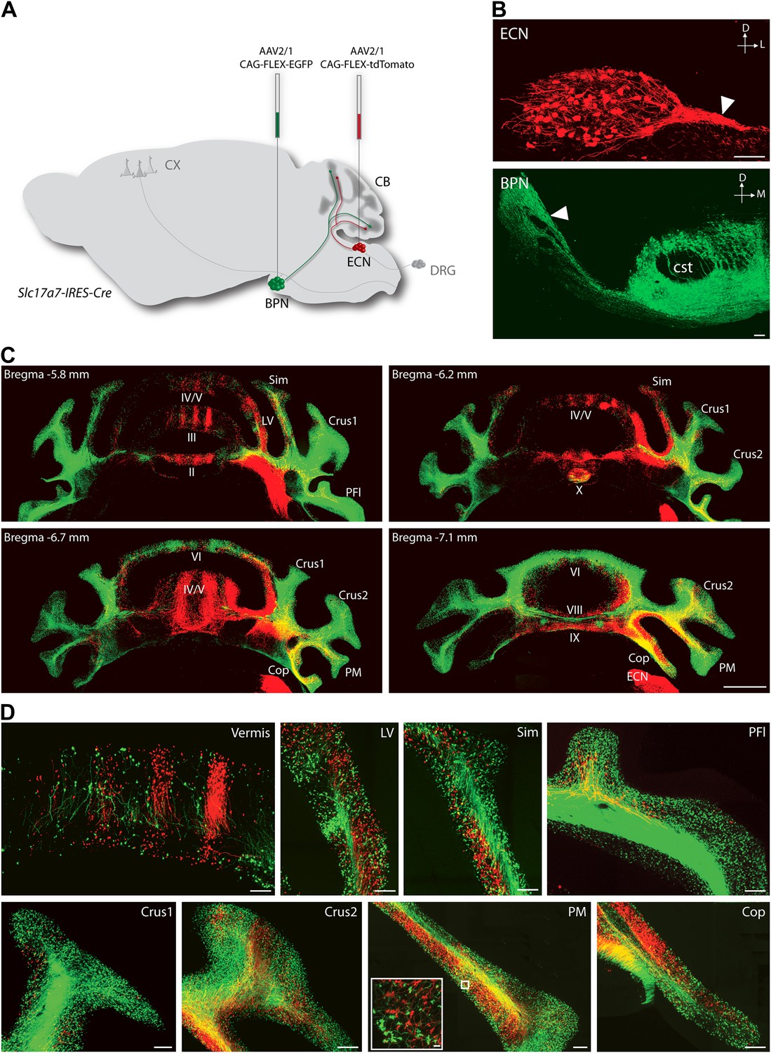
Termination patterns of ECN and BPN mossy fibers in the cerebellum.
(A) Genetic and viral scheme to specifically label ECN and BPN mossy fibers. Cre-dependent AAVs expressing tdTomato and EGFP are stereotaxically injected into ECN and BPN respectively in the Slc17a7-IRES-Cre mouse brain. CX, cortex; CB, cerebellum; DRG, dorsal root ganglia. (B) Confocal images of viral injection sites (ECN and BPN). White arrowheads, ECN and BPN axonal tracts; cst, corticospinal tract. D: dorsal; L: lateral; M: medial. Scale bars, 100 µm. (C) Projection pattern of ECN (red) and BPN (green) mossy fibers in the cerebellum. Montage confocal images of the cerebellum from rostral to caudal (Bregma −5.8 to −7.1 mm) positions. Vermis (II, III, IV/V, VI VIII, IX, X); Copula of the pyramis (Cop); lateral vermis (LV); Paraflocculus (PFl); Paramedian lobule (PM); simple lobule (Sim). Scale bar, 1 mm. (D) Magnified co-termination fields of ECN (red) and BPN (green) mossy fibers in selected cerebellar lobules. Boxed area shows high density of ECN and BPN mossy fiber terminations in the paramedian lobule. Scale bars, 100 µm; 10 µm in boxed area.
Figure 1—figure supplement 1
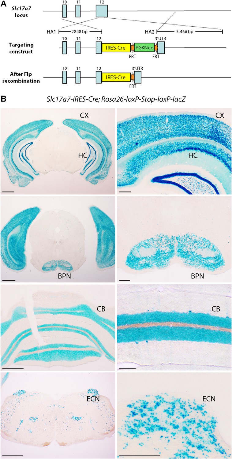
Expression pattern of Slc17a7-IRES-Cre mouse line.
(A) Targeting strategy for generating Slc17a7-IRES-Cre mice. Exons: blue boxes. HA1 and HA2: homology arms; FRT: flippase recognition target; PGKneo: neomycin resistance cassette; UTR: untranslated region. (B) β-Galactosidase activity in four coronal levels (low and high magnification) of a Slc17a7-IRES-Cre; Rosa26-loxP-Stop-loxP-lacZ animal. Left scale bars, 1 mm; right scale bars, 250 µm. CX: cortex; CB: cerebellum; HC: hippocampus.
Figure 2
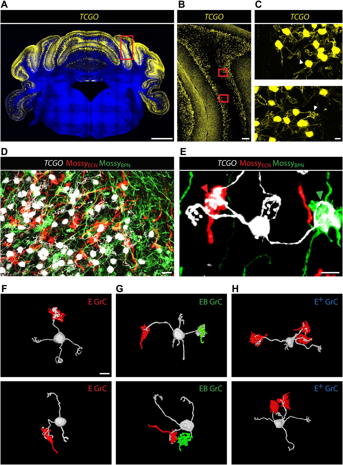
Convergence of ECN and BPN mossy fibers on cerebellar granule cells.
(A) TCGO transgene expression in a representative section of cerebellum. Scale bar, 1 mm. (B) TCGO mCitrine expression in boxed area of (A), simple lobule. Scale bar, 100 µm. (C) Maximum projection of labeled granule cells in TCGO mice (white arrowhead: dendritic arborization) in boxed areas of (B). Scale bar, 5 µm. (D) Co-termination of ECN (red) and BPN (green) mossy fibers in paramedian lobule of a Slc17a7-IRES-Cre; TCGO mouse. Scale bar, 10 µm. (E) Maximum projection of a labeled granule cell that receives mossy fiber inputs from ECN (red arrowhead) and BPN (green arrowhead) in a Slc17a7-IRES-Cre; TCGO mouse. Scale bar, 5 µm. (F)–(H) 3D reconstruction of granule cells with associated mossy fiber terminations. E granule cell (GrC), granule cell with one ECN input and one other traceable dendrite; EB GrC, granule cell with ECN and BPN input(s); E+ GrC, granule cell with two or more ECN inputs but no BPN input. Scale bar, 5 µm.
Figure 3

Cerebellar areas not exhibiting convergence of sensory and pontine inputs.
Survey of ECN and BPN convergence in the anterior vermis. (A) Vermis III. (B) Vermis IV/V. (i) Granule cell (GrC) classification and distribution. Red cross, E GrC; green cross, EB GrC; blue cross, E+ GrC. Scale bars, 100 µm. (ii) density contour map of E, EB and E+ granule cells. D: dorsal; V: ventral; M: medial; L: lateral. Red, green and blue lines in the contour map represent density of E, EB, and E+ granule cells respectively. (iii) upper, percentage of E, EB and E+ granule cells of two Slc17a7-IRES-Cre; TCGO cerebella. Lower, comparison between annotators in percentage of E, EB and E+ granule cells in a selected section. (C) Pontocentric view of vermis III. (D) Pontocentric view of vermis IV/V. (C and D) Same organization as in (A and B) but B replaces E and B+ replaces E+ granule cells. B GrC: granule cell with one BPN input and one other traceable dendrite; B+ GrC: granule cell with two or more BPN inputs but no ECN input. EB GrC is the same as in (A and B)
Figure 4
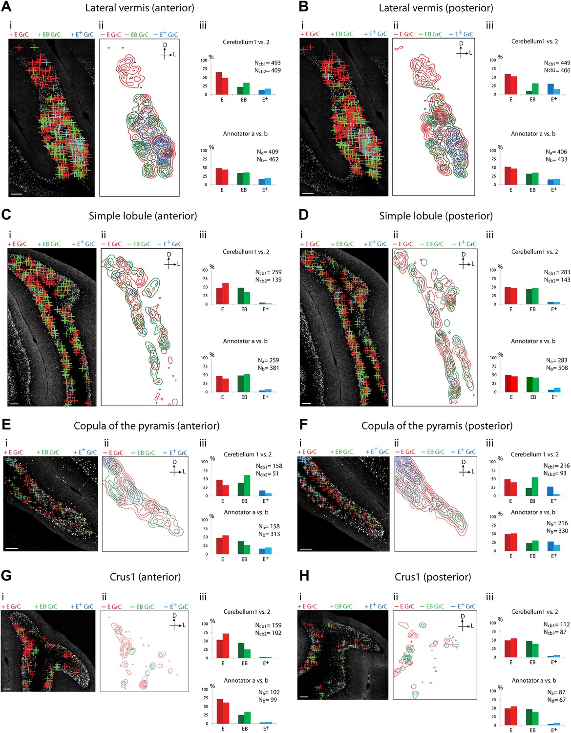
Cerebellar areas exhibiting mixtures of convergence and separation of sensory and pontine inputs.
Survey of ECN and BPN convergence in the lateral vermis, simple lobule, copula of the pyramis and Crus1. (A) Lateral vermis, anterior section. (B) Lateral vermis, posterior section. (C) Simple lobule, anterior section. (D) Simple lobule, posterior section. (E) Copula of the pyramis, anterior section. (F) Copula of the pyramis, posterior section. (G) Crus1, anterior section. (H) Crus1, posterior section. (i) Granule cell (GrC) classification and distribution. Red cross: E GrC; green cross: EB GrC; blue cross: E+ GrC. Scale bars, 100 µm. (ii) Density contour map of E, EB and E+ granule cells. D: dorsal; L: lateral. Red, green and blue lines in the contour map represent density of E, EB, and E+ granule cells, respectively. (iii) Upper, percentage of E, EB and E+ granule cells of two Slc17a7-IRES-Cre; TCGO cerebella. Lower, comparison between annotators in percentage of E, EB and E+ granule cells.
Figure 5
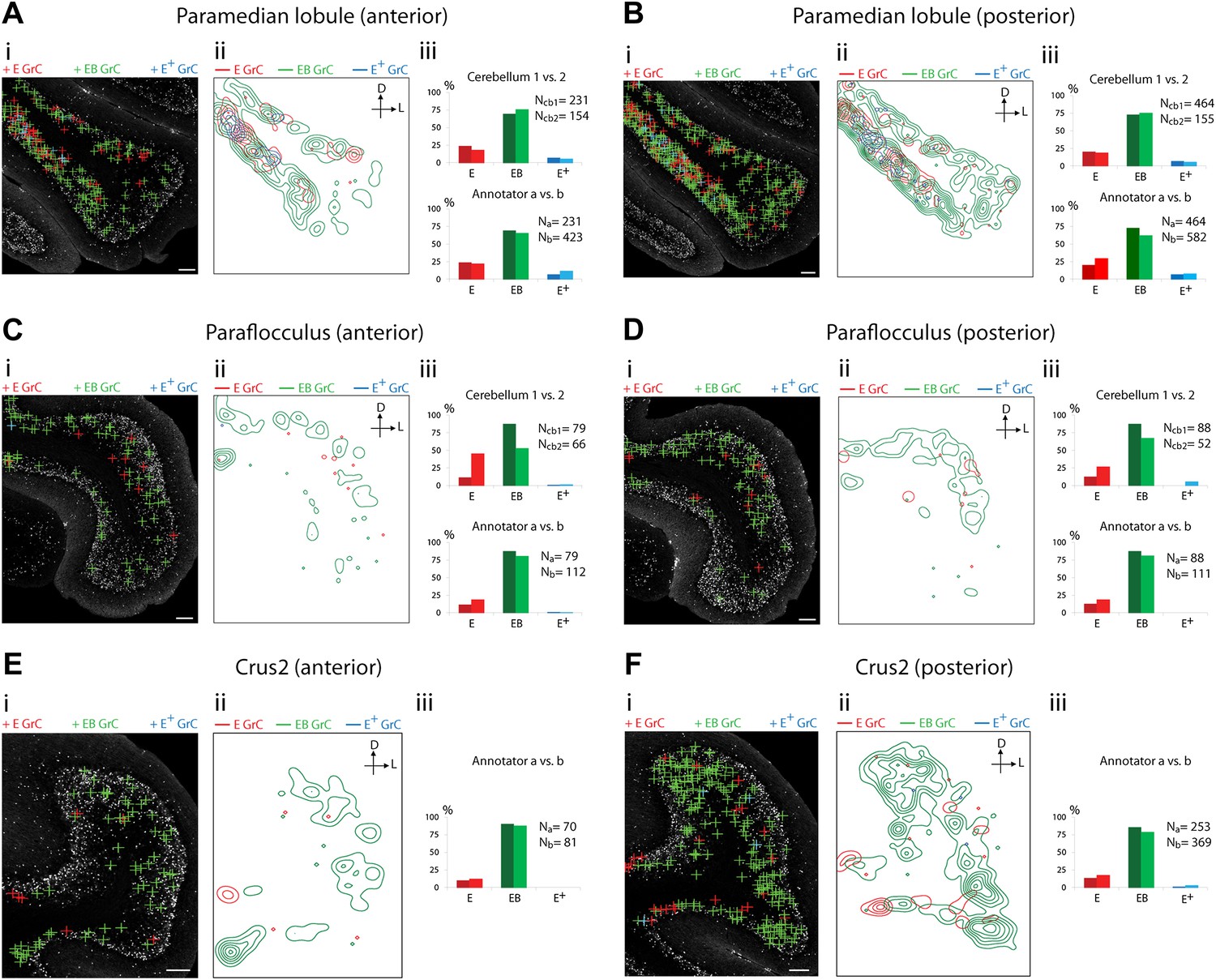
Cerebellar areas exhibiting abundant convergence of sensory and pontine inputs.
Survey of ECN and BPN convergence in the hemispheric regions. (A) Paramedian lobule, anterior section. (B) Paramedian lobule, posterior section. (C) Paraflocculus, anterior section. (D) Paraflocculus, posterior section. (E) Crus2, anterior section. (F) Crus2, posterior section. (i) Granule cell (GrC) classification and distribution. Red cross: E GrC; green cross: EB GrC; blue cross: E+ GrC. Scale bars, 100 µm. (ii) Density contour map of E, EB and E+ granule cells. D: dorsal; L: lateral. Red, green and blue lines in the contour map represent density of E, EB, and E+ granule cells respectively. (iii) Upper, percentage of E, EB and E+ granule cells from two Slc17a7-IRES-Cre; TCGO cerebella. Lower, comparison between annotators in percentage of E, EB and E+ granule cells in a selected section. (E and F) do not have comparisons across the two cerebella in (iii).
Figure 6
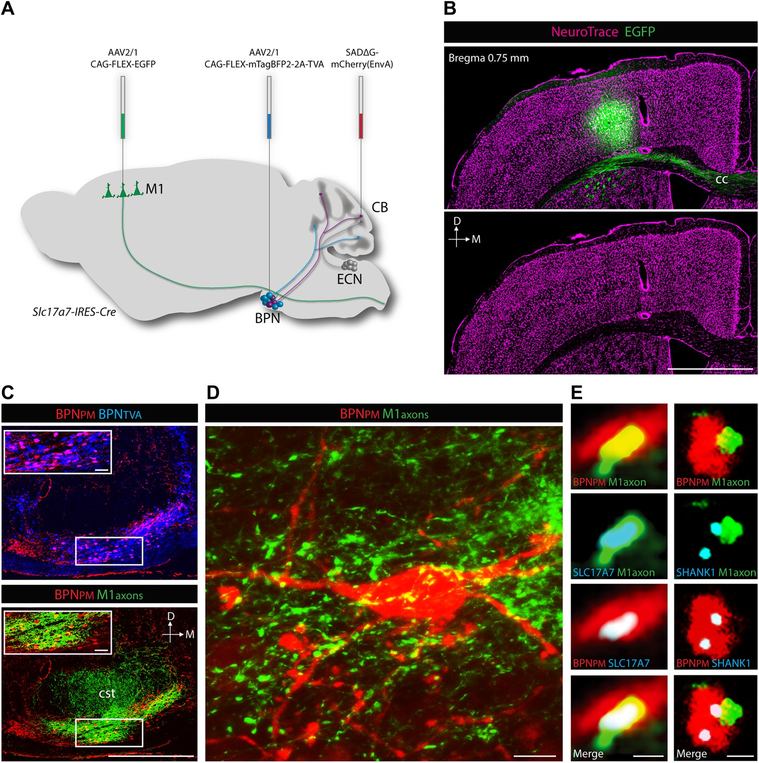
Cortical inputs to paramedian-projecting BPN neurons.
Combining M1 anterograde tracing and paramedian lobule retrograde tracing. (A) Scheme to anterogradely label forelimb/upper body M1 cortical axons and retrogradely label paramedian-projecting BPN neurons (BPNPM). Cre-dependent AAVs expressing EGFP and mTagBFP2-2A-TVA were stereotaxically injected into M1 and BPN respectively in the Slc17a7-IRES-Cre mouse and SADΔG-mCherry(EnvA) rabies virus was injected into the paramedian lobule of the cerebellum. CB, cerebellum; M1, primary motor cortex. (B) Location of EGFP-expressing neurons with relation to cortical cytoarchitecture. cc: corpus callosum. D: dorsal; M: medial. Scale bar, 1 mm. (C) Upper, relationship of BPNPM and TVA-expressing BPN neurons (BPNTVA) sensitive to rabies infection. Lower, colocalization of M1 axons and BPNPM neurons. D: dorsal; M: medial. Scale bars, 500 µm; 50 µm in magnified areas. (D) High-magnification image of BPNPM neurons and the M1 axon termination field. Scale bar, 10 µm. (E) Synaptic arrangement between M1 axons and BPNPM. Left, apposition of a M1 axon expressing the presynaptic marker SLC17A7 and a dendrite of a BPNPM in a single confocal slice. Right, apposition of a M1 axon and a SHANK1-containing postsynaptic density of a BPNPM in a single confocal slice. Scale bars, 1 µm.
Download links
A two-part list of links to download the article, or parts of the article, in various formats.
Downloads (link to download the article as PDF)
Open citations (links to open the citations from this article in various online reference manager services)
Cite this article (links to download the citations from this article in formats compatible with various reference manager tools)
Convergence of pontine and proprioceptive streams onto multimodal cerebellar granule cells
eLife 2:e00400.
https://doi.org/10.7554/eLife.00400


