Prefrontal dopamine regulates fear reinstatement through the downregulation of extinction circuits
- University of Tokyo, Japan
- Center for Information and Neural Networks, Japan
Figures
Figure 1 with 4 supplements
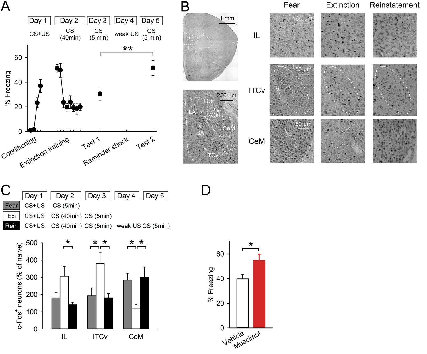
Reinstatement is associated with low IL activity.
(A) A reminder shock reinstated extinguished fear (n = 10 mice; paired t-test, t(9) = 3.6, p = 0.0059). (B) Representative images of the infralimbic cortex (IL), the ventral intercalated amygdala neurons (ITCv), and the central nucleus of the amygdala (CeM) in the Fear, Extinction, and Reinstatement groups. (C) c-Fos+ cell density decreased in the IL and the ITCv and increased in the CeM with reinstatement (n = 8–11 mice; F(2,27) = 4.3, p = 0.023 [IL]; F(2,26) = 4.8, p = 0.0016 [ITCv]; F(2,26) = 6.3, p = 0.0058 [CeM]; Tukey's test, PExtinction vs Reinstatement = 0.029 [IL], 0.035 [ITCv], 0.013 [CeM]). (D) IL muscimol infusions resulted in high freezing (n = 10 mice; t(18) = 2.4, p = 0.030). **p < 0.01, *p < 0.05. Data represent mean ± standard error.
Figure 1—figure supplement 1
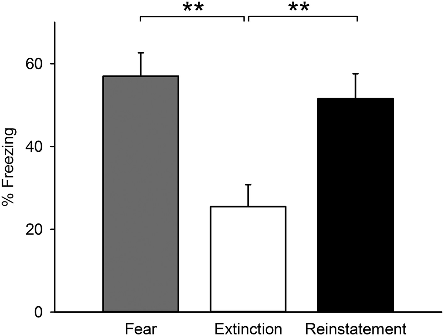
Freezing behaviour of the mice subjected to c-Fos activity mapping.
Mice of the Reinstatement group showed greater freezing behaviour relative to the Extinction group (F(2,25) = 8.7, p = 0.0013; Tukey's test, PFear vs Extinction = 0.0023, PExtinction vs Reinstatement = 0.0073). **p < 0.01. Data represent mean ± standard error.
Figure 1—figure supplement 2
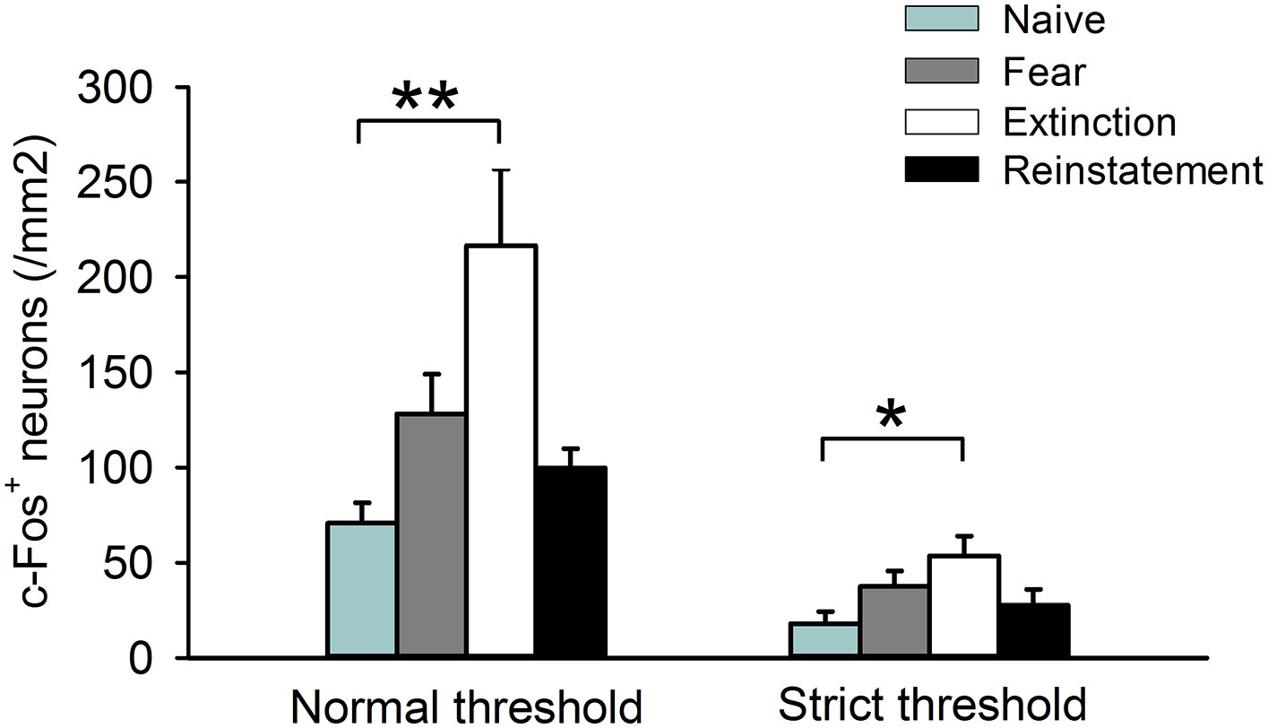
c-Fos+ cell density in the IL calculated in the analysis using a normal threshold and in an additional analysis using a strict threshold.
(n = 8–11 mice; F(3,35) = 5.9, p = 0.0023 [Normal threshold]; F(3,35) = 3.0, p = 0.042 [Strict threshold]; Tukey's test, PNaive vs Extinction = 0.0017 [Normal threshold], 0.032 [Strict threshold]). **p < 0.01, *p < 0.05. Data represent mean ± standard error.
Figure 1—figure supplement 3
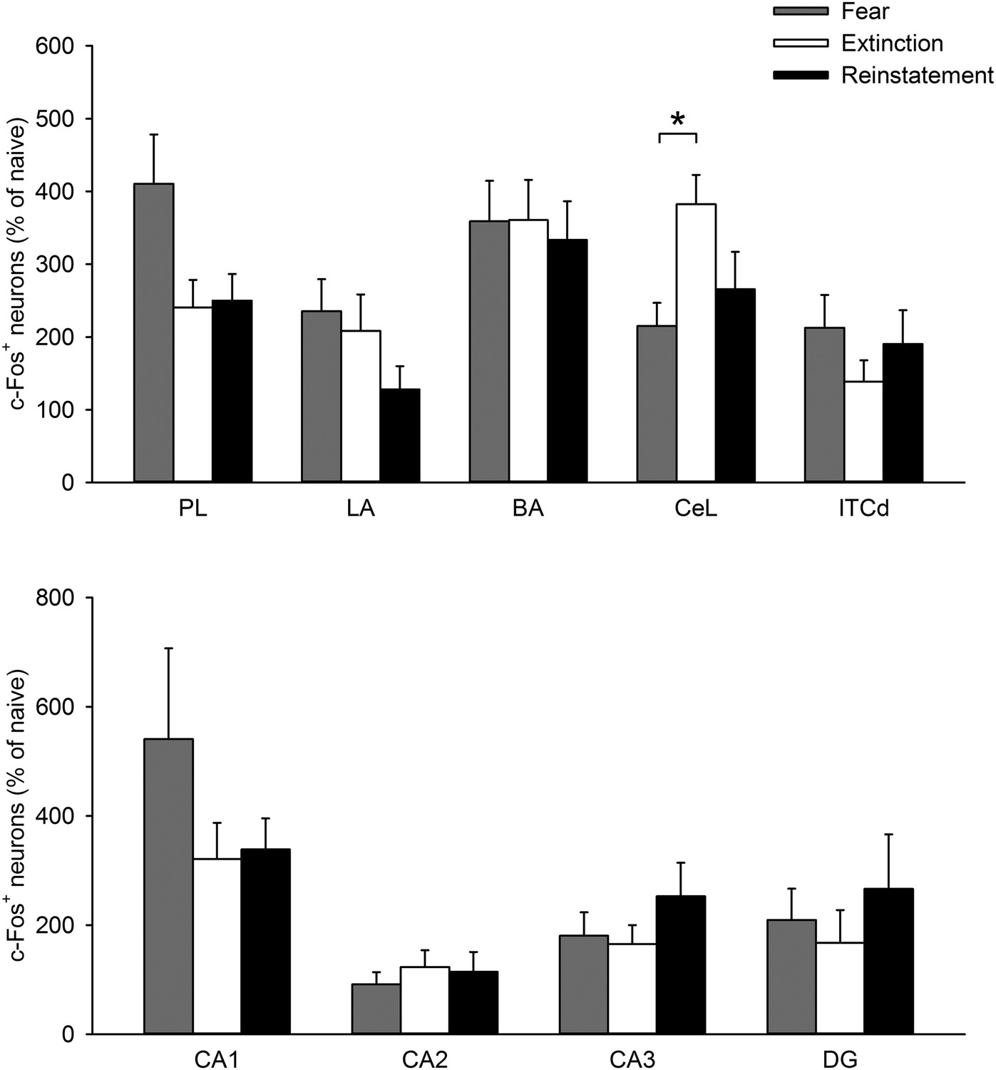
The density of c-Fos+ cells in the PL, LA, BA, CeL, ITCd, CA1, CA2, CA3, and DG was comparable between the Extinction and Reinstatement groups (PL, F(2,28) = 3.6, p = 0.041; LA, F(2,26) = 1.4, p = 0.27; BA, F(2,26) = 0.068, p = 0.93; CeL, F(2,26) = 4.8, p = 0.017; ITCd, F(2,27) = 0.97, p = 0.39; CA1, F(2,22) = 1.3, p = 0.29; CA2, F(2,22) = 0.29, p = 0.75; CA3, F(2,22) = 1.0, p = 0.38; DG, F(2,22) = 0.46, p = 0.64; Tukey's test, CeL: PFear vs Extinction = 0.015).
PL: prelimbic cortex, LA: lateral amygdala, BA: basal amygdala, CeL: lateral subdivision of central nucleus of the amygdala, ITCd: dorsal intercalated amygdala neurons. *p < 0.05. Data represent mean ± standard error.
Figure 1—figure supplement 4
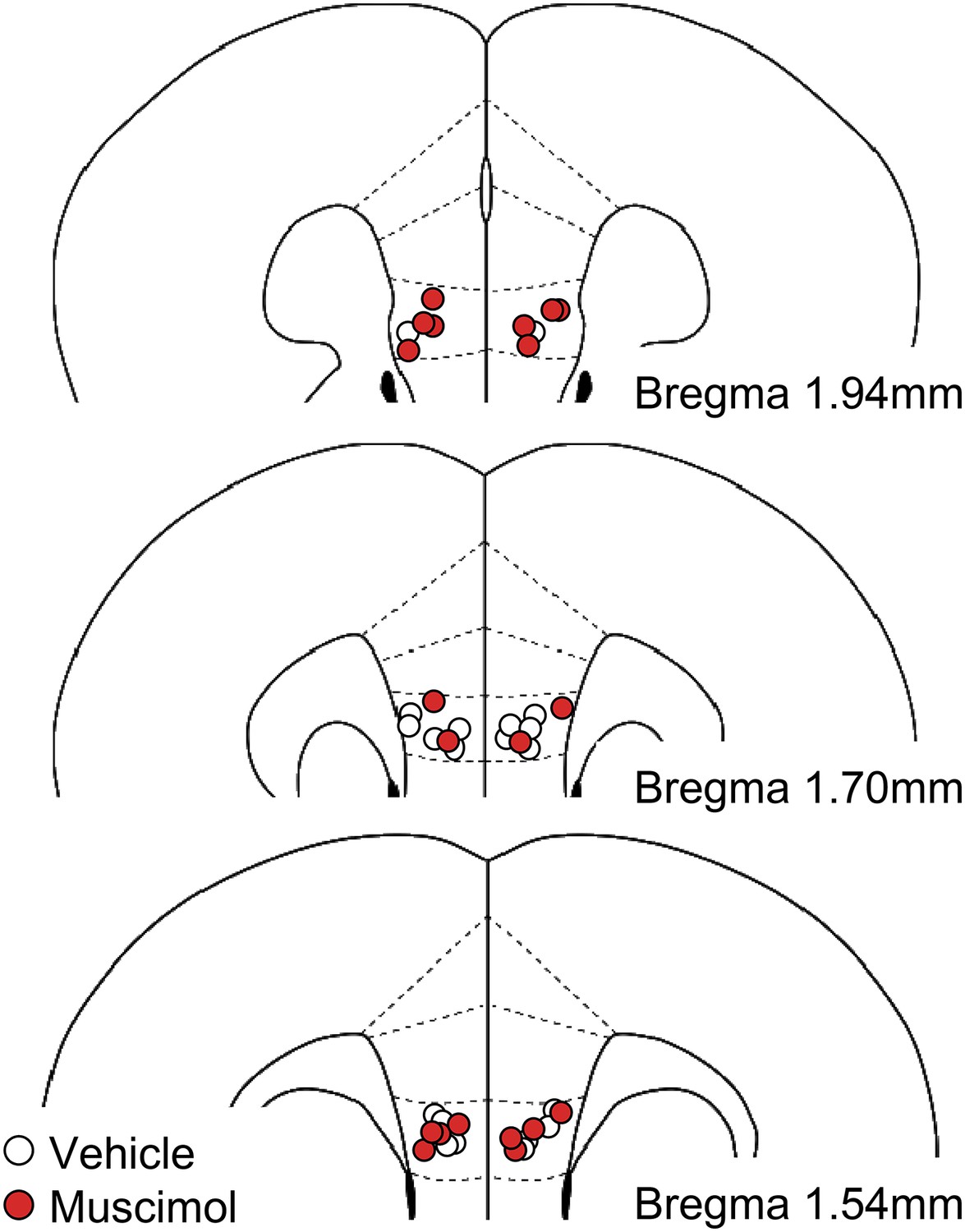
Histological verification of cannula placements in the experiment with muscimol infusions into the IL.
https://doi.org/10.7554/eLife.08274.007
Figure 2 with 1 supplement

Reinstatement is associated with presynaptic depression in the IL.
(A) Representative miniature excitatory postsynaptic current (mEPSC) traces. (B) IL neurons had lower mEPSC frequency in the Reinstatement group (n = 8 neurons from 6 mice) than the Extinction group (n = 8 neurons from 4 mice) (F(2,21) = 3.9, p = 0.037; PExtinction vs Reinstatement = 0.030). (C) mEPSC amplitude did not differ across groups (F(2,21) = 1.9, p = 0.38). (D) mEPSC frequency negatively correlated with Δfreezing (different degrees of freezing time between tests 1 and 2) in the Reinstatement group (r = −0.83, p = 0.040). (E) Representative traces of EPSCs evoked by paired-pulse stimulation. (F) IL neurons had a higher paired-pulse ratio (PPR) in the Reinstatement group (n = 8 neurons from 5 mice) than the Extinction group (n = 8 neurons from 4 mice) (t(14) = 2.2, p = 0.049). (G) PPR positively correlated with Δfreezing in the Reinstatement group (r = 0.95, p = 0.012). *p < 0.05. Data represent mean ± standard error.
Figure 2—figure supplement 1
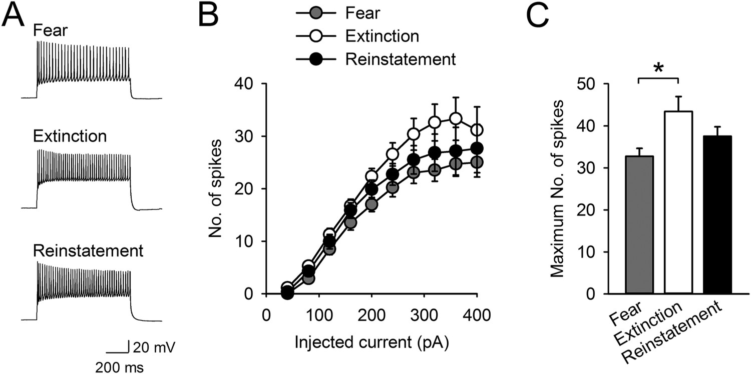
Intrinsic excitability of infralimbic neurons did not change with fear reinstatement.
(A) Representative traces showing spikes in response to a 400-pA current pulse. (B) The number of action potentials in response to depolarizing steps at different current intensities (n = 21, 20, 20 neurons). (C) The maximum number of evoked spikes at any current step increased with fear extinction (F(2,58) = 4.1, p = 0.022; Tukey's test, p = 0.016) and did not change with fear reinstatement. *p < 0.05. Data represent mean ± standard error.
Figure 3
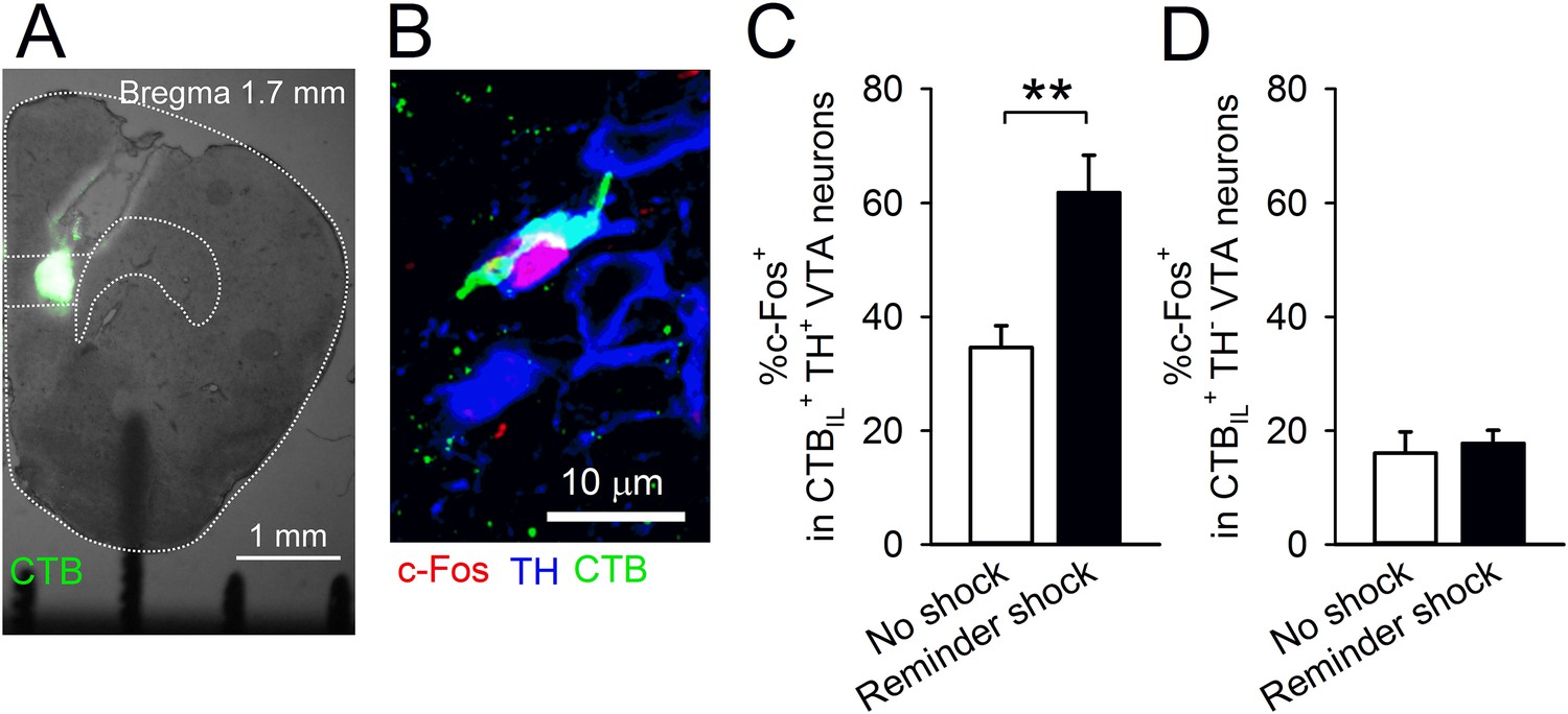
A reminder shock activates dopaminergic VTA neurons projecting to the IL.
(A) Coronal brain section of a mouse with Alexa 488-conjugated cholera toxin subunit B (CTB) infusion into the IL. (B) A representative immunofluorescence image of the ventral tegmental area (VTA) neurons with c-Fos, tyrosine hydroxylase (TH), and CTB. (C) A reminder shock increased the proportion of c-Fos+ neurons in IL-projecting TH+ VTA neurons (no shock: n = 7, reminder shock: n = 6 mice; t(11) = 4.3, p = 0.0012). (D) A reminder shock did not increase the proportion of c-Fos+ neurons in IL-projecting TH− VTA neurons (no shock: n = 7, reminder shock: n = 6 mice). **p < 0.01, Data represent mean ± standard error.
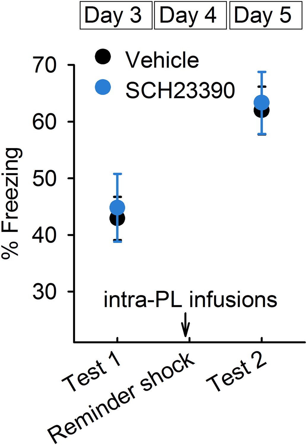
Figure 4 with 2 supplements
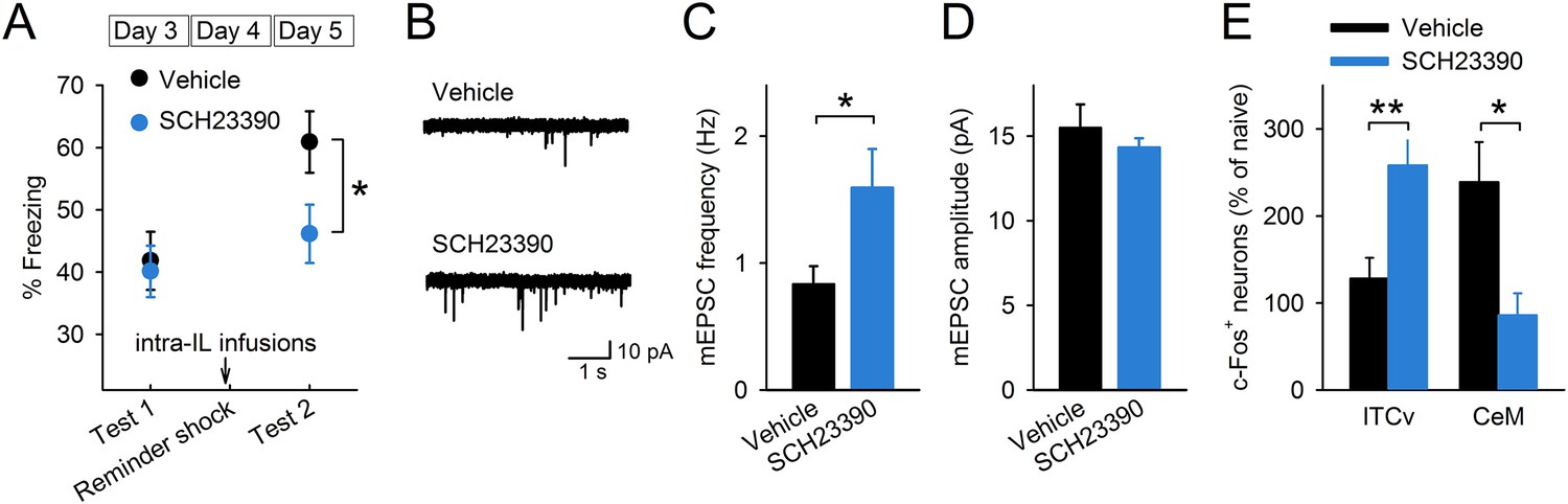
D1Rs in the IL mediate reinstatement.
(A) SCH23390 infusions into the IL before a reminder shock suppressed reinstatement (phosphate-buffered saline [PBS]: n = 15, SCH23390: n = 14 mice; t(27) = 2.2, p = 0.039). (B) Representative mEPSC traces. (C) SCH23390-infused mice demonstrated higher mEPSC frequency (n = 9, 10 neurons; t(17) = 2.2, p = 0.044). (D) SCH23390 infusions had no effects on mEPSC amplitude. (E) SCH23390-infused mice demonstrated higher and lower c-Fos+ cell density in the ventral intercalated amygdala neurons (ITCv) and the central nucleus of the amygdala (CeM), respectively (n = 7–8 mice; ITCv: t(13) = 3.0, p = 0.0093; CeM: t(14) = 2.9, p = 0.011). **p < 0.01, *p < 0.05. Data represent mean ± standard error.
Figure 4—figure supplement 1
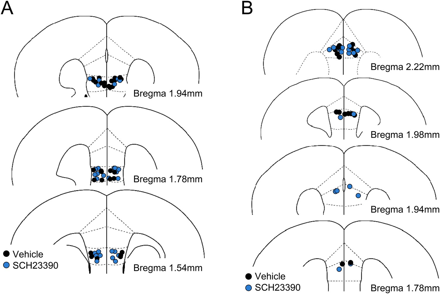
Histological verification of cannula placements in the experiment with SCH23390 infusions into the infralimbic (A) and the prelimbic (B) cortices.
https://doi.org/10.7554/eLife.08274.013
Figure 4—figure supplement 2
SCH23390 infusions into the prelimbic cortex had no effects on reinstatement (n = 9 mice).
Data represent mean ± standard error.
Tables
Table 1
Electrophysiological properties of IL neurons
| Fear | Extinction | Reinstatement | |
|---|---|---|---|
| Resting potential (mV) | −70.7 ± 1.1 | −72.0 ± 1.0 | −70.6 ± 0.7 |
| Input resistance (MΩ) | 276.5 ± 24.6 | 391.9 ± 32.4* | 362.6 ± 30.9 |
| Spike amplitude (mV) | 75.5 ± 1.5 | 72.9 ± 2.0 | 76.3 ± 1.2 |
| First interspike interval (ms) | 7.9 ± 0.5 | 8.8 ± 0.6 | 8.9 ± 0.4 |
| Rheobase (pA) | 78.1 ± 5.7 | 65.5 ± 4.9 | 70.0 ± 6.1 |
| Spike threshold (mV) | −37.3 ± 0.7 | −37.2 ± 1.0 | −35.1 ± 0.6 |
| Voltage sag (mV) | −3.0 ± 0.3 | −3.1 ± 0.2 | −3.8 ± 0.4 |
| Half width of spike (ms) | 1.01 ± 0.03 | 0.98 ± 0.02 | 1.04 ± 0.02 |
| fAHP (mV) | −17.3 ± 0.7 | −16.7 ± 0.7 | −16.7 ± 0.7 |
| mAHP (mV) | −1.6 ± 0.5 | −0.9 ± 0.4 | −1.0 ± 0.5 |
-
*
p < 0.05 vs Fear, Tukey's test.
-
fAHP, fast afterhyperpolarization; mAHP, medium afterhyperpolarization.
Download links
A two-part list of links to download the article, or parts of the article, in various formats.
Downloads (link to download the article as PDF)
Open citations (links to open the citations from this article in various online reference manager services)
Cite this article (links to download the citations from this article in formats compatible with various reference manager tools)
Prefrontal dopamine regulates fear reinstatement through the downregulation of extinction circuits
eLife 4:e08274.
https://doi.org/10.7554/eLife.08274


