Oldest skeleton of a fossil flying squirrel casts new light on the phylogeny of the group
- Universitat Autònoma de Barcelona, Spain
- Muséum national d’Histoire naturelle, France
- University of Kansas, United States
- Ludwig-Maximilians-Universität München, Germany
- Bayerische Staatssammlung für Paläontologie und Geologie, Germany
- Bavarian State Collection of Zoology, Germany
- Museo Storico della Fisica e Centro Studi e Ricerche Enrico Fermi, Italy
- The ‘Abdus Salam’ International Centre for Theoretical Physics, Italy
Figures
Figure 1
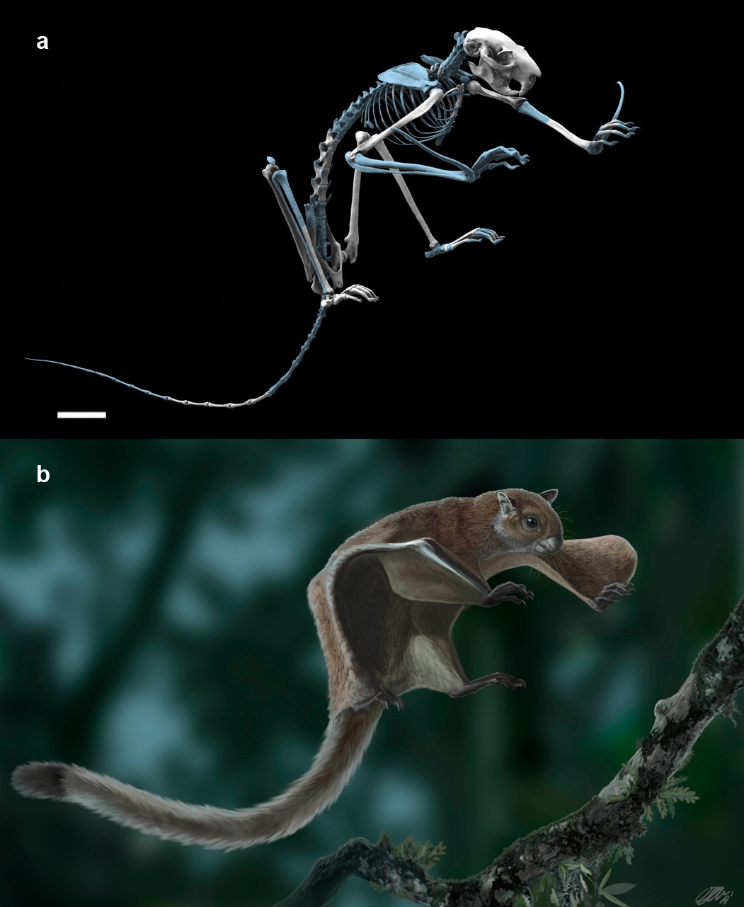
The fossil flying squirrel Miopetaurista neogrivensis.
(a) Reconstruction of the skeleton based in the partial skeleton IPS56468 from Abocador de Can Mata. Missing elements are based on extant giant flying squirrel Petaurista petaurista and are colored in blue. (b) Life appearance of Miopetaurista neogrivensis showing the animal ready to land on a tree branch. Coat pattern and color are based in extant Petaurista species, the sister taxon of Miopetaurista (see Figure 7). See Video 1 for an animated version of this reconstruction and 3D model in Supplementary file 1 to view and manipulate a low-quality model of the skeleton. For recovered elements of the postcranial skeleton see Figures 2 and 4 and Table 1. For a description and comparison of the postcranial bones, see Appendix 3.3. See Figure 6 and Video 3 for a more detailed cranial reconstruction. 3D models generated from µCT scan data and photogrammetry. Scale bar is 4 cm.
Figure 2 with 1 supplement
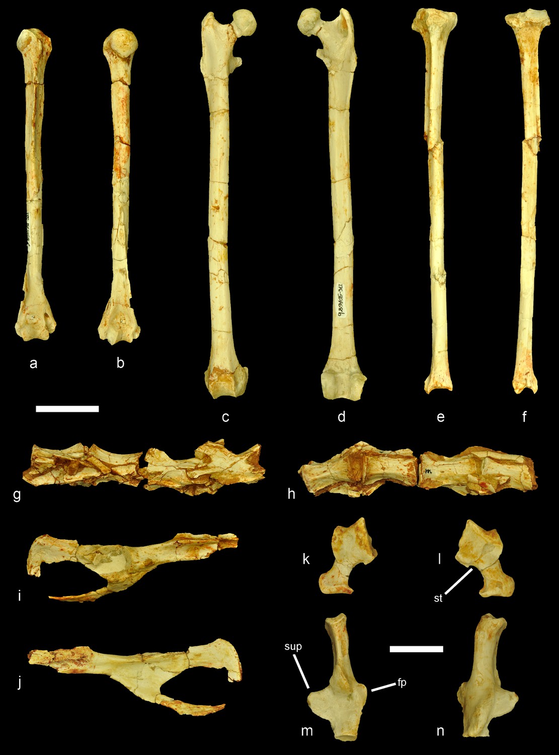
Selected postcranial elements of the partial skeleton of Miopetaurista neogrivensis.
(a–b) Right humerus (IPS56468f) in cranial and caudal views. (c–d) Right femur (IPS56468b) in cranial and caudal views. (e–f) Right tibia (IPS56468a) in cranial and caudal views. (g–h) Lumbar vertebrae L3–L6 (IPS56468m–n) in dorsal and ventral views. Note that vertebrae are in anatomical connection. (i–j) Partial right coxal (IPS56468k) in lateral and medial views. The proximal end of the ilium is not preserved and part of the pubis is damaged. (k–l) Left astragalus (IPS56478t). (m–n) Left calcaneus (IPS56468s). fp, fibular process; st, sulcus tali; sup, sustentacular process. Scale bar is 2 cm in figures (a–j) and 1 cm in figures (k–n). For a reconstruction of the skeleton see Figure 1, Videos 1 and 3D model in Supplementary file 1. Details of particular bones are shown in Figure 5; Figure 2—figure supplement 1. For a detailed description and comparison of the postcranial bones of M. neogrivensis see Appendix 3.3.
Figure 2—figure supplement 1
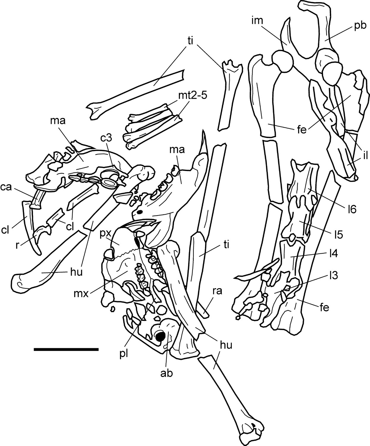
Schematic drawing of the recovered skeletal elements of Miopetaurista neogrivensis from Abocador de Can Mata locality ACM/C5-D1 (IPS56468) in the position that they were found.
Note that some elements, such as the femora, pelvis and tail vertebrae are in anatomical connection. This scheme has been drawn based on multiple pictures taken during the preparation process. Not all the elements shown here were visible at the same time, many of them being unearthed during the separation of the specimen from the matrix. ab, auditory bulla; c3, cervical vertebra 3; ca, caudal vertebra; cl, clavicle; fe, femur; hu, humerus; il, ilium; im, ischium, l3–l6, lumbar vertebrae 3 to 6; ma, mandible; mt2–5, metatarsals 2 to 5 in anatomical connection; mx, maxilla; pb, pubis; pl, palatine; px, premaxilla; r, rib; ra, radius; ti, tibia. For a complete catalogue of the recovered elements of the partial skeleton IPS56468 see Table 1. Scale bar is 5 cm.
Figure 3
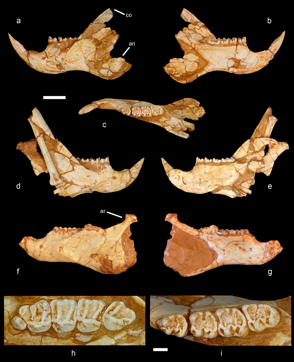
Mandible and cheek teeth of Miopetaurista neogrivensis.
(a to c) Partial left hemimandible (IPS56468j) in lateral, medial and dorsal views. (d to e) Partial right hemimandible (IPS56468i) in lateral and medial views. A caudal vertebra and a bone fragment are attached to the lateral side of the mandibular ramus. Both hemimandibles were associated to the partial skeleton IPS56468 from ACM/C5-D1. (f to g) Partial hemimandible (IPS87560) from ACM/C8-B sector in lateral and medial views. (h) Left upper cheek teeth series (P3–M3) of IPS56468h (Figure 6—Figure supplement 1 ). (i) Left lower cheek teeth series (p4–m3) of IPS56468j. Cheek teeth measurements are given in Supplementary file 4 whereas mandibular measurements are given in Supplementary file 6. For a detailed description and comparisons of cheek teeth and mandible morphology see Appendix 3.1 and 3.2. an, angular process; ar, articular process; co, coronoid process. Scale bar is 1 cm in figs. a to g; 2 mm in (h to i).
Figure 4
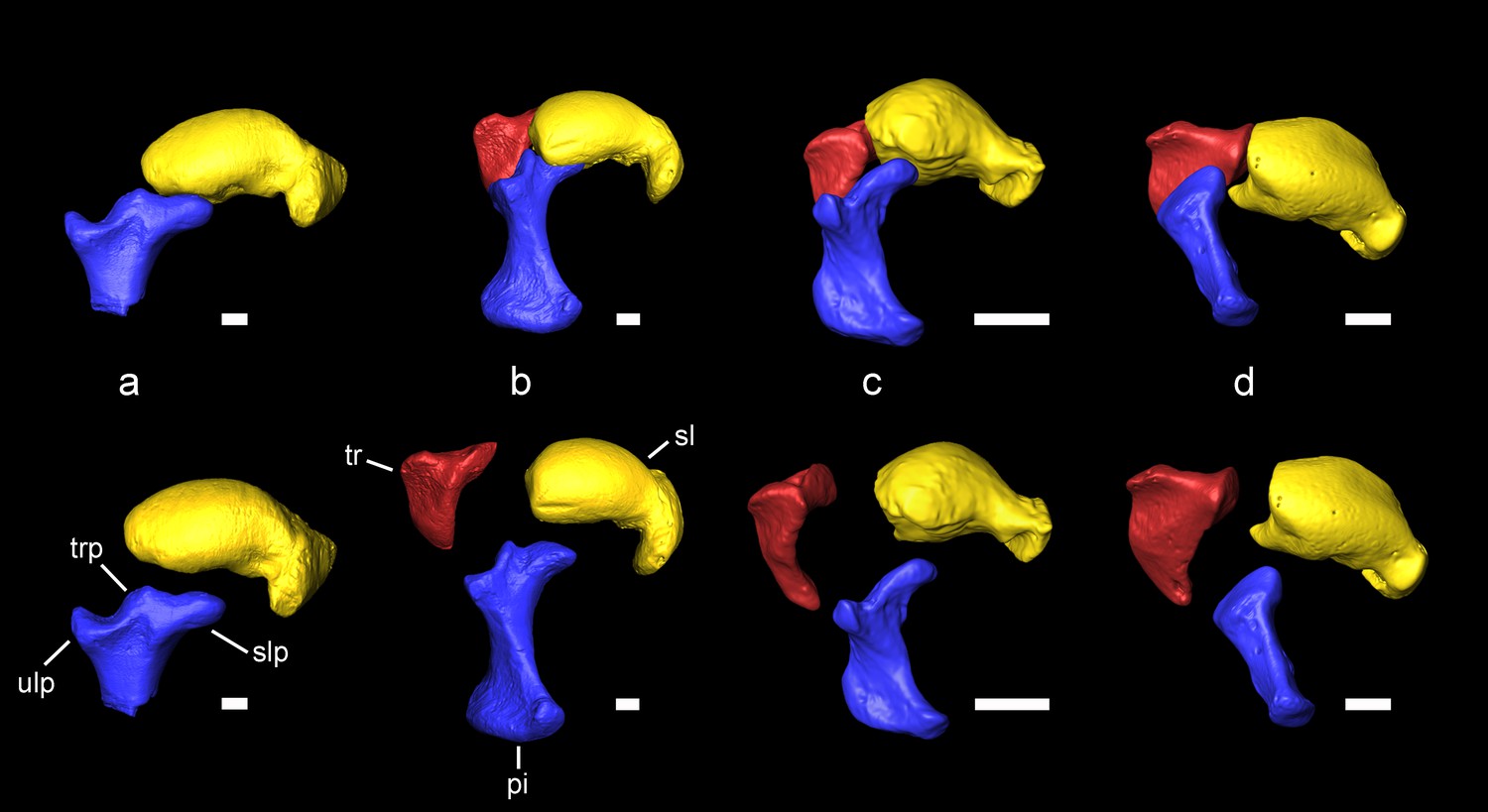
Carpal bones associated with the extension of the patagium of Miopetaurista neogrivensis as compared to extant squirrels.
Articulated bones are shown on top and disarticulated ones are shown below. (a) Miopetaurista neogrivensis. (b) Petaurista petaurista, large-sized flying squirrel, subtribe Pteromyina. (c) Hylopetes sagitta, small-sized flying squirrel, subtribe Glaucomyina. (d) Sciurus vulgaris, tree squirrel, tribe Sciurini. The patagium is supported by the styliform cartilage which is attached to the pisiform bone. Flying squirrels present an elevated process for articulation with the scapholunate in the pisiform, whereas in tree squirrels this bone only articulates with the triquetrum and the ulna. In addition, note the presence of a triquetral process in Miopetaurista and Petaurista, characteristic of the Pteromyina. All extant specimens are kept in the collections of the Naturalis Biodiversity Center (Leiden, the Netherlands). See Video 2 for an animated version of this figure. Collection numbers of the scanned specimens and computed tomography parameters used are given in Table 5. Tridimensional models generated from µCT scan data. pi, pisiform (in blue); sl, scapholunate (in yellow); slp, scapholunate process of the pisiform; tr, triquetrum (in red); trp, triquetral processes of the pisiform; ulp, ulnar process of the pisiform. Scale bar is 1 cm.
Figure 5 with 1 supplement
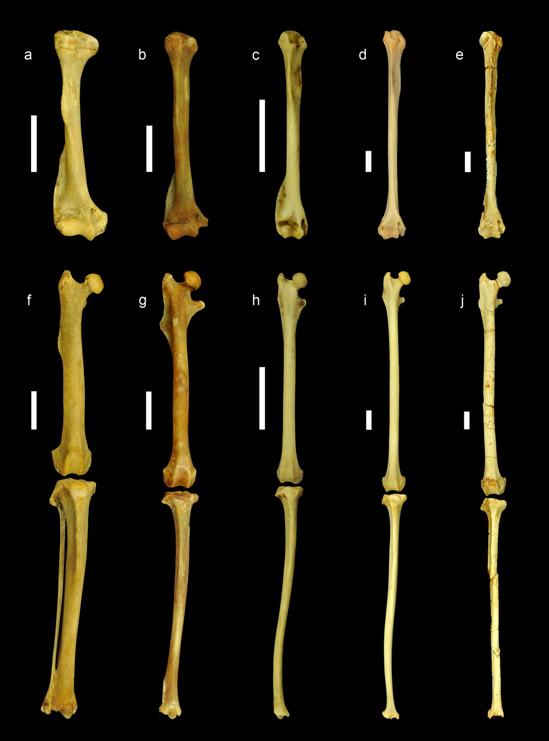
Comparison of the limb bones of extant ground, tree and flying squirrels with Miopetaurista neogrivensis.
All elements are scaled to femur length and shown in anterior view. Humerus (a–e) and articulated femur and tibia (f–j) of: (a,f) the xerin ground squirrel Xerus erythropus; (b,g) the callosciurin tree squirrel Callosciurus prevostii; (c,h) the small-sized flying squirrel (subtribe Glaucomyina) Hylopetes sagitta; (d,i) the large-sized fying squirrel (subtribe Pteromyina) Petaurista petaurista; (e,j) Miopetaurista neogrivensis. Note that limb bones of flying squirrels and M. neogrivensis are much longer and more slender than those of tree and ground squirrels. Furthermore, processes and areas for the insertion of the main limb muscles are reduced. For a description and comparison of the postcranial bones of M. neogrivensis, see Appendix 3.3. See Supplementary file 7 for the collection numbers of the figured specimens and postcranial measurements. All bones are right elements, except for a–b and f–g, which are reversed left elements. Scale bar is 1 cm.
Figure 5—figure supplement 1
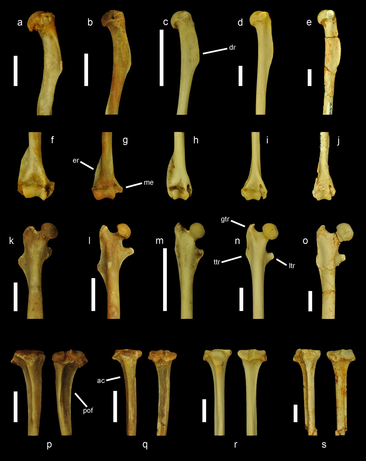
Comparison of the proximal and distal ends of the humerus, femur and tibia of extant ground, tree and flying squirrels with Miopetaurista neogrivensis.
(a to e) Proximal end of the humerus in lateral view. (f to j) Distal end of the humerus in anterior view. (k to o) proximal end of the femur in anterior view. (p to s) Proximal end of the tibia in anterior (left) and posterior (right) views. Figured specimens belong to: the marmotin ground squirrel Urocitellus paryii (a, f, k, p); the callosciurin tree squirrel Callosciurus prevostii (b, g, l, q); the small-sized flying squirrel (subtribe Glaucomyina) Hylopetes sagitta (c, h, m); the large-sized flying squirrel (subtribe Pteromyina) Petaurista petaurista (d, i, n, r); and Miopetaurista neogrivensis (e, j, o, s). All bones are right elements, except for b, g, l and q, which are reversed left elements. ac, anterior crest; dr, deltoid ridge; er, epicondylar ridge; gtr, greater trochanter; ltr, lesser trochanter; me, medial epicondyle; pof, popliteal fossa; ttr, third trochanter. Scale bar is 1 cm.
Figure 6 with 2 supplements
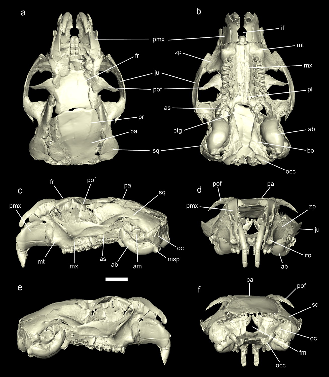
Reconstruction of the cranium of Miopetaurista neogrivensis.
Virtual reconstruction based on µCT scan data from specimens IPS56468h (see Figure 6—figure supplement 1) and IPS88677 (see Figure 6—figure supplement 2). Specimen IPS56468h was used as the basis for the reconstruction, with missing elements taken from IPS88677 (Table 3. (a) Dorsal view. (b) Ventral view. (c) Lateral (left) view. (d) Anterior view. (e) Lateral (right) view. (f) Posterior view. See Video 3 for an animated version of the skull reconstruction. For a detailed description of skull morphology see Appendix 3.2. Cranial measurements for original fossil specimens (IPS56468h, IPS88677) as well as for the virtually reconstructed cranium are given in Supplementary file 5. ab, auditory bulla; am, auditory meatus; as, alisphenoid; bo, basioccipital; fm, foramen magnum; fr, frontal; if, incisive foramen; ifo, infraorbital foramen; ju, jugal; msp, mastoid process; mt, masseteric tubercle; mx, maxillary; oc, occipital; occ, occipital condyle; pa, parietal; pl, palatine; pmx, premaximallary; pof, postorbital process of the frontal; pr, parietal ridges; ptg, pterygoid; sq, squamosal; zp, zygomatic plate. Scale bar is 1 cm.
Figure 6—figure supplement 1
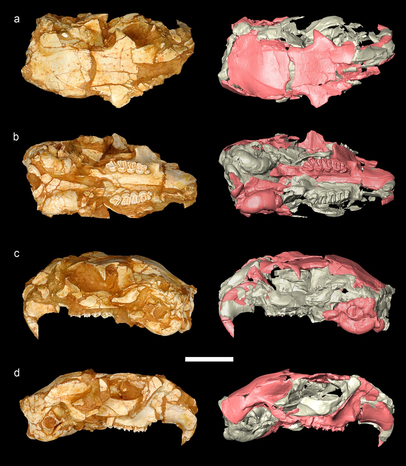
Cranium of Miopetaurista neogrivensis associated with partial skeleton IPS56468.
The partial skull (IPS56468h) belongs to the same individual as the partial skeleton IPS56468 from Abocador de Can Mata locality ACM/C5-D1. Fossil specimens are shown on the left, whereas tridimensional models based on µCT scan data are shown on the right. (a) Dorsal view. (b) Ventral view. (c) Lateral (left) view. (d) Lateral (right) view. Note that the cranium is obliquely crushed but not plastically deformed. Parts used in the reconstruction of the skull of M. neogrivensis are colored in pink. Cranial measurements are given in Supplementary file 5. Scale bar is 1 cm.
Figure 6—figure supplement 2
Cranium of Miopetaurista neogrivensis (IPS88677) from Abocador de Can Mata locality ACM/C8-Af.
Fossil specimens are shown on the left, whereas tridimensional models based on µCT scan data are shown on the right. (a) Dorsal view. (b) Ventral view. (c) Lateral (left) view. (d) Lateral (right) view. Note that the skull is dorsoventrally crushed and most of the premaxillary and frontal bones are not preserved. Parts used in the reconstruction of the skull of M. neogrivensis are colored in pink. Cranial measurements are given in Supplementary file 5. Scale bar is 1 cm.
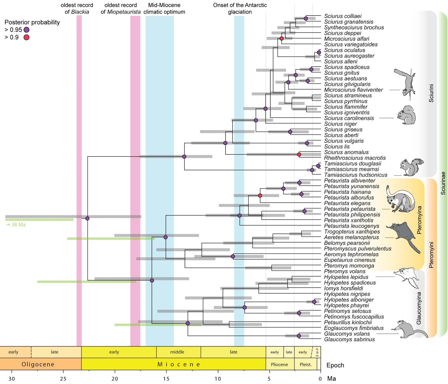
Figure 7 with 1 supplement

Flying squirrel phylogeny and node dating estimates based on a Bayesian total evidence analysis including Miopetaurista neogrivensis.
The analysis is based on 38 taxa, 105 morphological characters and 3345 base pairs (see Materials and Methods and Appendix 1.1). Purple circles at the nodes indicate posterior probabilities higher than 0.95. Error bars (gray shading) at the nodes are 95% highest posterior density (HPD) intervals for divergence dates. For selected nodes, 95% HPD intervals derived from an independent node dating analysis using BEAST (Figure 7—figure supplement 1) are also shown as green bars. Note the position of Miopetaurista neogrivensis as sister taxon of extant Petaurista. The age of the oldest purported flying squirrels in the fossil record as well as that of the earliest representatives of the genera Blackia and Miopetaurista is indicated for comparison (see also Figure 8). Two major global climatic events are also indicated. The morphological character list is given in Appendix 2. Genbank accession numbers for all the sequences used in phylogenetic analyses are given in Supplementary file 2 and morphological character matrix is given in Supplementary file 3.
Figure 7—figure supplement 1
Sciurinae phylogeny and node dating estimates using BEAST.
Based on 58 extant taxa and 3345 base pairs (see Materials and Methods and Appendix 1.2). The extinct Miopetaurista neogrivensis and Douglassciurus jeffersoni are used as calibration points. Purple and red circles at the nodes indicate posterior probabilities higher than 0.95 and 0.9, respectively. Error bars (grey shading) at the nodes are 95% highest posterior density (HPD) intervals for divergence dates. For selected nodes, 95% HPD intervals derived from an independent total evidence analysis (Figure 7) are also shown as blue bars. The age of the oldest purported flying squirrels in the fossil record as well as that of the earliest representatives of the genus Miopetaurista is indicated for comparison (see also Figure 8). Two major global climatic events are also indicated. Fossils used in the calibration are given in Table 4 (see also Appendix 1.2). Genbank accession numbers for all the sequences used in phylogenetic analyses are given in Supplementary file 2.
Figure 8

Fossil record of ‘flying squirrels’ and paleoclimatic data.
Temporal ranges of purported flying squirrel genera in Europe, Asia and North America. The 95% highest posterior density (HPD) intervals for flying squirrel divergence as derived from total evidence and node dating analyses are indicated in orange shading (see Figure 7 and Figure 7—figure supplement 1 ). Darker shading indicates the time interval where both independently calculated estimates overlap, thus defining the most likely time interval for flying squirrel divergence. Global paleoclimatic data are taken from Zachos et al., 2001.
Figure 9
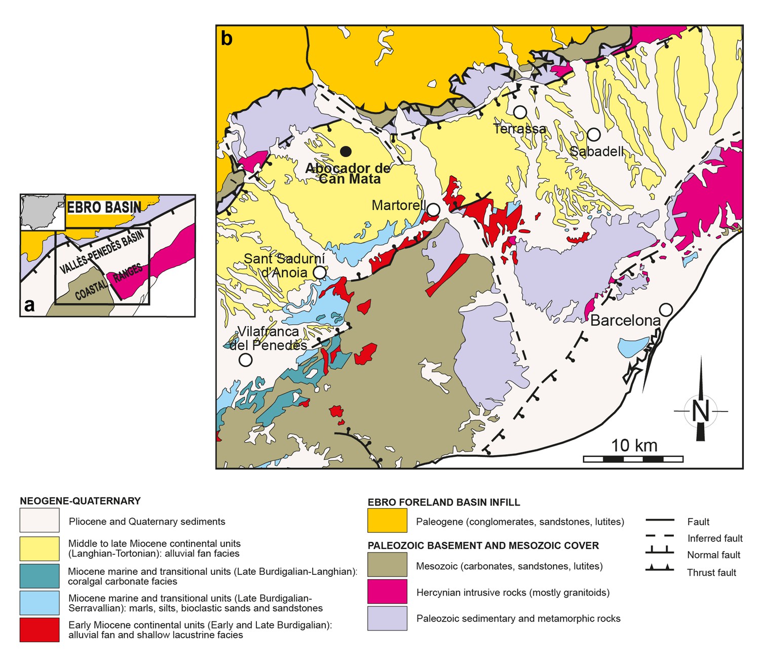
Geological map of the Vallès-Penedès Basin and situation of the fossil site.
(a) Schematic geological map of the Vallès-Penedès Basin (Catalonia, Spain) showing the area enlarged in Figure 9b. The inset shows its location within the Iberian Peninsula. (b) Situation of the Abocador de Can Mata series, located in distal alluvial fan facies of middle to late Miocene age. See Alba et al., 2017 for further details on the stratigraphy and chronology of the Abocador de Can Mata series and main sites. Map modified from Casanovas-Vilar et al., 2016.
Videos
Video 1
Reconstruction of the skeleton and life appearance of Miopetaurista neogrivensis.
The squirrel is shown reducing speed just before landing on a tree branch. Skeleton reconstruction based in the partial skeleton IPS56468 from Abocador de Can Mata. Coat pattern and color are based in extant Petaurista species, the sister taxon of Miopetaurista. A high-quality 3D surface model of the reconstructed skeleton is available at MorphoBank https://morphobank.org/index.php/Projects/ProjectOverview/project_id/3108
Video 2
Proximal carpal bones of Miopetaurista neogrivensis as compared to tree and flying squirrels.
Miopetaurista neogrivensis is compared to Petaurista petaurista (large-sized flying squirrel; subtribe Pteromyina); Hylopetes sagitta (small-sized flying squirrel; subtribe Glaucomyina); and Sciurus vulgaris (tree squirrel; tribe Sciurini). These bones form the morphofunctional complex associated with extension of the patagium. The flying membrane is supported by the styliform cartilage which is attached to the pisiform bone. High-quality 3D surface models of the carpal bones of Miopeataurista and other squirrels are available at MorphoBank https://morphobank.org/index.php/Projects/ProjectOverview/project_id/3108
Video 3
Cranial reconstruction of Miopetaurista neogrivensis.
Virtual reconstruction based on µCT scan data from specimens IPS56468h and IPS88677. Specimen IPS56468h was used as the basis for the reconstruction, with missing elements taken from IPS88677. A high-quality 3D surface model of the reconstructed cranium is available at MorphoBank https://morphobank.org/index.php/Projects/ProjectOverview/project_id/3108.
Video 4
Middle ear anatomy of Miopetaurista neogrivensis as compared to Petaurista petaurista.
Middle ear cavity anatomy in Miopetaurista neogrivensis (left; IPS88677) and the extant giant flying squirrel Petaurista petaurista (right; ZMA13418) reconstructed from µCT scan data. Note the presence of two ventral transbullar septa. Most flying squirrels show just two septa except, for a few Glaucomyina (Petinomys, Trogopterus, Pteromyscus) that exhibit a more complex pattern (see Appendix 3.2). Scale bar is 1 cm.
Tables
Table 1
Catalogue of bones and bone fragments composing the partial skeleton of Miopetaurista neogrivensis.
This list includes the catalogue numbers (preceded by the acronym ‘IPS’) of the various bones and bone fragments belonging to the partial skeleton of a single individual of Miopetaurista neogrivensis (IPS56468) from locality ACM/C5-D1. IPS, acronym for the collections of the Institut Català de Paleontologia Miquel Crusafont.
| Catalogue no. | Region | Description |
|---|---|---|
| IPS56468a | leg | right tibia |
| IPS56468b | leg | right femur |
| IPS56468c | leg | left femur |
| IPS56468d | leg | distal half of the left tibia |
| IPS56468e | arm | left humerus |
| IPS56468f | arm | right humerus |
| IPS56468g | arm | distal half of the right radius, with damaged epiphysis |
| IPS56468h | cranium | almost complete cranium, laterally compressed in an oblique angle |
| IPS56468i | cranium + tail | partial right mandible (angular process broken), caudal vertebra (probably corresponding to the mid part of the tail) |
| IPS56468j | cranium | partial left mandible (articular process broken, all other processes with minor damage) |
| IPS56468k | pelvic girdle | partial right coxal (proximal end of the ilium and part of the pubis damaged) |
| IPS56468l | pelvic girdle | partial left coxal (missing most of the pubis and ischium, extensive damage in the ilium) |
| IPS56468m | trunk | lumbar vertebrae (L3–L4) in anatomical connection |
| IPS56468n | trunk | lumbar vertebrae (L5–L6) in anatomical connection |
| IPS56468o | neck | partial axis (only part of the vertebral body is preserved) and partial cervical vertebra (C3) |
| IPS56468p | trunk | thoracic vertebra (T1?) |
| IPS56468q | tail | four caudal vertebrae (mid part of the tail) that articulate with one another |
| IPS56468r | trunk | seven rib fragments |
| IPS56468s | ankle | left calcaneus and left navicular |
| IPS56468t | ankle | left astragalus |
| IPS56468u | trunk | three partial thoracic vertebrae (T2–T4?) that articulate with one another |
| IPS56468v | indeterminate | associated bone fragments (may not belong to M. neogrivensis) |
| IPS56468w | indeterminate | associated bone fragments (may not belong to M. neogrivensis) |
| IPS56468x | foot? | six distal phalanges which are not assigned to any particular ray or side; attribution to the foot is tentative |
| IPS56468y | trunk | two sternebrae that articulate with one another |
| IPS56468z | foot | left metatarsals 2–4 in anatomical connection |
| IPS56468aa | thoracic girdle | right clavicle (with minor damage in its acromial end) and partial left clavicle (acromial end missing) |
| IPS56468ab | foot | complete right metatarsal 3, and partial rigth metatarsals 4 (proximal end missing), 2 and 4 (only distal half preserved) |
| IPS56468ac | foot | four proximal phalanges and one partial proximal phalanx (distal half); they are not assigned to any particular ray or side |
| IPS56468ad | foot? | seven intermediate phalanges and five fragments; they are not assigned to any particular ray or side and attribution to the foot is tentative |
| IPS56468ae | hand | four proximal phalanges; they are not assigned to any particular ray or side |
| IPS56468af | ankle | left intermediate and medial cuneiform |
| IPS56468ag | ankle | right navicular |
| IPS56468ah | wrist | right scapholunate and dorsal end of the right pisiform |
Table 2
Catalogue of additional material of Miopetaurista neogrivensis.
This list includes the additional material of Miopetaurista neogrivensis from locality ACM/C5-D1 and the approximately stratigraphicaly equivalent localities ACM/C8-Af and ACM/C6-A5, as well as from sector ACM/C8-B. IPS, acronym for the collections of the Institut Català de Paleontologia Miquel Crusafont.
| Catalogue no. | Locality | Anatomical element |
|---|---|---|
| IPS43480 | ACM/C5-D1 | L m1 |
| IPS43505 | ACM/C5-D1 | L m3 |
| IPS43675 | ACM/C5-D1 | R P4 |
| IPS43677 | ACM/C5-D1 | partial left mandible with p4–m3 (only part of the mandibular body preserved) |
| IPS43724 | ACM/C5-D1 | right maxillary fragment with partial P4-M2 |
| IPS77856 | ACM/C5-D1 | R P4 |
| IPS77857 | ACM/C5-D1 | R P4 |
| IPS77858 | ACM/C5-D1 | R M1/M2 |
| IPS77859 | ACM/C5-D1 | R M1/M2 |
| IPS77860 | ACM/C5-D1 | fragment of R M1/M2 |
| IPS77861 | ACM/C5-D1 | fragment of R M1/M2 |
| IPS77862 | ACM/C5-D1 | L M1/M2 |
| IPS77863 | ACM/C5-D1 | broken L M1/M2 |
| IPS77864 | ACM/C5-D1 | fragment of L M1/M2 |
| IPS77865 | ACM/C5-D1 | fragment of L M1/M2 |
| IPS77866 | ACM/C5-D1 | L M3 |
| IPS77867 | ACM/C5-D1 | R M3 |
| IPS77868 | ACM/C5-D1 | L dp4 |
| IPS77869 | ACM/C5-D1 | fragment of L dp4 |
| IPS77870 | ACM/C5-D1 | R dp4 |
| IPS77871 | ACM/C5-D1 | L p4 |
| IPS77872 | ACM/C5-D1 | R p4 |
| IPS77873 | ACM/C5-D1 | R p4 |
| IPS77874 | ACM/C5-D1 | L m1 |
| IPS77875 | ACM/C5-D1 | L m1 |
| IPS77876 | ACM/C5-D1 | L m1 |
| IPS77877 | ACM/C5-D1 | L m2 |
| IPS77878 | ACM/C5-D1 | L m2 |
| IPS77879 | ACM/C5-D1 | broken L m1/m2 |
| IPS77880 | ACM/C5-D1 | abraded L m1/m2 |
| IPS77881 | ACM/C5-D1 | fragment of L m1/m2 |
| IPS77882 | ACM/C5-D1 | fragment of L m1/m2 |
| IPS77883 | ACM/C5-D1 | R m1 |
| IPS77884 | ACM/C5-D1 | lingually abraded R m1 |
| IPS77885 | ACM/C5-D1 | broken R m1/m2 |
| IPS77886 | ACM/C5-D1 | R m3 |
| IPS77887 | ACM/C5-D1 | fragment of upper molar |
| IPS77888 | ACM/C5-D1 | fragment of lower molar |
| IPS78179 | ACM/C5-D1 | L M1/M2 |
| IPS85340 | ACM/C5-D1 | fragment of left mandible with m1–m3 and associated p4 |
| IPS85410 | ACM/C6-A5 | partial cranium (includes the dorsal half of the skull as well as part of the right zygomatic arch and maxillary bone with damaged P4–M3) |
| IPS87560 | ACM/C8-B sector | partial left mandible with p4–m3 (incisor and coronoid process broken, p4 damaged) |
| IPS88677 | ACM/C8-Af | partial cranium (dorsoventrally crushed) |
Table 3
Cranial reconstruction of Miopetaurista neogrivensis.
List of cranial elements used in the reconstruction of the complete skull indicating catalog no. of each bone or bone fragment and, when appropriate, which side (left or right) was mirrored.
| Cranial element | Side | Catalog. no |
|---|---|---|
| alisphenoid | left | IPS88677 |
| auditory bulla | left | IPS56468h |
| basisphenoid | left | IPS56468h |
| basioccipital and occipital condyle | left | IPS56468h |
| cheek teeth | right | IPS56468h |
| frontal | – | IPS56468h |
| frontal (orbital part) | left | IPS88677 |
| hamular process of pterygoid | right | IPS56468h |
| incisor | left | IPS56468h |
| jugal | left | IPS88677 |
| jugal (posterior half) | right | IPS88677 |
| maxillary | right | IPS56468h |
| occipital (right half) | right | IPS88677 |
| palatines | – | IPS88677 |
| parietal | – | IPS56468h |
| postorbital process of frontal | right | IPS88677 |
| pterygoid | left | IPS56468h |
| pterygoid fossa (alisphenoid) | – | IPS56468h, IPS88677 |
| premaxillary | right | IPS56468h |
| premaxillary (dorsal fragments) | right, left | IPS88677 |
| squamosal | left | IPS56468h |
| squamosal (zygomatic process) | right | IPS88677 |
| zygomatic plate (maxillary) | left | IPS88677 |
Table 4
Fossils used in the total evidence and node dating phylogenetic analyses.
Miopetaurista neogrivensis is considered as a calibration point for the divergence between Petaurista and all the other Pteromyina in node dating analysis and is considered as a tip date in the total evidence analysis. Root age in the total evidence analysis is calibrated considering the age of the oldest Aplodontiidae (sister group of Sciuridae), the oldest Gliridae (sister group of Aplodontiidae + Sciuridae), the oldest Myodonta, and the oldest-known rodent, the ischyromyid Acritoparamys atavus. The oldest sciurid that is reasonably complete (Douglassciurus jeffersoni) is used as tip date in the total evidence analysis and is also considered as a calibration point in the node dating analysis. See Appendix 1 for details on the calibration of the molecular clock using fossils.
| Fossil | Locality | Node on phylogeny | Max. age | Min. age | Taxonomic refs. | Age refs. |
|---|---|---|---|---|---|---|
| Acritoparamys atavus | Bear Creek, Montana, USA | maximum age for the root (oldest-known rodent) | 56.0 Ma | 55.8 Ma | Janis et al., 2008; Korth, 1994 | Janis et al., 2008 |
| Erlianomys combinatus | AS-1, Nuhetingboerhe, Erlian Basin, Inner Mongolia, China | maximum age for the root (oldest-known Myodonta) | 54.8 Ma | 53.9 Ma | Li and Meng, 2010; Wu et al., 2012 | Wu et al., 2012 |
| Eogliravus wildi | Mas de Gimel, Hérault, France | maximum age for the root (oldest-known Gliridae) | 50.7 Ma | 47.4 Ma | Daams and Bruijn, 1995; Dawson, 2003; Hartenberger J-L and Hartenberger, 1971; Storch and Seiffert, 2007; Vianey-Liaud, 1985 | Daams and Bruijn, 1995; Dawson, 2003 |
| Spurimus selbyi | Upper Teppe Formation, Wyoming, USA | minimum age for the root (oldest-known Aplodontiidae) | 46.3 Ma | 45.7 Ma | Flynn and Jacobs, 2008; Korth, 1994 | Flynn and Jacobs, 2008 |
| Douglassciurus jeffersoni | Renova Formation, Pipestone Springs, Montana, USA | Douglassciurus/other Sciuridae | 36.6 Ma | 35.8 Ma | Emry and Korth, 1996; Emry and Thorington, 1982; Goodwin, 2008 | Goodwin, 2008 |
| Heteroxerus sp. | Canales, Loranca Basin, Spain | Xerinae/Sciurinae | 26.4 Ma | 25.0 Ma | Álvarez-Sierra et al., 1999 | Álvarez-Sierra et al., 1999 |
| Sciurus olsoni | NB29A, Truckee Formation, Nevada, USA | Sciurus/ Tamiasciurus | 13.6 Ma | 10.3 Ma | Emry et al., 2005 | Goodwin, 2008; Emry et al., 2005; Woodburne, 2004 |
| Miopetaurista neogrivensis | Abocador de Can Mata ACM/C5- D1, Vallès-Penedès Basin, Spain | Petaurista/other Pteromyina | 11.6 Ma | this work | this work |
Table 5
μCT scan parameters used for Miopetaurista neogrivensis and additional comparative material.
IPS, acronym for the collections of the Institut Català de Paleontologia Miquel Crusafont. All other specimens are kept in the collections of the Naturalis Biodiversity Center (Leiden, the Netherlands). When a filter was used, it was aluminum. For additional details see Materials and Methods.
| Catalogue no. | Taxon | Element | Voltage (kV) | Current (µA) | Filter | Projections | Voxel size (µm) |
|---|---|---|---|---|---|---|---|
| IPS56468h | Miopetaurista neogrivensis | skull | 130 | 72 | 1 mm | 1880 | 39.71 |
| IPS88677 | Miopetaurista neogrivensis | skull | 130 | 72 | 1 mm | 1880 | 39.71 |
| ZMA13418 | Petaurista petaurista | skull | 100 | 100 | 0.5 mm | 1290 | 13.17 |
| IPS56468ah | Miopetaurista neogrivensis | carpals | 80 | 124 | not used | 651 | 10.67 |
| 056.054.b | Hylopetes sagitta | carpals | 100 | 100 | not used | 558 | 11.32 |
| RMNH.MAM.130 | Petaurista petaurista | carpals | 79 | 125 | not used | 482 | 13.17 |
| 27291 | Sciurus vulgaris | carpals | 59 | 167 | not used | 550 | 13.04 |
Table 6
Model specifications for phylogenetic analyses.
Molecular model specifications for total evidence and node dating phylogenetic analyses.
| Analysis software | Genes | Model | Gamma distribution | Invariant |
|---|---|---|---|---|
| BEAST | cytb codon 1, 16S, 12S | GTR | yes | yes |
| BEAST | cytb codon 2, irbp codon 2 | HKY | no | yes |
| BEAST | cytb codon 3 | GTR | yes | yes |
| BEAST | irbp codon 1 | HKY | no | no |
| BEAST | irbp codon 3 | HKY | yes | no |
| MrBayes | cytb codon 1, 16S, 12S | GTR | yes | yes |
| MrBayes | cytb codon 2, irbp codon 2 | HKY | yes | yes |
| MrBayes | cytb codon 3 | GTR | yes | yes |
| MrBayes | irbp codon 1 | HKY | yes | no |
| MrBayes | irbp codon 3 | GTR | yes | no |
Additional files
-
Supplementary file 1
Reconstruction of the skeleton of Miopeaturista neogrivensis.
- https://doi.org/10.7554/eLife.39270.027
-
Supplementary file 2
GenBank accession numbers for all the sequences used in phylogenetic analyses.
Accession numbers for all the species considered in total evince analysis and/or node dating analysis. We retrieved the four genes more frequently used in previous phylogenetic studies of the Sciuridae: 12S, 16S, cytochrome b, and irbp (Mercer and Roth, 2003).
- https://doi.org/10.7554/eLife.39270.028
-
Supplementary file 3
Morphological character matrix for total evidence phylogenetic analysis.
See Appendix 2 for character description and coding.
- https://doi.org/10.7554/eLife.39270.029
-
Supplementary file 4
Cheek tooth measurements of Miopetaurista neogrivensis.
Cheek tooth measurements of Miopetaurista neogrivensis from localities ACM/C5-D1, ACM/C8-Af, ACM/C6-A5, as well as sector ACM/C8-B (material is listed in Tables 1–2). Measurement method follows Casanovas-Vilar et al., 2015: L, mesiodistal length; W, buccolingual width; w1, buccolingual width measured at the mesial margin of the tooth (only for m1 and m2); w2, buccolingual width measured at the distal margin of the tooth (only for m1 and m2). Estimated measurements (due to minor damage to the cheek teeth) are within parentheses. Abbreviations for dental elements follow Smith and Dodson, 2003. IPS, acronym for the collections of the Institut Català de Paleontologia Miquel Crusafont. All measurements are in mm.
- https://doi.org/10.7554/eLife.39270.030
-
Supplementary file 5
Cranial measurements of Miopetaurista neogrivensis and extant flying squirrels.
Cranial measurements for Miopetaurista neogrivensis and some extant flying squirrels. The specimens of the extant species are housed in the Naturalis Biodiversity Center (Leiden, the Netherlands), except for Glaucomys sabrinus, which comes from the collections of the Institut Català de Paleontologia Miquel Crusafont. Cranial measurements for the virtually reconstructed skull of M. neogrivensis are also included. Estimated measurements are in parentheses. Acronyms and definitions of the measurements follow Bertrand et al., 2016 and Nicolas et al., 2008: BNAS, maximum breadth of the nasals; BRCA, maximum breadth of the braincase; BULL, maximum length of the auditory bulla; CTL, length of the upper cheek tooth row; DIA, length of the diastema (from the alveolus of the incisor to the alveolus of the P3); HEBA, henselion–basion length; HEPA, henselion–palation length; IF, length of the incisive foramen; INTE, smallest interorbital breadth; LNAS, maximum length of the nasals; PALA, smallest palatal breadth; RB, maximum rostrum breadth; RH, mediosagittal projection of rostrum height at the anterior border of the M1; SL, maximum length of the skull. The virtual reconstruction of the cranium of M. neogrivensis is shown in Figure 6, for the original crania see Figure 6—figure supplements 1–2. All measurements are in mm.
- https://doi.org/10.7554/eLife.39270.031
-
Supplementary file 6
Mandibular measurements of Miopetaurista neogrivensis and extant flying squirrels.
Mandibular material of Miopetaurista neogrivensis is listed in Tables 1–2. The specimens of the extant species are housed in the Naturalis Biodiversity Center (Leiden, the Netherlands), except for Glaucomys sabrinus which comes from the collections of the Institut Català de Paleontologia Miquel Crusafont. Estimated measurements are in parentheses. Acronyms and definitions of the measurements are as follows: DIA1, length of the diastema measured from the alveolus of the incisor to the anterior alveolus of the p4; DIA2, length of the diastema from the anterior alveolus of the p4 to the projection of the incisor; DDIA, depth of the mandibular corpus at the lowermost point of the diastema; DM1, depth of the mandibular corpus below the anterior border of the m1; DM3, depth of the mandibular corpus below the anterior border of the m3; LAJ, length from the most ventral point of the angular apophysis to the anterior margin of the articular condyle; LCJ, length from the tip of the coronoid apophysis to the anterior margin of the articular condyle; LTL, length of the lower cheek tooth row; ML, maximum length of the mandible; RH, maximum height of the mandibular ramus. The mandibular remains of M. neogrivensis are shown in Figure 3. All measurements are in mm.
- https://doi.org/10.7554/eLife.39270.032
-
Supplementary file 7
Postcranial measurements of Miopetaurista neogrivensis and extant squirrels.
Postcranial measurements for Miopetaurista neogrivensis (for material list see Table 1) and some extant squirrels. The specimens of the extant species are housed in the Naturalis Biodiversity Center (Leiden, the Netherlands), except for Glaucomys sabrinus and Sciurus carolinensis which come from the collections of the Institut Català de Paleontologia Miquel Crusafont. Only right elements were measured. Acronyms and definitions of the measurements are based in Samuels and Van Valkenburgh, 2008: BI, brachial index (RL/HL), indicating the proportions of the distal and proximal elements of the forelimb; CI, crural index (TL/FL), indicating the proportions of the distal and proximal elements of the forelimb; FEB, epicondylar mediolateral breadth of the distal femur; FGT, mediolateral breadth of the greater trochanter of the femur; FL, femoral length from the tip of the femoral head to the tip of the medial epicondyle; FMD, greater diameter of the femur at midshaft; FRI, femoral robusticidty index (FMD/FL); HEB, epicondylar mediolateral breadth of the distal humerus; HL, humeral length from the tip of the head to the tip of the most distal point of the trochlea; HMD greater diameter of the humerus at midshaft; HRI, humeral robusticity index (HMD/HL); IM, intermembral index [(HL +RL)/(FL +TL)], indicating the length of the forelimb relative to the hindlimb; TL, tibial length from the medial condyle to the tip of the medial malleolus; TMD, greater diameter of the tibia at midshaft; TRI, tibial robusticity index (TMD/TL); RL, radius lenght. All measurements are in mm. Specimens shown in Figure 5 are: Miopetaurista neogrivensis IPS56468; Petaurista petaurista RMNH.MAM.130; Hylopetes sagitta 056.054.b; Callosciurus prevostii 056.099 .j; Urocitellus paryii 065.238a; Xerus erythropus 26483.
- https://doi.org/10.7554/eLife.39270.033
-
Transparent reporting form
- https://doi.org/10.7554/eLife.39270.034
Download links
A two-part list of links to download the article, or parts of the article, in various formats.
Downloads (link to download the article as PDF)
Open citations (links to open the citations from this article in various online reference manager services)
Cite this article (links to download the citations from this article in formats compatible with various reference manager tools)
Oldest skeleton of a fossil flying squirrel casts new light on the phylogeny of the group
eLife 7:e39270.
https://doi.org/10.7554/eLife.39270


