Gαq-PKD/PKCμ signal regulating the nuclear export of HDAC5 to induce the IκB expression and limit the NF-κB-mediated inflammatory response essential for early pregnancy
- Xiamen Key Laboratory of Reproduction and Genetics, Department of Reproductive Medicine, Women and Children's Hospital, School of Medicine, Xiamen University, China
- Xiamen Key Library of Rheumatology and Clinical Immunology, Department of Rheumatology and Clinical Immunology, The First Affiliated Hospital of Xiamen University, School of Medicine, Xiamen University, China
- Fujian Provincial Key Laboratory of Reproductive Health Research, Department of Obstetrics and Gynecology, The First Affiliated Hospital of Xiamen University, School of Medicine, Xiamen University, China
- Department of Pathology, Women and Children's Hospital, School of Medicine, Xiamen University, China
- Department of Obstetrics and Gynecology, Key Laboratory for Major Obstetric Diseases of Guangdong Province, The Third Affiliated Hospital of Guangzhou Medical University, China
Figures
Figure 1 with 2 supplements
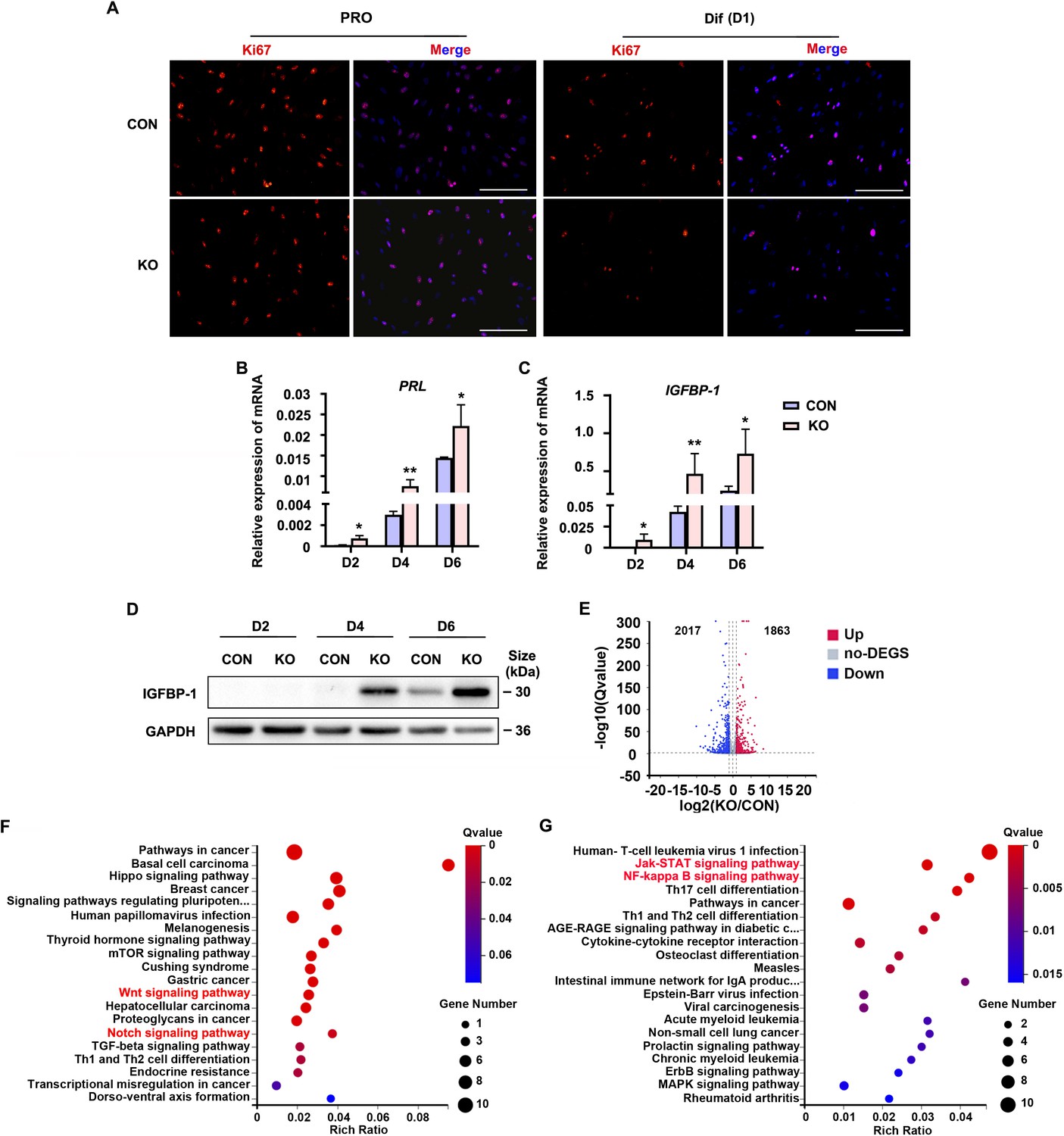
Human endometrial stromal cell (HESC) decidualization was advanced in the absence of Gαq.
(A) Immunofluorescence staining of Ki67 in control (CON) and GNAQ knockout (KO) proliferative HESCs (D0) and decidual HESC treated with differential culture medium (Dulbecco’s modified Eagle’s medium [DMEM]/F12 with 2% charcoal-stripped fetal bovine serum [CS-FBS], dibutyryl cAMP [dbcAMP], and medroxyprogesterone 17-acetate (MPA)) for 1 day. Scale bars, 100 μm. (B, C) Quantitative RT-PCR (qRT-PCR) analysis of prolactin (PRL) and insulin-like growth factor-binding protein-1 (IGFBP-1) mRNA expression levels in CON and KO HESC treated with differential culture medium for 2, 4, and 6 days (n = 5). (D) Western blot analysis of the IGFBP-1 protein levels in CON and KO decidual cells treated as in (B). GAPDH was used as the loading control. (E) Volcano plots showing significantly altered genes of KO/CON cells treated with differential culture medium for 3 days, UP, significant up regulated genes (Q value ≤0.05, red); Down, significant downregulated genes (Q value ≤0.05, blue); no-DEGS, no significant changed genes (gray). (F, G) Kyoto Encyclopedia of Genes and Genomes (KEGG) pathway analysis upregulated genes of in knockout cells during decidualization (Q value ≤0.05). Representative data are shown from three to five independent experiments. (B) and (C) were analyzed with unpaired Student’s t-test. *p < 0.05 and **p < 0.01.
-
Figure 1—source data 1
Original blot images of Figure 1D.
- https://cdn.elifesciences.org/articles/83083/elife-83083-fig1-data1-v1.zip
Figure 1—figure supplement 1
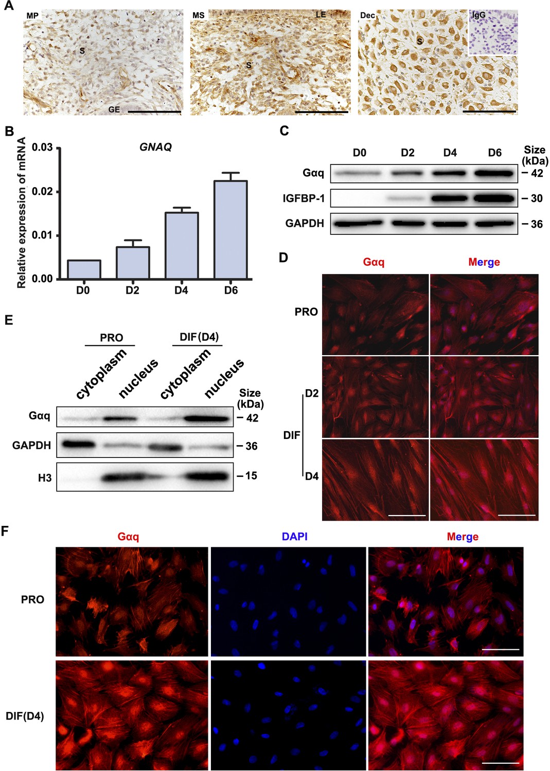
Gαq is expressed in both human endometria in vivo and decidual stromal cells in vitro.
(A) Gαq expression in the mid-proliferative and mid-secretory phases of the menstrual cycle and in the deciduas of early pregnancy was revealed by immunohistochemical staining. MP, mid-proliferative phase; MS, mid-secretory phase; Dec, decidual cells of early pregnancy; S, stroma; LE, luminal epithelium; GE, glandular epithelium. IgG was used for the negative control (inset). Scale bars, 100 μm. (B) qRT-PCR analysis of GNAQ mRNA expression levels in proliferative human endometrial stromal cell (HESC) (D0) and HESC treated with differential culture medium for 2, 4, and 6 days (n = 3). (C) Western blot analysis of the Gαq and IGFBP-1 protein expression in proliferative HESC (D0) and HESC treated as in (B). GAPDH was used as the loading control. (D) Immunofluorescence staining of Gαq in proliferative HESC and HESC treated with differential culture medium for 2 and 4 days. Scale bars, 100 μm. (E) Western blot analysis of the Gαq protein expression in the cytoplasm and nucleus of proliferative HESC (D0) and HESC treated with differential culture medium for 6 days. GAPDH and histone 3 (H3) were used as the loading controls. (F) Immunofluorescence staining of Gαq in proliferative and decidual primary ESCs treated with differential culture medium for 2 and 4 days. PRO, proliferation; DIF, differentiation. Scale bars, 100 μm. Representative data are shown from three independent experiments.
-
Figure 1—figure supplement 1—source data 1
Original blot images of Figure 1—figure supplement 1C and E.
- https://cdn.elifesciences.org/articles/83083/elife-83083-fig1-figsupp1-data1-v1.zip
Figure 1—figure supplement 2
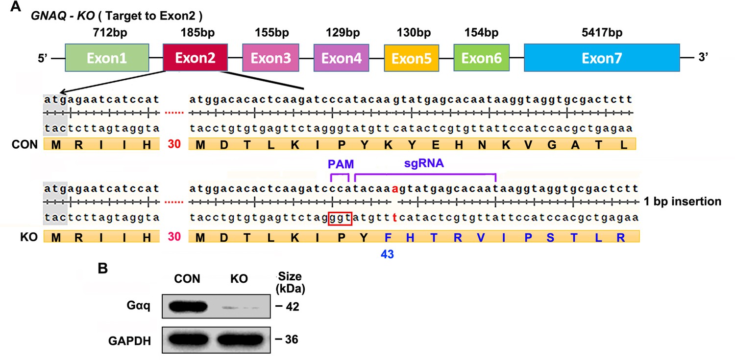
Generation of GNAQ knockout human endometrial stromal cell (HESC) lines.
(A) Sanger-sequencing analysis of genomic DNA from GNAQ knockout (KO) cells. The sequencing results indicate the formation of frameshift mutations in the shown KO cells lines. (B) Western blot analysis Gαq protein levels in control (CON) and KO HESC cells. GAPDH was used as the loading controls.
-
Figure 1—figure supplement 2—source data 1
Original blot images of Figure 1—figure supplement 2B.
- https://cdn.elifesciences.org/articles/83083/elife-83083-fig1-figsupp2-data1-v1.zip
Figure 2 with 2 supplements
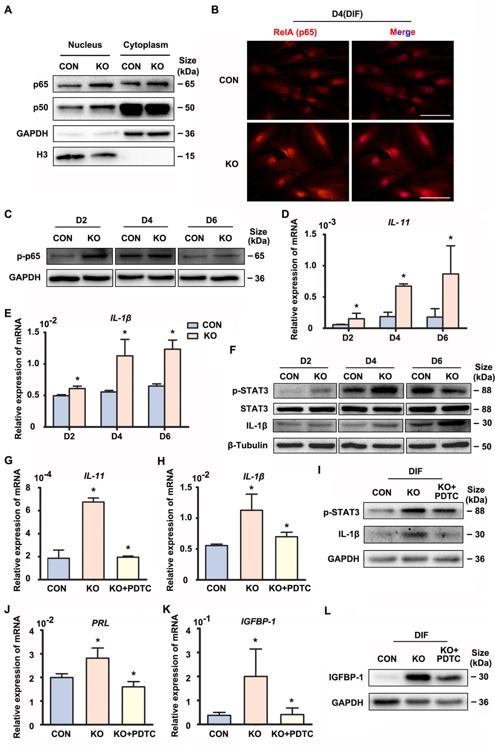
Gαq deficiency increased inflammatory cytokine expression in decidual stromal cells.
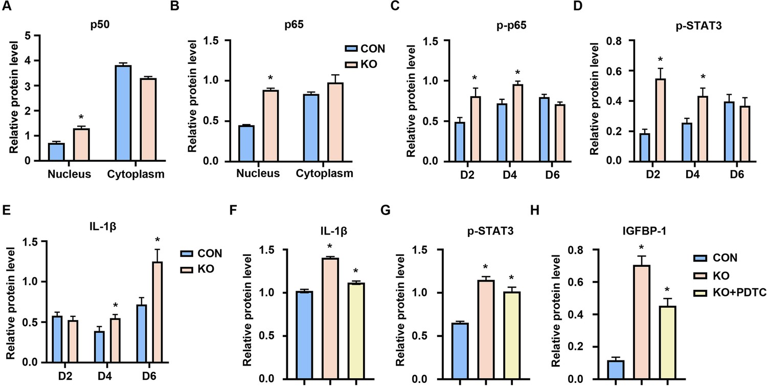
(A) Western blot analysis of the p65 and p50 expression in cytoplasm and nucleus of CON and KO human endometrial stromal cell (HESC) treated with differential culture medium for 4 days, nuclear translocation of p65 and p50 were higher in GNAQ-KO decidual cells than in control. GAPDH and histone 3 (H3) were used as the loading controls. (B) Immunofluorescence staining of p65 in CON and KO stromal cells and treated with differential culture medium for 4 days, nuclear translocation p65 were higher in GNAQ-KO decidual cells than in control. Scale bars, 100 μm. (C) Western blot analysis of p-p65 expression in whole-cell lysates of CON and KO cells treated with differential culture medium for 2, 4, and 6 days, phosphorylation of the nuclear factor (NF)-κB subunit p65 were higher in GNAQ-KO decidual cells than in control. GAPDH was used as the loading controls. (D, E) qRT-PCR analysis of IL-11 and IL-1β mRNA expression levels in CON and KO HESC treated as in (C) (n = 5). (F) Western blot analysis of p-STAT3, STAT3, and IL-1β expression in whole-cell lysates of CON and KO HESC treated as in (C). β-Tubulin was used as the loading control. (G, H, J, K) qRT-PCR analysis of IL-11, IL-1β, PRL, and IGFBP-1 mRNA after treatment with NF-κB inhibitor, pyrrolidinedithiocarbamate ammonium (PDTC, 10 μM), in CON and KO HESC treated with differential culture medium for 4 days (n = 3). (I, L) Western blot analysis of p-STAT3, IL-1β, and IGFBP-1 expression in whole-cell lysates of differentiated CON and KO HESC treated as in (G). GAPDH was used as the loading control. Representative data are shown from three to five independent experiments. (D and E) were analyzed with unpaired Student’s t-test. *p < 0.05 . (G, H, J, and K) were calculated with one-way analysis of variance (ANOVA) with Bonferroni’s multiple comparison tests. *p < 0.05.
-
Figure 2—source data 1
Original blot images of Figure 2.
- https://cdn.elifesciences.org/articles/83083/elife-83083-fig2-data1-v1.zip
Figure 2—figure supplement 1
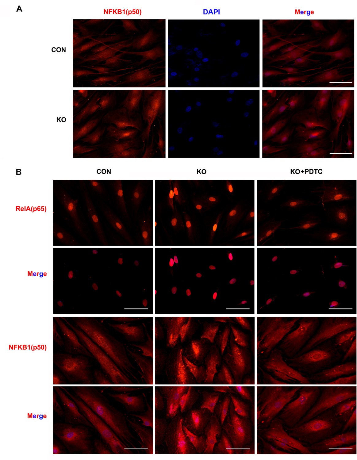
Immunofluorescence staining of p50 and p65 in CON, KO or PDTC treated differentiated stromal cells.
(A) Immunofluorescence staining of p50 in CON and KO differentiated stromal cells and treated with differential culture medium for 4 days, nuclear translocation of the p50 were higher in GNAQ-KO decidual cells than in control. Scale bars, 100 μm. (B) Immunofluorescence staining of p65 and p50 after treatment with nuclear factor (NF)-κB inhibitor, pyrrolidinedithiocarbamate ammonium (PDTC, 10 μM), in differentiated CON and KO human endometrial stromal cell (HESC) treated with differential culture medium for 4 days, PDTC blocked p65 and p50 translocate to the nucleus. Scale bars, 100 μm.
Figure 2—figure supplement 2
Relative of western blot analysis of Figure 2.
(A, B) Relative of western blot analysis of the p65 and p50 expression in cytoplasm and nucleus of CON and KO. The values are normalized to the GAPDH or H3 expression level and indicated as the mean ± standard error of the mean (SEM) of three independent experiments (n = 3, *p < 0.05, versus controls). (C–E) Relative of western blot analysis of p-p65, p-STAT3, and IL-1β expression in whole-cell lysates of CON and KO cells. The values are normalized to the GAPDH or β-Tubulin expression level and indicated as the mean ± SEM of three independent experiments (n = 3, *p < 0.05, versus controls). (F-H) Relative of western blot analysis of p-STAT3, IL-1β, and IGFBP-1 expression in whole-cell lysates of differentiated CON and KO human endometrial stromal cell (HESC) treated with nuclear factor (NF)-κB inhibitor, pyrrolidinedithiocarbamate ammonium (PDTC, 10 μM). The values are normalized to the GAPDH or β-Tubulin expression level and indicated as the mean ± SEM of three independent experiments (n = 3, *p < 0.05, versus controls).
Figure 3 with 1 supplement
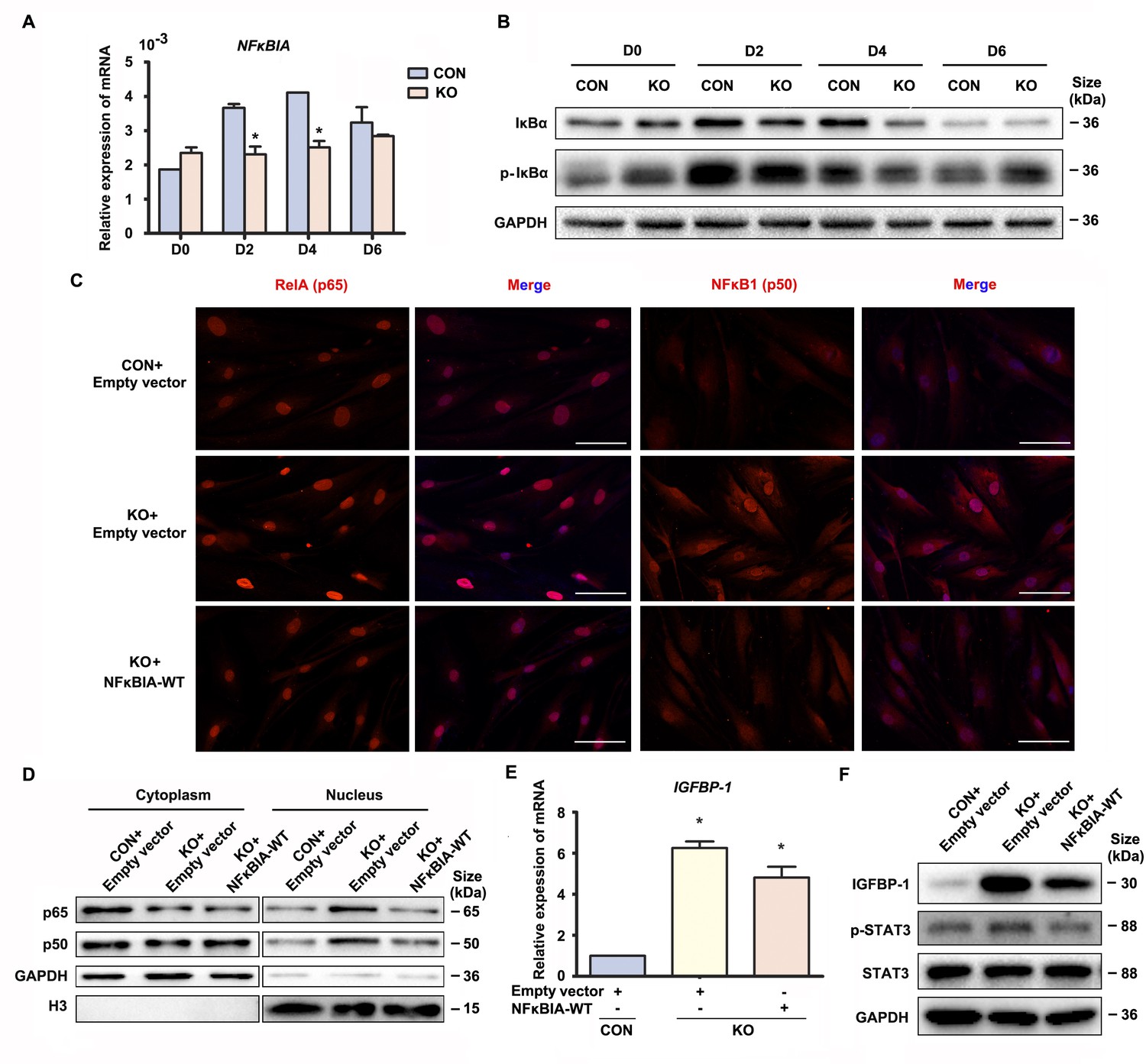
Gαq deficiency led to over-activated nuclear factor (NF)-κB signaling pathway during decidualization.
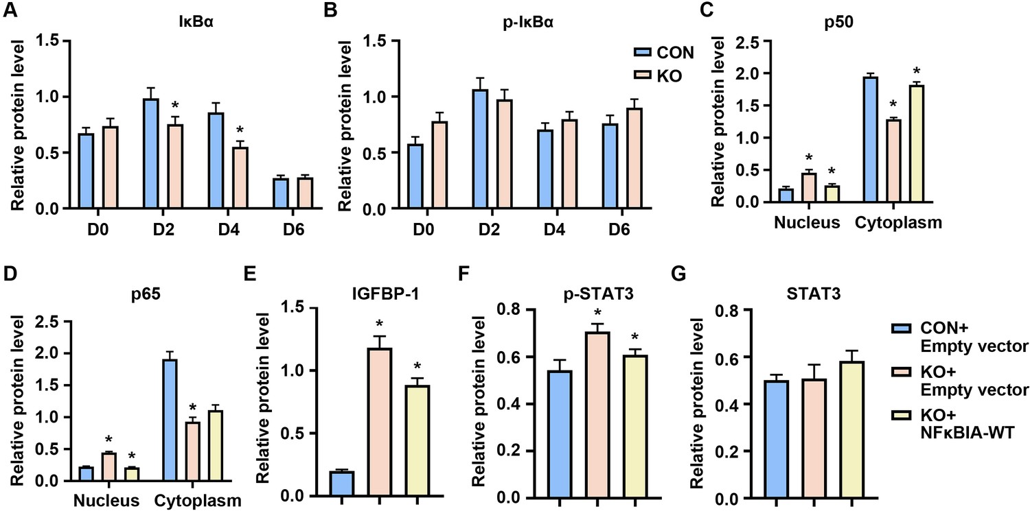
(A) qRT-PCR analysis of NFκBIA mRNA expression in proliferative (D0) and decidualized human endometrial stromal cell (HESC) treated with differential culture medium for 2, 4, and 6 days (n = 3). (B) Western blot analysis IκBα and p-IκBα expression in whole-cell lysates of proliferative (D0) and decidualized HESC treated as in (A). GAPDH was used as the loading control. (C) Immunofluorescence staining of p65 and p50 in CON and KO stromal cells and treated with lentivirus transfected with empty vector or NFκBIA overexpression lentivirus for 3 days. Scale bars, 100 μm. (D) Western blot analysis of the p65 and p50 expression in cytoplasm and nucleus of decidualized HESC treated with lentivirus transfected with empty vector or NFκBIA overexpression lentivirus for 3 days. GAPDH and H3 were used as the loading controls. (E) qRT-PCR analysis of IGFBP-1 mRNA expression levels in CON and KO HESC treated as in (C) (n = 3). (F) Western blot analysis of the IGFBP-1, p-STAT3, and STAT3 expression in whole-cell lysates of decidualized HESC treated as in (C). GAPDH was used as the loading controls. Representative data are shown from three independent experiments. (A) was analyzed with unpaired Student’s t-test. (E) was calculated with one-way analysis of variance (ANOVA) with Bonferroni’s multiple comparison tests. *p < 0.05.
-
Figure 3—source data 1
Original blot images of Figure 3.
- https://cdn.elifesciences.org/articles/83083/elife-83083-fig3-data1-v1.zip
Figure 3—figure supplement 1
Relative of western blot analysis of Figure 3.
(A, B) Relative of western blot analysis of IκBα and p-IκBα expression in whole-cell lysates of CON and KO cells. The values are normalized to the GAPDH expression level and indicated as the mean ± standard error of the mean (SEM) of three independent experiments (n = 3, *p < 0.05, versus controls). (C, D) Relative of western blot analysis of the p65 and p50 expression in cytoplasm and nucleus of CON and KO treated with lentivirus transfected with empty vector or NFκBIA overexpression lentivirus for 3 days. The values are normalized to the GAPDH or H3 expression level and indicated as the mean ± SEM of three independent experiments (n = 3, *p < 0.05, versus controls). (E–G) Relative of western blot analysis of IGFBP-1, p-STAT3, and STAT3 expression in whole-cell lysates of CON and KO treated as in (C). The values are normalized to the GAPDH expression level and indicated as the mean ± SEM of three independent experiments (n = 3, *p < 0.05, versus controls).
Figure 4
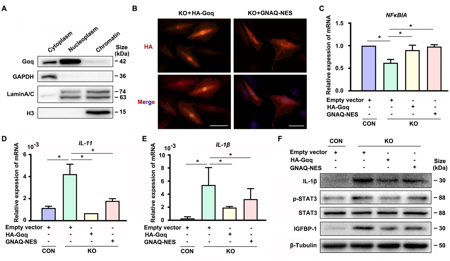
Cytoplastic localized Gαq played the main role in regulating the expression of NFκBIA during decidualization.
(A) Western blot analysis of the Gαq expression in cytoplasm, nucleoplasm, and chromatin of WT human endometrial stromal cell (HESC) treated with differential medium for 4 days. GAPDH, Lamin A/C, and H3 were used as the loading controls for different cellular compartments. (B) Immunofluorescence staining of hemagglutinin (HA) in KO differentiated stromal cells infected with lentivirus encoding HA-tagged HA-Gαq or GNAQ-NES for 2 days. Scale bars, 100 μm. (C–E) qRT-PCR analysis of NFκBIA, IL-11, and IL-1β mRNA expression levels in CON and KO HESC treated with control HA-Gαq or GNAQ-NES overexpression lentivirus for 3 days (n = 3). (F) Western blot analysis of the IL-1β, p-STAT3, STAT3, and IGFBP-1 expression in whole-cell lysates of CON and KO HESC treated as in (C). β-Tubulin was used as the loading controls. Representative data are shown from three to five independent experiments. (C–E) were calculated with one-way analysis of variance (ANOVA) with Bonferroni’s multiple comparison tests. *p < 0.05.
-
Figure 4—source data 1
Original blot images of Figure 4.
- https://cdn.elifesciences.org/articles/83083/elife-83083-fig4-data1-v1.zip
Figure 5 with 2 supplements
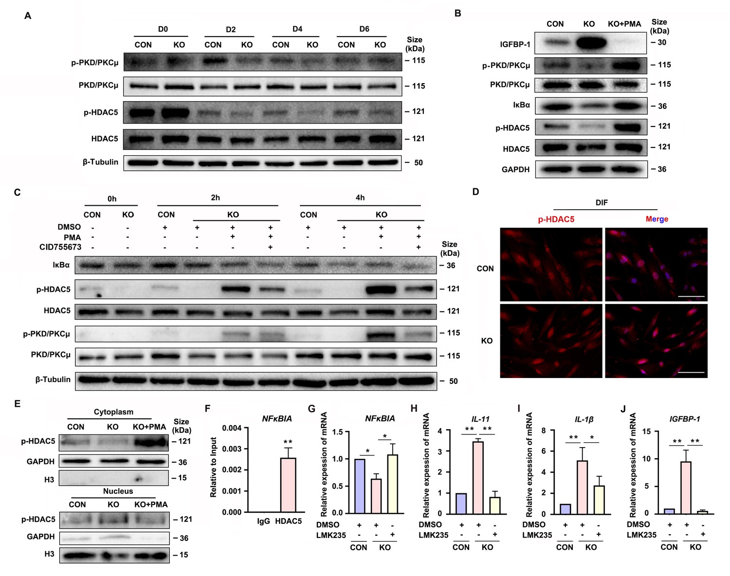
Gαq deficiency attenuated PKD/PKCμ activation and reduced HDAC 5 phosphorylation level to inhibit NFκBIA expression during decidualization.
(A) Western blot analysis of the p-PKD/PKCμ, PKD/PKCμ, p-HDAC5, and HDAC5 expression in whole-cell lysates of proliferative (D0) and decidualized human endometrial stromal cell (HESC) treated with differential culture medium for 2, 4, and 6 days. β-Tubulin was used as the loading controls. (B) Western blot analysis of the IGFBP-1, p-PKD/PKCμ, PKD/PKCμ, IκBα, p-HDAC5, and HDAC5 expression in whole-cell lysates after treatment with PKD/PKCμ activator, Phorbol 12-myristate 13-acetate (PMA, 1 nM) in CON and KO HESC treated with differential culture medium for 4 days. GAPDH was used as the loading controls. (C) Western blot analysis of the IκBα, p-HDAC5, HDAC5, p-PKD/PKCμ, and PKD/PKCμ expression in whole-cell lysates after treatment with PMA (1 nM) and or PKD/PKCμ inhibitor CID755673 for 0, 2, and 4 hr in CON and KO HESC treated with differential culture medium for 4 days. β-Tubulin was used as the loading controls. (D) Immunofluorescence staining of p-HDAC5 in CON and KO decidualized stromal cells treated with differential culture medium for 4 days. Scale bars, 100 μm. (E) Western blot analysis of the p-HDAC5 expression in cytoplasm and nucleus after treatment with PMA (1 nM) in CON and KO HESC treated with differential culture medium for 4 days. GAPDH and H3 were used as the loading controls. (F) Chromatin immunoprecipitation (ChIP)-qRT-PCR analysis of enrichment of HDAC5 on promoters of NFκBIA in WT HESC treated with differential culture medium for 4 days (n = 3). (G–J) qRT-PCR analysis of NFκBIA, IL-11, IL-1β, and IGFBP-1 mRNA expression levels after treatment with HDAC5 inhibitor LMK235 (2 μM), in CON and KO HESC treated with differential culture medium for 4 days. Representative data are shown from three independent experiments (n = 3). (F) was analyzed with unpaired Student’s t-test. (G–J) were calculated with one-way analysis of variance (ANOVA) with Bonferroni’s multiple comparison tests. *p < 0.05 and **p < 0.01.
-
Figure 5—source data 1
Original blot images of Figure 5.
- https://cdn.elifesciences.org/articles/83083/elife-83083-fig5-data1-v1.zip
Figure 5—figure supplement 1
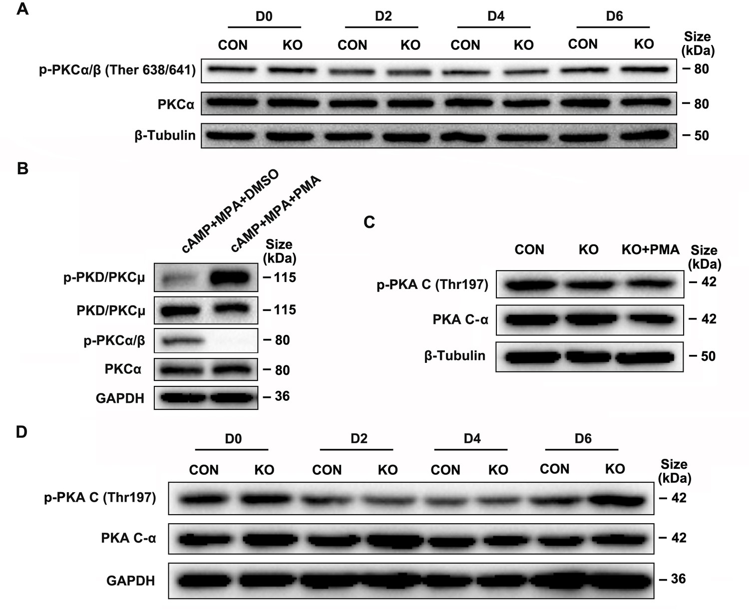
Gαq deficiency has no significant affection in PKCα/β and PKA C phosphorylation during decidualization.
(A) Western blot analysis of the p-PKCα/β and PKCα expression in whole-cell lysates of proliferative (D0) and CON and KO human endometrial stromal cell (HESC) treated with differential culture medium for 2, 4, and 6 days. β-Tubulin was used as the loading controls. (B) Western blot analysis of the p-PKD/PKCμ, PKD/PKCμ, p-PKCα/β, and PKCα expression in whole-cell lysates after treatment with PKD/PKCμ activator, Phorbol 12-myristate 13-acetate (PMA, 1 nM) in normal HESC treated with differential culture medium for 4 days. GAPDH was used as the loading controls. (C) Western blot analysis of the p-PKA C and PKA C-α expression in whole-cell lysates after treatment with PMA (1 nM) in CON and KO treated with differential culture medium for 4 days. β-Tubulin was used as the loading controls. (D) Western blot analysis of the p-PKA C and PKA C-α expression in whole-cell lysates of proliferative (D0) and CON and KO HESC treated with differential culture medium for 2, 4, and 6 days. GAPDH was used as the loading controls.
-
Figure 5—figure supplement 1—source data 1
Original blot images of Figure 5—figure supplement 1.
- https://cdn.elifesciences.org/articles/83083/elife-83083-fig5-figsupp1-data1-v1.zip
Figure 5—figure supplement 2
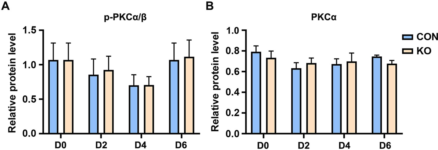
Relative of western blot analysis of Figure 5—figure supplement 1A.
(A, B) Relative of western blot analysis of p-PKCα/β and PKCα expression in whole-cell lysates of CON and KO cells. The values are normalized to the β-Tubulin expression level and indicated as the mean ± standard error of the mean (SEM) of three independent experiments (n = 3).
Figure 6
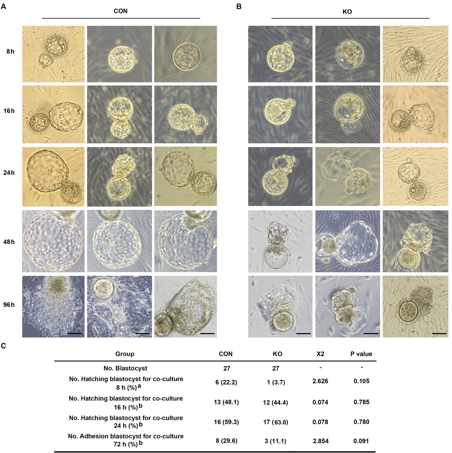
Gαq deficiency in decidual cells inhibits the blastocyst hatch and adhesion in the embryo-endometrial decidual cells co-culture assay.
(A, B) The process of blastocysts hatching, expansion, and adhesion progressively with outgrowth into the stromal cell layer. Images captured from typical day 5–6 blastocysts co-cultured on a layer of CON or KO stromal cells treated with differential culture medium. Scale bar, 100 μm. (C) Table of statistical analyses for the role of stromal Gαq for blastocyst hatching and adhesion in the co-culture assay. n = 5 independent experiments. (C) analyzed with correction Chi-square test (a) and Chi-square test (b).
-
Figure 6—source data 1
Table of statistical analyses for the role of stromal Gaq for blastocyst hatching and adhesion in the co-culture assay.
- https://cdn.elifesciences.org/articles/83083/elife-83083-fig6-data1-v1.doc
Figure 7
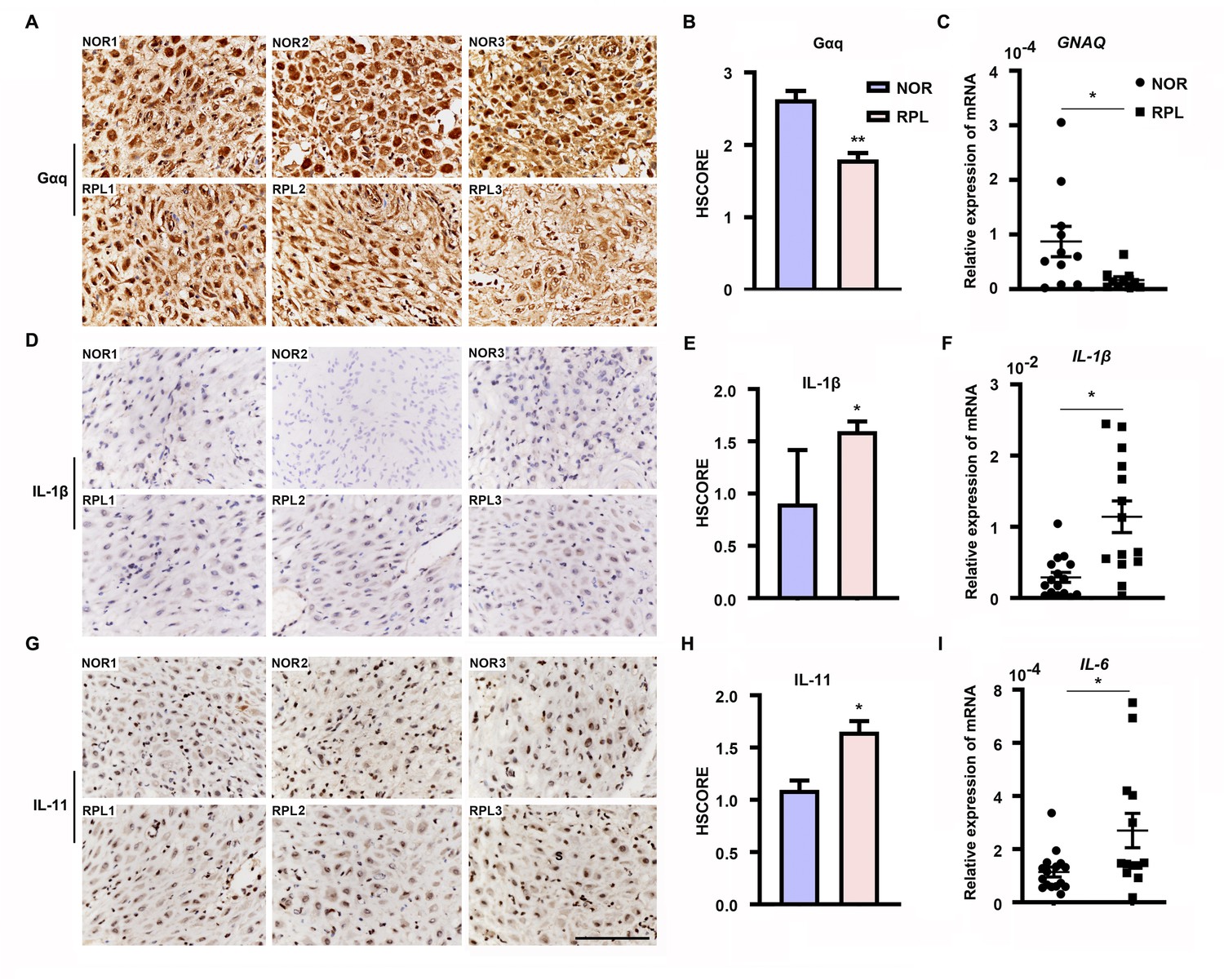
Expression of decidual Gαq is decreased in women with recurrent pregnancy loss (RPL).
(A, D, G) Immunohistochemical staining of Gαq, IL-1β, and IL-11 in the deciduas of women with normal early pregnancies (n = 13) and those with RPL (n = 13). Scale bar, 100 μm. S, stroma cells; NOR, normal. (B, E, H) HSCORE of immunostaining for Gαq, IL-1β, and IL-11 in decidual cells. (C, F, I) qRT-PCR analysis of GNAQ, IL-1β, and IL-6 mRNA expression in the deciduas of women with normal early pregnancies and women with RPL. Representative data were analyzed with unpaired Student’s t-test. *p < 0.05 and **p < 0.01.
Figure 8
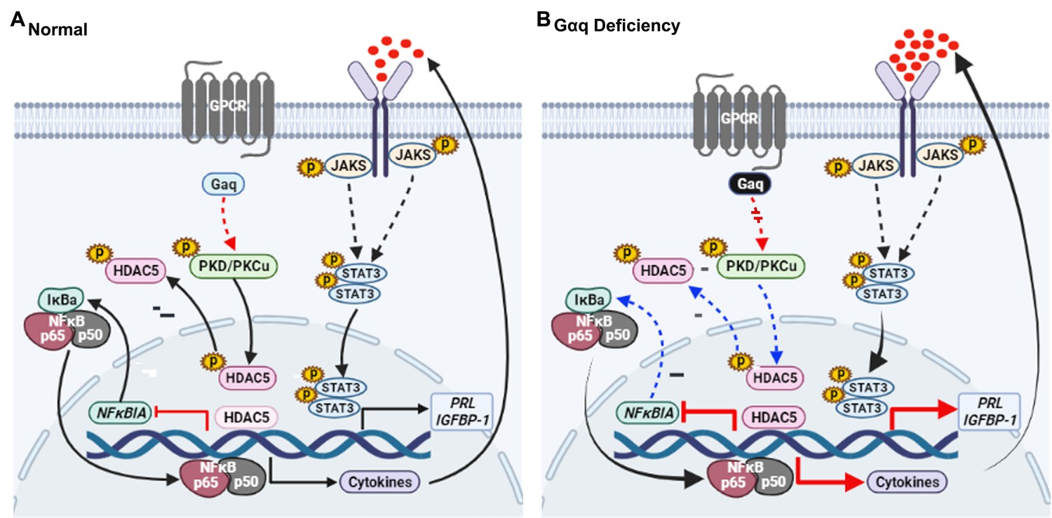
Model outlining the functions of Gαq during decidualization.
(A) During normal decidualization, Gαq-PKD/PKCμ signal inactive HDAC5 through phosphorylation to induce the IκB expression and limit the nuclear factor (NF-κB-mediated inflammatory response for endometrial decidual homeostasis). (B) In Gαq deficiency decidual cells, Gαq-PKD/PKCμ signal is defective, and excessive nuclear location of HDAC5 reduce the IκB expression and over-active NF-κB pathway-mediated inflammatory response result in enhanced inflammatory decidualization.
Additional files
-
MDAR checklist
- https://cdn.elifesciences.org/articles/83083/elife-83083-mdarchecklist1-v1.pdf
-
Supplementary file 1
Tables of characteristics of all patients and controls, all primers, inhibitors and activators used in this study.
- https://cdn.elifesciences.org/articles/83083/elife-83083-supp1-v1.docx
Download links
A two-part list of links to download the article, or parts of the article, in various formats.
Downloads (link to download the article as PDF)
Open citations (links to open the citations from this article in various online reference manager services)
Cite this article (links to download the citations from this article in formats compatible with various reference manager tools)
Gαq-PKD/PKCμ signal regulating the nuclear export of HDAC5 to induce the IκB expression and limit the NF-κB-mediated inflammatory response essential for early pregnancy
eLife 12:e83083.
https://doi.org/10.7554/eLife.83083


