Plasticity of both planar cell polarity and cell identity during the development of Drosophila
- University of Cambridge, United Kingdom
- MRC National Institute for Medical Research, United Kingdom
Figures
Figure 1
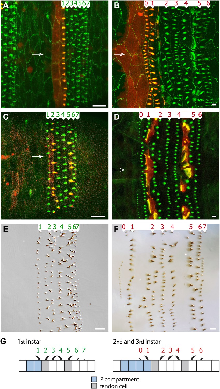
The arrangement of epithelial cells differs between embryo and larva.
Anterior is to the left in all figures. (A and B) An embryo is shown in (A) and the pre-L3 (i.e., the new third instar epidermis developing under the second instar cuticle) in (B). The seven rows of pre-denticles are numbered and arrows indicate the ventral midline. The pre-denticles are labelled with utrp::GFP and the cell outlines with DE-cad::GFP (both in green). Posterior cells and pre-denticles (rows 1 in (A) and rows 0 and 1 in (B)) show red because en.GAL4 is driving expression of UAS.cherry::moesin in the entire P compartment. (C and D) Embryo (C) and pre-L3 (D). Pre-denticles labelled with utrp::GFP as above. The muscle-attaching tendon cells are marked by sr.GAL4 driving expression of UAS.cherry::moesin (red). In the embryo (C) sr.GAL4 marks the pre-denticles of rows 2 and 5, made by the two lines of tendon cells in the embryo. In the pre-L3 larva (D) note the actin palisades in the tendon cells that are labelled in both green and red. In the larva, no pre-denticles are made by these two lines of cells. (E and F) show the cuticular denticles of the L1 (E) and L3 (F) larvae. Scale bars are 10 µm. (G) Diagrams of the embryonic and larval ventral epithelium. The green numbers indicate rows of denticles in L1, the red numbers in L2 and L3. Their polarities are indicated. Note the many changes between embryo and larvae (see also Figure 2).
Figure 2
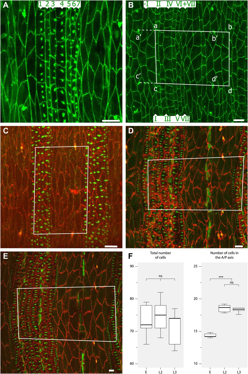
Convergent extension in the anteroposterior axis between pre-L1 and L2.
(A) Mid stage embryo with pre-denticles. The seven rows of pre-denticles (1–7) are indicated. At first all pre-denticles are found on the posterior boundaries of the appropriate rows. However, in later embryos pre-denticle rows 1 and 4, the two rows that will make denticles pointing forwards, are now situated near the middle of the cells. This suggests that some movement of the pre-denticles may be part of polarity reversal. Also it may be relevant that cell lines I and IV of the embryo are the only lines that make extra lines of cells in the larva, and therefore contribute to convergent extension. Labelling for (A–E): The pre-denticles are labelled with utrp::GFP and the cell outlines with DE-cad::GFP (A and B) or DE-cad::tomato (C–E). (B) Late stage embryo, before moulting to L1 but after actin pre-denticles have gone. The pattern is similar to the earlier embryo, with two lines of cells between the tendon cells (II and V). (C) Mid stage embryo showing the marked portion of the epidermis. The rectangle demarcates a segment in the anteroposterior axis and the region between the pair of ventral sensorial papillae (p1) (Dambly-Chaudière and Ghysen, 1986) in the mediolateral axis. The total number of cells and the numbers along the axes were counted, see (F). (D) The pre-L2 stage. By this stage the cells have rearranged and extended in the anteroposterior axis: the number of cells in that axis has increased from ca 14 to 18 cells per segment. The fixed rectangle has changed shape dramatically but contains the same number of cells as in the embryo (Table 1). This change of dimensions is due to convergent extension that involves cell rearrangement as well as alterations in the shapes of cells. (E) The pre-L3 stage. The pattern of cells and the shape of the fixed rectangle resembles that in the pre-L2. (F) Quantitation of the evidence for convergent extension: Boxplots (Frigge et al., 1989) of the number of cells enclosed by fixed pattern landmarks remains the same while these cells extend in the anteroposterior axis and narrow in the mediolateral axis. Pairwise comparisons using t tests with Bonferroni adjustment (ns = not significant, *** = p<0.001).
Figure 3
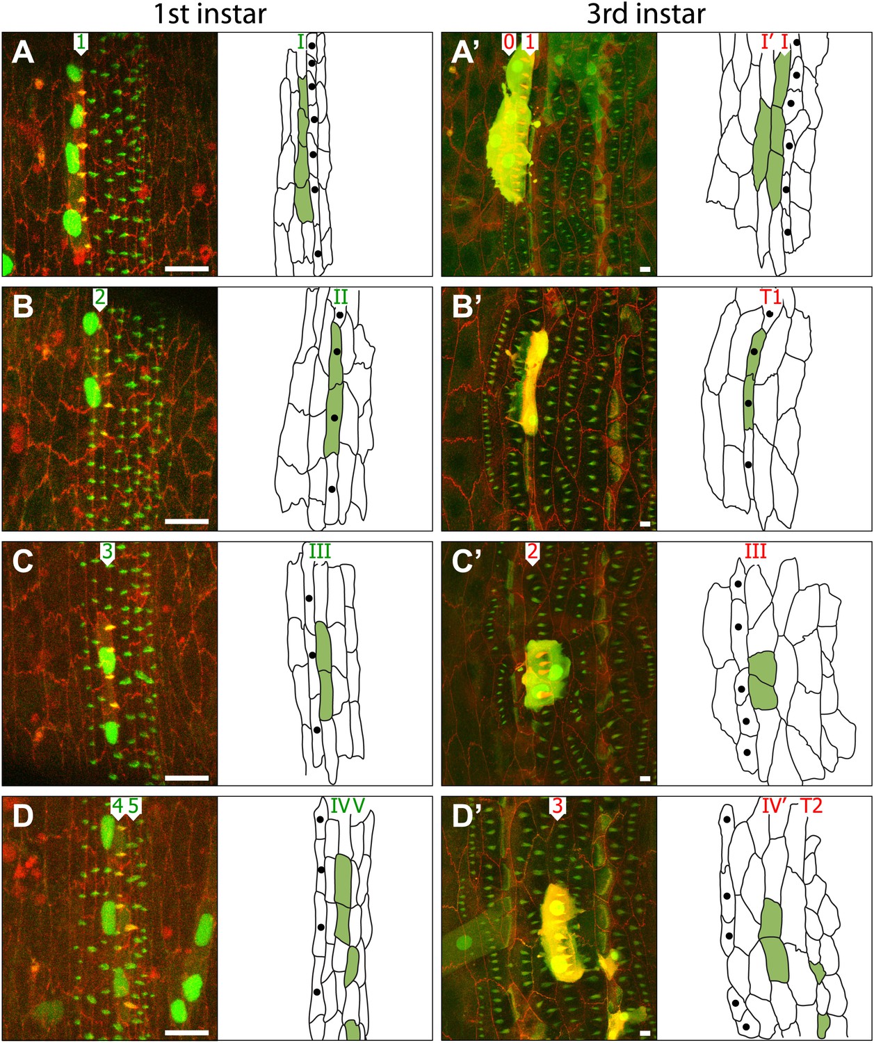
Cells rearrange and change both identity and polarity during larval development.
(A–D) Four individuals are shown; on the left as embryos (A–D) while the right column shows the same four individuals as pre-L3 larvae (A’–D’). The drawings indicate the disposition of the cells of each marked clone (apical profiles filled in green) in the individual embryo and larva respectively. Clones were induced with sry.FLP in the blastoderm stage, studied in the later embryo in the pre-L1 stage and then revisited in the pre-L3. The cells of the clones are marked with cherry::moesin (red cell membranes and pre-denticles), stinger::GFP and Cd8::GFP. All the cells, clone and non-clone are marked with utrp::GFP (pre-denticles are labelled in green) and DE-cad::tomato (cell outlines labelled in red). Numbers I–VII indicate the lines of cells while numbers 1–7 mark the rows of pre-denticles in the embryo (green digits) and the larva (red digits). The cells labelled with single black dots are the T1 tendon cells. Scale bars are 10 µm. (A–A’) Clone of four cells. In the embryo, cells of the clone mark pre-denticles of row 1. In the larva (A’), cells mark pre-denticles of both row 0 (1 cell) and row 1 (3 cells). (B–B’) Clone of two cells. In the pre-L1 embryo, cells in the clone mark pre-denticles of row 2. In the pre-L3 larva (B’), the same cells are the tendon cells. No pre-denticles are marked in the larva. (C–C’) Clone of two cells. In the embryo, cells in the clone mark pre-denticles of row 3 while in the larva the same two cells mark pre-denticles of row 2. (D–D’) Four marked cells in the denticle belt. In the embryo, two cells mark pre-denticles of row 4 and in the larva these same cells mark pre-denticles of row 3. In the embryo two cells make pre-denticles of row 5 and are, presumably, tendon cells. In the larva these same cells make frank T2 tendon cells. Note the tendon cells are small with smaller nuclei, presumably of lower polytenic values than the epidermal cells. Note in Figure D’ that there is a muscle from the adjacent more anterior segment labelled with utrp::GFP and that this muscle attaches to a T1 cell, the most anterior cell of the segment—exactly as in the adult (see Krzemień et al., 2012). See further cases of clones in Figure 4.
Figure 4
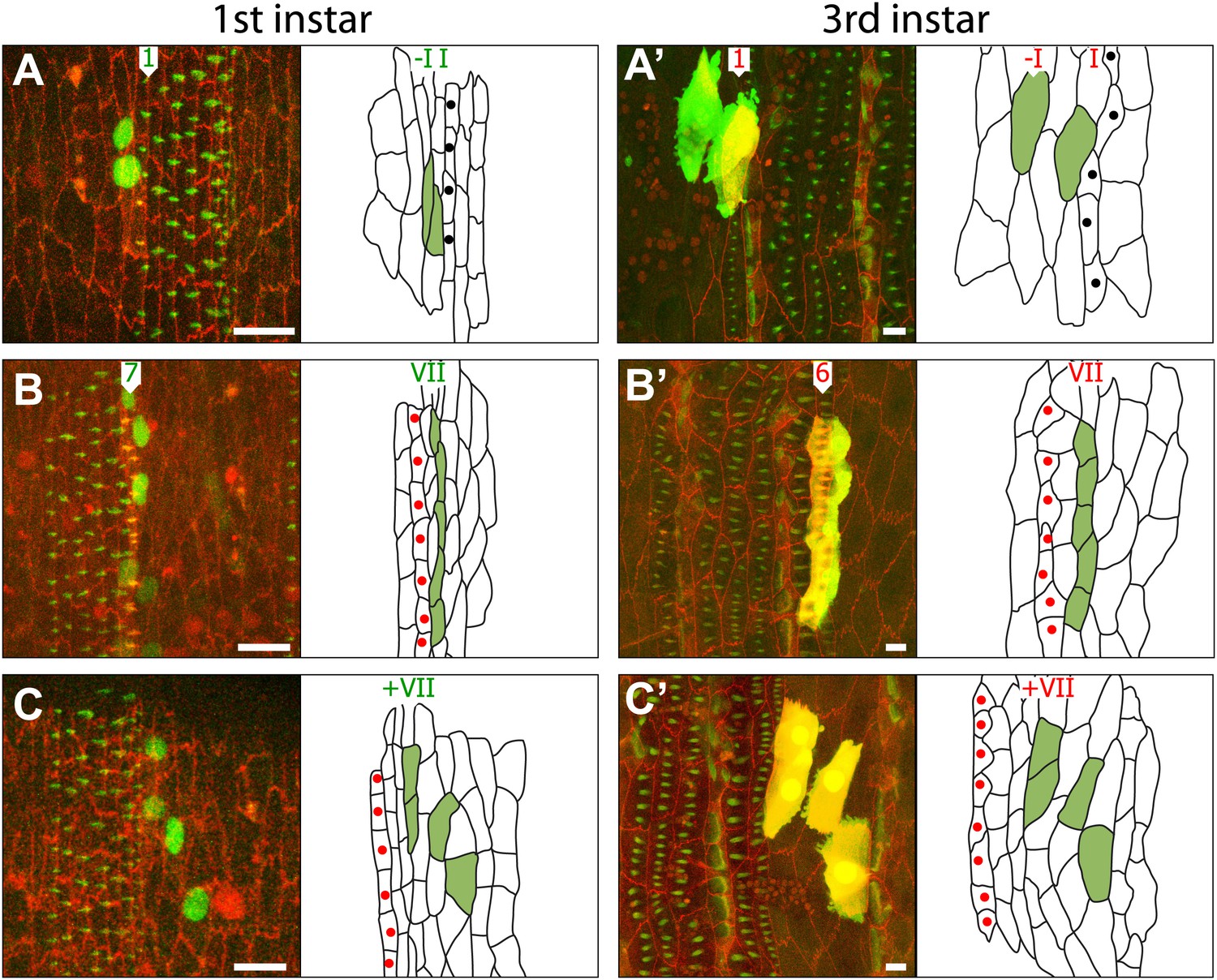
Some informative clones shown in embryo and pre-L3.
(A) A two-celled clone in lines–I and I of the embryo. Only the larval cell I exhibits pre-denticles. There is a line of cells I’ (unlabelled) in between lines–I and I in the larva. (B) A five-celled clone in line VII, it makes row 7 denticles in the embryo and row 6 denticles in the larva. (C) A four-celled clone that is in the naked cuticle posterior to line VII; this clone is not stretched out in the mediolateral axis, as the denticulate clones are. T1 tendon cells are labelled with black dots and T2 with red dots.
Figure 5

Summary of the embryo-to-larva transition.
(A) Diagram showing the studied 9 lines of cells (green roman numerals) and rows of denticles (green arabic numerals) in the embryo and the rows of larval cells and denticles they produce in the larva (numerals in red). Both denticle rows 0 and 1 in the larva arise from line I cells of the embryo. Also, a small percentage of these line I cells contribute to line I’ but make no denticles. Both rows 3 and 4 of the larva come from line IV cells in the embryo. II and V lines of cells are the tendon cells responsible for muscle attachments in the embryo that make tendon cell lines T1 and T2 in the larva. Cells of line VI and VII in the embryo produce denticles of row 5 and 6 respectively in the larva. The diameter of the circles indicates the proportion of the cells involved, for example 30% of all line I cells contribute to row 0 in the larva. (B) The changes between embryo and larva. Lines of cells both in front of and behind the denticles are shown. Cells shown in blue belong to the posterior compartment. Some cells of lines I and IV cells move anteriorly to form lines I’ and IV’, respectively. Line VII cells produce row 6 denticles in the larva.
Figure 6
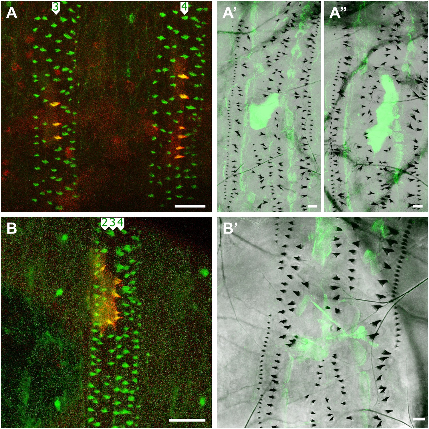
Clones expressing the stripe gene block L2 and L3 denticle formation.
(A–A’’) Two clones (labelled with cherry::moesin and Cd8::GFP) in adjacent segments in one individual make denticles in pre-L1 but no denticles in L3. The B isoform of sr is expressed in these clones which are found in the row 3 of the fourth and the row 4 of the fifth abdominal segments. (B–B’) One clone expressing the A isoform of sr in the embryo makes denticles in rows 2 to 4 of the sixth abdominal segment, but makes no denticles in the larva. The denticle rows are disturbed in this case.
Tables
Table 1
Summary of all clones analysed in both embryo and L3 of the same individual
| Clone # | ||||||||||||||||||||
|---|---|---|---|---|---|---|---|---|---|---|---|---|---|---|---|---|---|---|---|---|
| 1 | 2 | 3 | 4 | 5 | 6 | 7 | 8 | 9 | 10 | |||||||||||
| E | L | E | L | E | L | E | L | E | L | E | L | E | L | E | L | E | L | E | L | |
| −I | 2 | 2 −I | 2 | 2 −I | 4 | 4 −I | 2 | 2 −I | 4 | 4 −I | 2 | 2 −I | 1 | 1 −I | 4 | 4 −I | 4 | 4 −I | 4 | 4 −I |
| I | 4 | 1 I’ | 4 | 1 I’ | 3 | 1 I’ | 3 | 1 I’ | 4 | 2 I’ * | 4 | 1 I’ | 3 | 1 I’ | 4 | 1 I’ | 4 | 1 I’ | 4 | 2 I’ * |
| 3 I | 3 I | 2 I | 2 I | 2 I | 3 I | 2 I | 3 I | 3 I | 2 I | |||||||||||
| II | 4 | 4 T1 | 3 | 3 T1 | 3 | 3 T1 | 3 | 3 T1 | 2 | 2 T1 | 2 | 2 T1 | 4 | 4 T1 | 2 | 2 T1 | 4 | 4 T1 | 2 | 2 T1 |
| III | 1 | 1 III | 2 | 2 III | 2 | 2 III | 3 | 3 III | 1 | 1 III | 4 | 4 III | 1 | 1 III | 2 | 2 III | 1 | 1 III | 2 | 2 III |
| IV | 4 | 3 IV’ | 4 | 2 IV’ | 3 | 1 IV’ | 2 | 2 IV’ | 4 | 3 IV’ | 1 | 1 IV’ | 4 | 2 IV’ | 3 | 1 IV’ | 3 | 1 IV’ | 3 | 2 IV’ |
| 1 IV | 2 IV | 2 IV | 1 IV | 2 IV | 2 IV | 2 IV | 1 IV | |||||||||||||
| V | 2 | 2 T2 | 2 | 2 T2 | 3 | 3 T2 | 2 | 2 T2 | 2 | 2 T2 | 3 | 3 T2 | 3 | 3 T2 | 2 | 2 T2 | 2 | 2 T2 | 2 | 2 T2 |
| VI | 2 | 2 VI | 1 | 1 VI | 1 | 1 VI | 2 | 2 VI | 1 | 1 VI | 1 | 1 VI | 2 | 2 VI | 1 | 1 VI | 2 | 2 VI | 2 | 2 VI |
| VII | 5 | 5 VII | 2 | 2 VII | 2 | 2 VII | 1 | 1 VII | 1 | 1 VII | 2 | 2 VII | 4 | 4 VII | 1 | 1 VII | 1 | 1 VII | 2 | 2 VII |
| +VII | 2 | 2 +VII | 1 | 1 +VII | 2 | 2 +VII | 2 | 2 +VII | 2 | 2 +VII | 4 | 4 +VII | 4 | 4 +VII | 4 | 4 +VII | 2 | 2 +VII | 5 | 5 +VII |
-
The row headers at the left show the line of cells in the embryo from which each clone originates. The numbers of cells in each clone are shown in arabic numerals. The locations of these same cells (within the lines of cells) in the larva are highlighted in bold roman numerals. Cells in lines I and IV of the embryo contribute to two separate lines of cells in the larva; within the denticulate region, apart from lines I and IV, each embryonic cell contributes to only one line of cells in the larva. Cells in line –I in the embryo never make denticles in embryo or larva, but we do not know if they contribute to two lines of cells in the larva. However, since the P compartment is made of two lines of cells in the embryo and four lines of cells in the larva, one extra row has to come from somewhere and line –I is the obvious suspect. (*) Only one these two cells showed denticles in the larva.
Download links
A two-part list of links to download the article, or parts of the article, in various formats.
Downloads (link to download the article as PDF)
Open citations (links to open the citations from this article in various online reference manager services)
Cite this article (links to download the citations from this article in formats compatible with various reference manager tools)
Plasticity of both planar cell polarity and cell identity during the development of Drosophila
eLife 3:e01569.
https://doi.org/10.7554/eLife.01569


