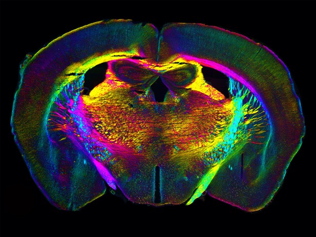
Density and orientation image of a section of mouse brain tissue acquired with QLIPP. Image credit: Syuan-Ming Guo, Li-Hao Yeh, and Shalin Mehta (CC-BY 4.0)
Microscopy is central to biological research and has enabled scientist to study the structure and dynamics of cells and their components within. Often, fluorescent dyes or trackers are used that can be detected under the microscope. However, this procedure can sometimes interfere with the biological processes being studied.
Now, Guo, Yeh, Folkesson et al. have developed a new approach to examine structures within tissues and cells without the need for a fluorescent label. The technique, called QLIPP, uses the phase and polarization of the light passing through the sample to get information about its makeup.
A computational model was used to decode the characteristics of the light and to provide information about the density and orientation of molecules in live cells and brain tissue samples of mice and human. This way, Guo et al. were able to reveal details that conventional microscopy would have missed. Then, a type of machine learning, known as ‘deep learning’, was used to translate the density and orientation images into fluorescence images, which enabled the researchers to predict specific structures in human brain tissue sections.
QLIPP can be added as a module to a microscope and its software is available open source. Guo et al. hope that this approach can be used across many fields of biology, for example, to map the connectivity of nerve cells in the human brain or to identify how cells respond to infection. However, further work in automating other aspects, such as sample preparation and analysis, will be needed to realize the full benefits.


