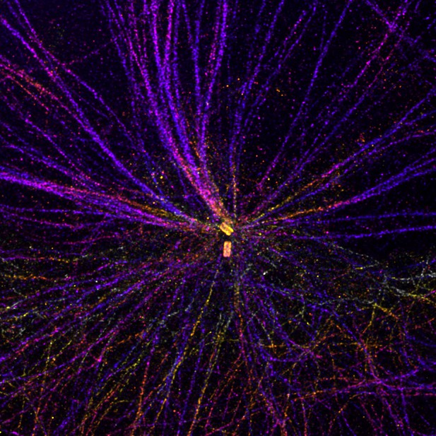
A human cell imaged using ultrastructure expansion microscopy, showing two centrioles (yellow) at the center of a network of microtubules (purple). Colors indicate the depth of the image in three-dimensions. Image credit: Marine Laporte (CC BY 4.0)
Cells are made up of compartments called organelles that perform specific roles. A cylindrical organelle called the centriole is important for a number of cellular processes, ranging from cell division to movement and signaling. Each centriole contains nine blades made up of protein filaments called microtubules, which link together to form a cylinder. This well-known structure can be found in a variety of different species. Yet, it is unclear how centrioles are able to maintain this stable architecture whilst carrying out their various different cell roles.
In early 2020, a group of researchers discovered a scaffold protein at the center of centrioles that helps keep the microtubule blades stable. Further investigation suggested that another protein called WDR90 may also help centrioles sustain their cylindrical shape. However, the exact role of this protein was poorly understood.
To determine the role of WDR90, Steib et al. – including many of the researchers involved in the 2020 study – used a method called Ultrastructure Expansion Microscopy to precisely locate the WDR90 protein in centrioles. This revealed that WDR90 is located on the microtubule wall of centrioles in green algae and human cells grown in the lab. Further experiments showed that the protein binds directly to microtubules and that removing WDR90 from human cells causes centrioles to lose their scaffold proteins and develop structural defects.
This investigation provides fundamental insights into the structure and stability of centrioles. It shows that single proteins are key components in supporting the structural integrity of organelles and shaping their overall architecture. Furthermore, these findings demonstrate how ultrastructure expansion microscopy can be used to determine the role of individual proteins within a complex structure.


