Correction: Drosophila PDGF/VEGF signaling from muscles to hepatocyte-like cells protects against obesity
Main text
Ghosh AC, Tattikota SG, Liu Y, Comjean A, Hu Y, Barrera V, Sui SJH, Perrimon N. 2020. Drosophila PDGF/VEGF signaling from muscles to hepatocyte-like cells protects against obesity. eLife 9:e56969. doi: 10.7554/eLife.56969.
Published 27, October 2020
While preparing Figure 5 for the manuscript, three Thin-layer Chromatography lanes in Figure 5E got inadvertently duplicated, a mistake that was brought to our notice by a reader of the published article. While revisiting our original TLC blots, we realized that in Figure 5C the labelling of our experimental TLC lanes were unintentionally swapped. Additionally, TLC data shown in Figure 5B, C, D and E were assembled by splicing out irrelevant lanes from the plates from which each of those figures were prepared, however, the junctions were not demarcated. The corrections do not affect or change the corresponding quantifications for the TLC lanes in Figure 5. Neither do they change or affect any of the conclusions of the manuscript.
The corrected text in the figure legend of Figure 5 (change is underlined):
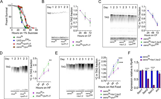
Figure 5: Muscle to oenocyte Pvf1 signaling suppresses lipid synthesis.
(A) Starvation resistance of adult male flies on 1% sucrose and 0.8% agar food. Males with muscle-specific knockdown of pvf1 (musts>pvf1-i1) and oenocyte-specific loss of PvR signaling (oenots>pvrDN) were significantly more resistant to starvation compared to respective controls flies (musts= muscle-Gal4Gal80ts and UAS-pvrDN/+). N = 100, Log rank (Mantel-Cox) test, p≤0.0001 for both comparisons, error bars = SEM.
(B) A representative TLC plate and corresponding quantification of the TLC bands showing rate of lipid mobilization in control (musts) and flies with muscle-specific loss of pvf1 (musts>pvf1-i1). N = 4, Multiple Student t-test, error bars = SEM. Note: The control and experimental TLC bands were rearranged for representation from the same TLC plate. Junctions are marked with a dotted line.
(C) A representative TLC plate and corresponding quantification of the TLC bands showing rate of lipid mobilization in control (oenots=oenocyte-Gal4Gal80ts) and flies with oenocyte-specific loss of TOR signaling (oenots>tsc1,tsc2) and PvR signaling (oenots>pvrDN). N = 4, Multiple Student t-test, error bars = SEM. Note: The control and experimental TLC bands were rearranged for representation from the same TLC plate. Junctions are marked with a dotted line.
(D) A representative TLC plate and corresponding quantification of the TLC bands showing rate of lipid synthesis and incorporation from [U-14C]-Sucrose in control (musts) and flies with muscle-specific loss of pvf1 (musts>pvf1-i1). Flies lacking Pvf1 in the muscle show a significantly faster rate of lipid incorporation compared to control animals. N = 4, ** denotes p≤0.01 at 72 hr on hot food. Multiple student t-test, error bars = SEM. Note: The control and experimental TLC bands were rearranged for representation from the same TLC plate. Junctions are marked with a dotted line.
(E) A representative TLC plate and corresponding quantification of the TLC bands showing rate of lipid synthesis and incorporation from [U-14C]-Sucrose in control flies (oenots) and flies with oenocyte-specific loss of TOR signaling (oenots>tsc1,tsc2) and PvR signaling (oenots>pvrDN). Flies lacking TOR or PvR signaling in the oenocytes show a significantly faster rate of lipid incorporation compared to control animals. N = 4, *** denotes p≤0.001 at 72 hr on hot food. Multiple student t-test, error bars = SEM. Note: The control and experimental TLC bands were rearranged for representation from the same TLC plate. Junctions are marked with a dotted line.
(F) Expression level of key lipid synthesis genes in control (oenots) and flies with oenocyte-specific loss of TOR signaling (oenots>tsc1,tsc2). Only oenocyte-specific fatty acid synthases, fasn2 and fasn3, show a significant reduction in expression in the experimental flies. N = 4, One-Way Anova followed by Tukey’s HSD test, **** denotes p≤0.0001, error bars = SEM.
The corrected Figure 5 is shown here:

The originally published Figure 5 is also shown here for reference:
The article has been corrected accordingly. We apologize for the error.
Article and author information
Author details
Sudhir Gopal Tattikota

Version history
Copyright
© 2021, Ghosh et al.
This article is distributed under the terms of the Creative Commons Attribution License, which permits unrestricted use and redistribution provided that the original author and source are credited.
Metrics
-
- 382
- views
-
- 4
- citations
Views, downloads and citations are aggregated across all versions of this paper published by eLife.
Citations by DOI
-
- 4
- citations for umbrella DOI https://doi.org/10.7554/eLife.66685
Download links
A two-part list of links to download the article, or parts of the article, in various formats.
Open citations (links to open the citations from this article in various online reference manager services)
Cite this article (links to download the citations from this article in formats compatible with various reference manager tools)
Correction: Drosophila PDGF/VEGF signaling from muscles to hepatocyte-like cells protects against obesity
eLife 10:e66685.
https://doi.org/10.7554/eLife.66685


