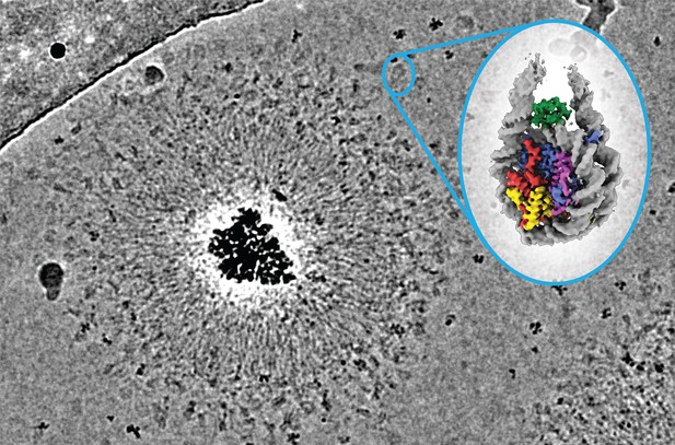
The MagIC-cryo-EM bead consist of 50 nm magnetic bead (black particle), spacer proteins (space round bead), and target-capturing proteins (anti-GFP nanobody) that capture H1.8-GFP-attached nucleosomes (in blue circle). Image credit: Yasuhiro Arimura (CC BY 4.0)
Whether a protein can properly carry out its role in a cell heavily depends on its unique three-dimensional shape. To understand the biological mechanisms underlying protein function – and how they can go awry in disease – scientists therefore need to understand the structure of each of these molecules with a high degree of detail. Researchers use computer-based systems, such as AlphaFold, to predict protein structures based on the properties of their building blocks. However, these predictions may not be accurate, and still need to be verified experimentally.
Cryo-electron microscopy (cryo-EM) is a technique that allows researchers to directly visualize proteins. The approach first requires flash-freezing a sample at extremely low temperatures, and then sending a beam of electrons through the specimen from several angles. The resulting series of two-dimensional images are analysed using advanced algorithms to construct a final three-dimensional protein model.
Despite its power, cryo-EM also has limitations, including requiring very large quantities of protein in the initial sample. The algorithms currently available for Cryo-EM analyses also struggle to determine the structure of smaller molecules.
To overcome these limitations, Arimura et al. developed MagIC-cryo-EM, a new method for preparing samples that relies on magnetic nano-beads. Proteins of interest are directly attached onto these particles using a short linking molecule, and the entire complex is then concentrated onto the imaging surface using a magnet. This greatly reduces the amount of protein needed in the initial sample. A refined analysis method, called DuSTER, was designed in parallel to help capture the structure of smaller molecules.
Arimura et al. successfully tested both approaches on the histone protein H1.8, which normally binds to cellular DNA and helps ‘package’ it stably. The experiments showed, for the first time, that H1.8 could exist in two distinct three-dimensional shapes. They also revealed the structure of a much smaller molecule associated with H1.8, NPM2.
MagIC-cryo-EM and DuSTER will allow scientists to use cryo-EM to examine the structure of proteins which are smaller, challenging to isolate or present in minute quantities inside cells. The knowledge gained from these findings could help us better understand a wide range of biological processes.


