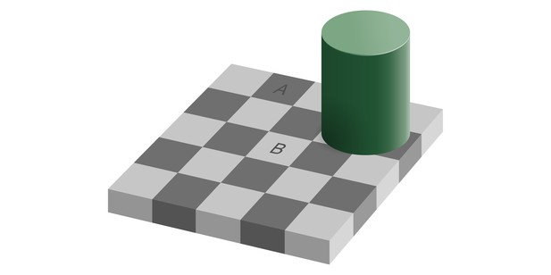
In this optical illusion, squares A and B seem to be a different color, while they are, in fact, exactly the same. Image credit: Edward H. Adelson, vectorized by Pbroks13 (CC BY 4.0)
Recent advances in brain imaging have made it possible to map brain activity in areas of tissue less than a millimeter in size. This resolution offers particular advantages for studying the brain’s outer surface, the cortex. The cortex is traditionally divided into several layers, each containing different types and arrangements of neurons. New high-resolution machines can now visualize the activity in individual layers of cortex, and this can reveal whether the layers also have different roles.
In humans, a large area in the cortex is devoted to vision. Our visual cortex receives sensory information that arrives from the eyes via the optic nerve. This is known as bottom-up processing. But what we see depends on more than just incoming sensory information: it also relies on where we focus our attention, and on our expectations about how things should look. Many optical illusions, for example, work because the brain attempts to decipher an ambiguous visual signal based on previous experiences. This use of existing knowledge to interpret sensory input is called top-down processing.
Using high-resolution brain scanning, Lawrence et al. show that bottom-up and top-down processing occur in different layers of visual cortex. Healthy volunteers viewed a series of images while lying inside a brain scanner. Lawrence et al. changed the contrast of the images to alter the volunteers’ bottom-up processing: this affected activity in the middle layer of visual cortex. To adjust their top-down processing, the volunteers were asked to attend to different features of the images on different trials: these changes in attention had more effect in the layers on either side of the middle layer. This suggests that bottom-up processing occurs in the middle layer of visual cortex, whereas top-down processing takes place in the layers above and below.
The findings by Lawrence et al. will help to better measure activity in cortical layers using modern brain imaging techniques. With further technological improvements, it may become possible to image each layer in the brain in more detail, in particular for other areas that support complex cognitive processes.


