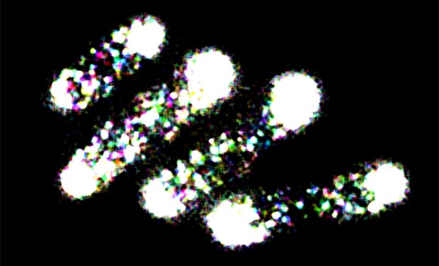
Microscopy image showing the protein concentration of Pom1 in the rod-shaped yeast, Schizosaccharomyces pombe. Image credit: Veneta Gerganova and Sophie G Martin (CC BY 4.0)
All organisms need to know how to arrange different cell types during the development of their organs and tissues. This information is provided by protein concentration patterns, or gradients, that tell cells how to behave based on where they are positioned. The same fundamental principles also work on a smaller scale. For example, although the rod-shaped yeast Schizosaccharomyces pombe is a single-celled organism, it uses protein concentration gradients to control its growth and timing of division.
Before S. pombe cells divide, they need to check that they have reached the right size. Several mechanisms contribute to this information. One of them involves a concentration gradient of a protein known as Pom1, which is found on the cell membrane, with more protein at the cell extremities and less towards the middle. Pom1 serves to block the activity of Cdr2 – an enzyme that localizes to the cell middle and controls cell division. An open question has been whether Pom1 levels at the center drop as the cell grows, coordinating growth and division.
One explanation for how the Pom1 gradient could be regulated is by the removal and addition of phosphate groups. At the cell’s tip, an enzyme removes phosphate groups from Pom1, causing it to bind to the membrane. As Pom1 diffuses along the membrane, it continuously ‘re-phosphorylates’ itself. This promotes Pom1 to gradually detach, restricting it from spreading along the membrane towards the cell middle. Another explanation is that clusters of Pom1, formed at the membrane, help establish a gradient by moving along the membrane at different rates: larger clusters, formed in high concentration areas, move slower than smaller clusters, causing levels of Pom1 to be higher at the tip, and lower towards the middle. Now, Gerganova et al. set out to find which of these two processes contributes more to shaping the Pom1 gradient, and determine where Pom1 acts on Cdr2.
Gerganova et al. used super resolution microscopy to track individual Pom1 molecules inside yeast cells. This revealed two findings. First, that individual Pom1 molecules do not travel all the way from the cell tip to the center, but ‘hop’ between clusters as they move towards the middle. Second, in longer cells levels of Pom1 on the membrane drop at the center, where Pom1 encounters Cdr2. As a result, Cdr2 will come across higher levels of Pom1 in short cells, but low levels of Pom1 in long cells. This allows Pom1 to act as a measure of cell size, preventing short cells from dividing too soon.
The role of clusters in creating gradients is not only relevant for yeast cell division. It could potentially apply to the gradients that organize cells and tissues in different organisms. Future work could examine whether similar principles apply in more complex systems.


