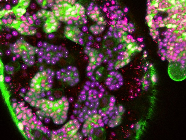
Three-dimensional image of a living fruit fly brain showing the cytoskeleton (green) and DNA (purple) of individual cells which have been detected (as white dots) by the CytoCensus tool. Image credit: Martin Hailstone (CC BY 4.0)
There are around 200 billion cells in the human brain that are generated by a small pool of rapidly dividing stem cells. For the brain to develop correctly, these stem cells must produce an appropriate number of each type of cell in the right place, at the right time. However, it remains unclear how individual stem cells in the brain know when and where to divide.
To answer this question, Hailstone et al. studied the larvae of fruit flies, which use similar genes and mechanisms as humans to control brain development. This involved devising a new method for extracting the brains of developing fruit flies and keeping the intact tissue alive for up to 24 hours while continuously imaging individual cells in three dimensions.
Manually tracking the division of each cell across multiple frames of a time-lapse is extremely time consuming. To tackle this problem, Hailstone et al. created a tool called CytoCensus, which uses machine learning to automatically identify stem cells from three-dimensional images and track their rate of division over time. Using the CytoCensus tool, Hailstone et al. identified a gene that controls the diverse rates at whichstem cells divide in the brain. Earlier this year some of the same researchers also published a study showing that this gene regulates a well-known cancer-related protein using an unconventional mechanism.
CytoCensus was also able to detect cells in other developing tissues, including the embryos of mice. In the future, this tool could aid research into diseases that affect complex tissues, such as neurodegenerative disorders and cancer.


