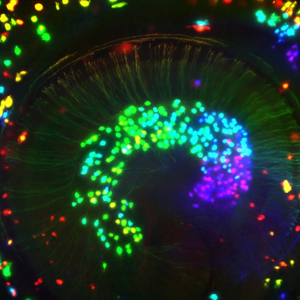
Microscopy image of nerve cells in the inner ear of a newborn mouse which are color-coded based on their depth within the tissue. Image credit: Travis Babola and YingXin Zhang-Hooks (CC BY 4.0)
As the brain develops, billions of cells respond to genetic and environmental cues to form the trillions of connections that make up its neural networks. However, before these brain circuits can respond to real life stimuli, their connections are refined by bursts of electrical activity. For example, sensory cells in the ear produce bursts of spontaneous electrical activity that mimic those made by sounds. This activity allows the neural network in the hearing system to ‘practice’ responding to sounds. However, the origin of these electrical bursts is unusual as they do not start in the sensory cells themselves, but are initiated by the non-sensory cells around them.
Past research has shown that as the ear develops these non-sensory cells, or supporting cells, release regular doses of a molecule called ATP. The supporting cells then detect their own ATP release using specialized receptor proteins on their surface. This self-stimulation causes the supporting cells to release potassium ions that interact with the sensory cells and trigger bursts of electrical activity. However, the identity of this ATP-detecting receptor was not known, and without this information it was unclear how the electrical activity starts and why it happens in rhythmic bursts.
To fill this knowledge gap, Babola et al. measured electrical activity in ear cells isolated from mice, and examined nerve cell activity in live mice during this critical stage of development. This revealed that the bursts of activity in the ear depend on a receptor called P2RY1 which can be found on the supporting cells located next to sensory cells. When P2RY1 is activated it triggers the release of calcium ions inside the supporting cells. This opens channels in the cell membrane, allowing the potassium ions to flow out and electrically activate the sensory cells.
But, when the potassium ions leave the supporting cells, water is drawn out with them, causing the cells to shrink and the space around the cells to get bigger. As a result, the released potassium ions disperse more quickly, moving away from the sensory cells and stopping the burst in electrical activity. Conversely, when P2RY1 is inhibited, this causes the supporting cells to swell, trapping potassium ions near the sensory cells and making them fire continuously. This indicates that bursts in electrical activity are controlled by the rhythmic swelling and shrinking of supporting cells.
Although supporting cells cannot detect sound themselves, they seem to play a crucial role in developing the hearing system. A better understanding of these cells could therefore aid research into hearing problems without a known cause such as hypersensitivity to sound, tinnitus, and complex auditory processing disorders in children.


