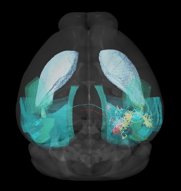
A three-dimensional image of a mouse brain generated using the new software program BIRDS, showing axons projecting from five single nerve cells. Image credit: Xuechun Wang (CC BY 4.0)
Mapping all the cells and nerve connections in the mouse brain is a major goal of the neuroscience community, as this will provide new insights into how the brain works and what happens during disease. To achieve this, researchers must first capture three-dimensional images of the brain. These images are then processed using computational tools that can identify distinct anatomical features and cell types within the brain.
Various microscopy techniques are used to capture three-dimensional images of the brain. This has led to an increasing number of computational programs that can extract data from these images. However, these tools have been specifically designed for certain microscopy techniques. For example, some work on whole-brain datasets while others are built to analyze specific brain regions. Developing a more flexible, standardized method for annotating microscopy images of the brain would therefore enable researchers to analyze data more efficiently and compare results across experiments.
To this end, Wang, Zeng, Yang et al. have designed an open-source software program for extracting features from three-dimensional brain images which have been captured using different microscopes. Similar to other tools, the program uses an ‘image registration’ method that is able to recognize and annotate features in the brain. These tools, however, are limited to whole-brain datasets in which the complete anatomy of each feature must be present in order to be recognized by the software.
To overcome this, Wang et al. combined the image registration method with a deep-learning algorithm which uses pixels in the image to identify features in isolated regions of the brain. Although these neural networks do not require whole-brain images, they do need large datasets to ‘learn’ from. Therefore, the image registration method also benefits the neural network by providing a dataset of annotated features that the algorithm can train on.
Wang et al. showed that their software program, named BIRDS, could accurately recognize pixel-level brain features within imaging datasets of brain regions, as well as whole-brain images. The deep-learning algorithm could also adapt to analyze various types of imaging data from different microscopy platforms. This open-source software should make it easier for researchers to share, analyze and compare brain imaging datasets from different experiments.


