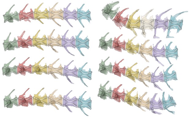
On the left, the spines of zebrafish with no mutations in Urp1 or Urp2. On the right, the spines of zebrafish lacking both genes, which show deformed spines. Image credit: Beth Bearce (CC BY 4.0)
The backbone, or spine, is an integral part of the human body, providing support to our torsos so that we can sit, stand, bend and twist. If this structure does not form correctly, it can lead to pain, neurologic problems, and mobility issues. The spine normally has curves, but these can become deformed for many reasons, including genetic and muscular factors. There are also cases in which the cause of a spine distortion is unknown, such as in scoliosis (where the spine twists to the side), lordosis (where the lower part of the spine curves excessively), and kyphosis (where the upper part of the spine shows extreme curvature).
The structure of the spine is laid out during embryonic development and maintained throughout life. Experiments in zebrafish have shown that a crucial element in preserving the shape of the spine is the flow of cerebrospinal fluid or CSF. Propelled by the movement of little ‘hairs’ at the surface of specialized cells, this liquid runs through our central nervous system along a cavity lined with neurons. These nerve cells produce Urp1 and Urp2, two short molecules (or peptides) built from the same components as proteins. In zebrafish embryos, lowering the levels of these peptides had previously been shown to cause early body deformities. But what role, if any, do Urp1 and Urp2 play in maintaining the shape of the spine in adult zebrafish?
Bearce et al. set out to answer this question. First, they generated mutant zebrafish which did not carry either Urp1, Urp2 or both peptides. Contrary to previous findings, all three of these mutants developed normally as embryos. Once they were adults, zebrafish lacking Urp1 exhibited normal spines, while those lacking Urp2 had slightly deformed curves. However, zebrafish lacking both peptides had prominent curves in the tail-region of their spines, somewhat akin to lordosis in humans. This indicates that both peptides are necessary for adult spine structure, but work in a semi-redundant manner. Interestingly, the defects observed first appeared in adolescent fish and gradually worsened as they grew; many forms of human spinal abnormalities follow a similar trajectory.
Bearce et al. also tested the role of the protein Uts2r3, a receptor for peptides which belong to the urotensin family (such as Urp1 and Urp2). Fish lacking this protein showed normal spine structure as embryos, but distorted spinal curves as adults, suggesting that Urp1 and Urp2 might control spine morphology by signaling via the Uts2r3 receptor.
Together, Bearce et al.’s observations show that disturbing urotensin signaling leads to a lordosis-like condition in adult zebrafish, with evident deformities in the tail-region of the spine. Considering the broad similarities in structures between the zebrafish and the human spine, these results point to a possible involvement of urotensin signaling in spine distortion in humans. More studies using zebrafish will likely provide further insights into the principles that control the shape of the spine and what goes wrong when it breaks down.


