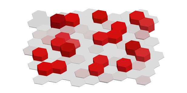
Illustration of 'salt and pepper' geometry of Tip and Stalk cells during angiogenesis. Image credit: Kang, Bocci et al. (CC BY 4.0).
Blood vessels are vital for transporting blood containing oxygen, nutrients and waste around the body. To maintain this function, new blood vessels are continually formed through a process called angiogenesis. Often triggered in areas requiring oxygen, new blood vessels form from existing vessels as ‘sprouts’ in response to elevated levels of a signaling molecule called vascular endothelial growth factor (or VEGF for short).
For ‘sprouting’ to occur, endothelial cells lining the parental blood vessel must become either ‘Tip’ or ‘Stalk’ cells. Tip cells lead the extension of the blood vessel sprouts, while Stalk cells proliferate rapidly, ensuring the growth of the sprout. Correct spatial arrangement of these different cell types is crucial for the development of functional blood vessels.
Previous work has shown that VEGF promotes differentiation of endothelial cells lining blood vessels into different cell types. In neighboring cells, a signaling pathway known as NOTCH is activated due to interactions between adjacent cells, promoting differentiation of Tip cells and Stalk cells. Ideally, Tip cells are spaced out by intervals of Stalk cells to allow separate sprouts to form. Throughout this process, a single cell can receive contradictory signals, with VEGF promoting Tip cell formation and NOTCH signaling promoting Stalk cell differentiation. It remained unclear how the right cells are formed in the right places when surrounded by these conflicting inputs.
To better understand these dynamics Kang, Bocci et al. combined a laboratory model of angiogenesis with mathematical modelling. Experiments using these approaches showed that the overall pattern of cell type specification induced by VEGF and NOTCH signaling is consistent with so-called order-disorder transition, commonly observed in crystals in other ordered structures. For blood vessel cells, this transition means that they can still robustly take on either the Tip or Stalk cell identities, but this fate selection is not stable in time. Additionally, the overall pattern is much more sensitive to additional cues and self-organization mechanisms. Further analysis revealed that one such cue can be local fluctuations the density of fibronectin, a key pro-angiogenic extracellular component, leading to formation of sprouts that tend to distance themselves as much as possible from other fully formed sprouts.
These findings provide a framework for understanding NOTCH-mediated patterning processes in the context of responding to a variety of environmental cues. This sensitivity in cell type specification is important for determining the dynamic nature of the initial steps of angiogenesis and may be crucial for understanding growth of new blood vessels in damaged organs, cancer and other diseases.


