Analysing the forces at work behind the obstructions that cause heart attacks is crucial for identifying patients at risk of these events, says a study published today in eLife.
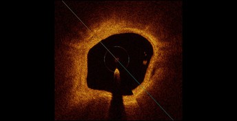
Image of the inside of an artery, obtained through an imaging method called optical coherence tomography. Image credit:
Milzi et al. (CC BY 4.0)
The findings suggest that bringing such biomechanical analyses into clinical practice could allow cardiologists to predict a future heart attack in patients by simulating the distribution of stress within diseased heart vessels.
A myocardial infarction, commonly known as a heart attack, occurs when the supply of blood to the heart is blocked by a blood clot or similar obstruction. A build-up of fatty deposits (lipids) over time forms plaques in the heart's arteries. If the plaque ruptures, it can form a blood clot that blocks the arteries and causes a heart attack.
Previous studies have identified some characteristics of these lipid deposits which put them at high risk of rupturing. These studies involved examining the obstructed coronary arteries of patients who died from infarction, or using special imaging techniques to view these vessels from the inside. But the exact mechanism of plaque rupture, and especially the forces at work behind it, are still not known.
“This may limit our ability to recognise and treat those high-risk lipid deposits before they cause a myocardial infarction,” says Andrea Milzi, Doctor of Medicine at the Department of Cardiology, University Hospital of the RWTH Aachen, Germany, and a co-first author of the study alongside Enrico Domenico Lemma (Karlsruhe Institute of Technology, Karlsruhe, Germany) and Rosalia Dettori (RWTH Aachen). “We wanted to analyse the forces that cause plaque rupture, taking into account a single patient heart vessel with all of its characteristics considered at once. This is important, because it may potentially allow the in-depth analysis of vessels which – even due to a combination of factors – are particularly at risk of developing a myocardial infarction.”
Milzi and the team began by gathering high-quality images of coronary plaques in 20 patients with type 2 diabetes and either stable coronary artery disease or acute myocardial infarction. “These analyses are particularly important in high-risk patients with type 2 diabetes,” says Nikolaus Marx, Head of the Department of Cardiology at the University Hospital of the RWTH Aachen. The images were taken using optical coherence tomography (OCT), an established intravascular imaging technique which, due to its extremely high resolution, can depict cardiac structures with high resolution.
The team carried out OCT on all 20 patients prior to coronary intervention at the Department of Cardiology, University Hospital of the RWTH Aachen. Based on these images, they then generated patient-specific reconstructions of coronary plaques, which they used as a basis to analyse stress concentration as a possible predictor of plaque rupture.
Analysing the biomechanics of these reconstructions allowed them to predict the presence of plaque rupture, as their OCT images showed significantly greater stress concentrations in ruptured plaques compared to non-ruptured plaques. Additionally, by highlighting stress concentrations within the arteries, their analyses also indicated the exact place where a rupture was likely to happen. “In other words, the force exerted on the superficial layers of the plaque plays a pivotal role in plaque rupture, which in turn causes myocardial infarction,” says Sebastian Reith, Consultant at the Department of Cardiology, University Hospital of the RWTH Aachen, and a co-senior author of the study.
“Our pilot study is hypothesis-generating and shows that OCT-based analysis of the forces within the vessel wall is a feasible tool to carry out patient-specific assessment of biomechanics in coronary lesions,” adds co-senior author Mathias Burgmaier, Consultant at the Department of Cardiology, University Hospital of the RWTH Aachen. “Including such analyses in clinical practice might eventually improve the treatment of coronary artery disease, allowing cardiologists to predict a future myocardial infarction by looking – among other factors – at the stress distributions in diseased vessels. However, this approach first needs to be automated and confirmed in larger, prospective studies before we can carry it into clinical practice.”
Media contacts
Emily Packer
eLife
e.packer@elifesciences.org
+441223855373
About
eLife is a non-profit organisation created by funders and led by researchers. Our mission is to accelerate discovery by operating a platform for research communication that encourages and recognises the most responsible behaviours. We aim to publish work of the highest standards and importance in all areas of biology and medicine, while exploring creative new ways to improve how research is assessed and published. eLife receives financial support and strategic guidance from the Howard Hughes Medical Institute, the Knut and Alice Wallenberg Foundation, the Max Planck Society and Wellcome. Learn more at https://elifesciences.org/about.
To read the latest research in Medicine published in eLife, visit https://elifesciences.org/subjects/medicine.


