Scientists have constructed a comprehensive set of functional maps of infant brain networks, providing unprecedented details on brain development from birth to two years old.
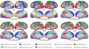
Image showing the infant-specific fine-grained functional parcellation maps for infants aged from three months to two years. Image credit: Gang Li (CC By 4.0)
The infant brain cortex parcellation maps, published today in eLife, have already provided novel insights into when different brain functions develop during infancy and provide valuable, publicly available references for early brain developmental studies.
Cortical parcellation is a means of studying brain function by dividing up cortical grey matter in different locations into ‘parcels’. Scans from functional magnetic resonance imaging (fMRI) are taken when the brain is in an inactive ‘resting’ state, alongside measurements of brain connectivity, to study brain function within each parcel.
Previous studies have created parcellation maps based on resting state fMRI connectivity in adult brains. However, these maps are unsuitable for studying infant brains because of significant differences in brain functional organisation between infants and adults.
“Creating infant-specific brain parcellation maps has been challenging, because of difficulties in acquiring high-resolution infant brain images and processing these images, which typically have a low and rapidly changing contrast between different brain tissues during this time of development,” explains first author Fan Wang, now Associate Professor at Xi’an Jiaotong University, China. “We developed a novel method that captures fine-grained functional patterns from individual infants, to generate a comprehensive set of age-specific and age-independent parcellation maps which will facilitate paediatric neuroimaging research.”
Wang and colleagues used 1,064 high-resolution functional MRI scans and 394 structural MRI scans of infants from birth to two years of age, collected as part of the UNC/UMN Baby Connectome Project Consortium.
To capture detailed patterns of sharp transition between areas of the brain, the team combined a conventional method of mapping the cortical folds (regions of the brain) across all individuals, with a novel algorithm that overlays gradients of functional connectivity for each region onto the brain scans from each infant. This allowed them to establish more accurate and meaningful connections between corresponding areas of functionality across individuals, resulting in a detailed characterisation of the brain’s functional boundaries. This was then used to generate infant-specific fine-grained functional parcellation maps for infants aged from three months to two years, as well as age-independent maps.
The age-independent infant parcellation was highly similar to some previously defined brain areas in adult parcellation maps, with parcel borders conforming to those seen in adult brains. But the infant maps revealed novel insights about the development of brain function.
First, the results suggest that a primitive form of brain functional networks is present at three months old, when the sensory system is more developed than higher-order systems such as cognition and behaviour. The team also identified a previously unreported trend of complex fluctuations in functional activity and network organisation across different ages as brain function develops, which are very different to patterns seen in early brain structure development. These fluctuations could reflect different milestones of behaviour and cognitive ability that emerge at different ages during infancy. Finally, there was an increase in local efficiency – the connection of parcels to neighbouring parcels – with age, reflecting increasing function maturity.
“Our method not only captured important coarse patterns discovered by previous methods but also revealed much more detailed functional boundaries at unprecedented resolution,” says senior author, Gang Li, Associate Professor of Radiology at the University of North Carolina at Chapel Hill, US. “The results suggest that although infant functional connectivity might not be as strong as in adults, the basic units of organisation are likely to be present in infant brains, and so the functional parcel units in infants could be comparable in scale to those in adults. These infant cortical parcellation maps are a powerful platform for analysing increasingly larger groups of infants and higher resolution paediatric neuroimaging data, providing greater accuracy for future studies in neurodevelopment.”
“It is worth noting that our method involves averaging across individuals. This inevitably introduces some registration errors into the parcellation, especially in regions with high topographic variation across subjects. This is an important issue that exists in most methods on population-level parcellations – the eventual solution might be individualised parcellation, and we are planning to work on this in the near future.” adds Li.
Media contacts
Emily Packer
eLife
e.packer@elifesciences.org
+441223855373George Litchfield
eLife
g.litchfield@elifesciences.org
About
eLife transforms research communication to create a future where a diverse, global community of scientists and researchers produces open and trusted results for the benefit of all. Independent, not-for-profit and supported by funders, we improve the way science is practised and shared. In support of our goal, we’ve launched a new publishing model that ends the accept/reject decision after peer review. Instead, papers invited for review will be published as a Reviewed Preprint that contains public peer reviews and an eLife assessment. We also continue to publish research that was accepted after peer review as part of our traditional process. eLife receives financial support and strategic guidance from the Howard Hughes Medical Institute, Knut and Alice Wallenberg Foundation, the Max Planck Society and Wellcome. Learn more at https://elifesciences.org/about.
To read the latest Neuroscience research in eLife, visit https://elifesciences.org/subjects/neuroscience.


