Researchers have created a detailed map of the circuits and more than 9,000 cells that make up the entire body and nervous system of a three-day-old larva of the sea worm Platynereis dumerilii.
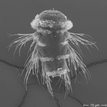
A scanning electron micrograph of the Platynereis dumerilii larva. Image credit: Gáspár Jékely (CC BY 4.0)
The study, published today as a Reviewed Preprint in eLife, is described by the editors as an important advancement towards the understanding of animal nervous system organisation and evolution. They note that this is only the third animal whole-body connectome to be assembled, and praise the wealth of high-quality data used which lays the foundation for future studies on the diversity and connectivity of different cell types, as well as other nervous system parts, their functions and evolution.
Platynereis dumerilii is a species of marine worm belonging to the polychaete class, and is increasingly used across laboratories to study development, behaviour and marine ecology. Its life cycle begins as a free-swimming larva, which then goes through several life stages before settling and transforming into an adult worm, with a simple, segmented body structure – a head featuring sensory organs, repeated body segments with bristle-bearing muscular appendages enabling movement, and a tail. Scientists have increasingly turned to these worms to study the origins of the central nervous system.
“Platynereis larvae are an excellent model for studying how circuits connecting nervous system cells help these creatures move,” explains lead author Csaba Verasztó from the École Polytechnique Fédérale de Lausanne . “They give us insights on the coordination of the nervous system that’s necessary for behaviours such as a startle response to water-borne vibrations or avoidance of UV light exposure.”
Jékely and his colleagues used high-powered electron microscopes to map more than 9,000 cells and the nervous system circuits that connect them in a three-day-old Platynereis larva. They identified 202 types of nervous system cells and more than 92 other cell types that interact with them. Their work describes how these circuits contribute to coordination between the body segments, help integrate multiple senses, connect the left and right sides of the body, and drive movement.
Already, the study has yielded important new insights into the evolution of complex nervous systems. For example, the team’s analysis suggests that some neural circuits in the sea worms that are connected by synapses evolved in parallel, by duplicating and differentiating over time. This process likely allowed the worms to develop more specialised sensory systems and behaviours, enhancing their ability to interact with their environment.
The findings also support a long-standing theory, called the Balfour-Sedgwick theory, which was first proposed in the late 19th century. This theory proposed that, during the formation of the embryo, repeated units called somites develop sequentially, which eventually gives rise to the characteristic segmented bodies seen in animals such as Platynereis dumerilii. In addition, it proposed that the nervous system of the ancestral species from which the sea worm evolved took the shape of a ring with sensory organs radiating out from it. Consistent with this, the authors find that the Platynereis dumerilii nervous system loops around all six of its body segments, and corresponds to radially-arranged mechanosensory organs.
eLife’s editors note that the volume of information in the study may make it challenging to digest, but that it provides a foundation for future studies to delve into more detail about the creatures’ cellular diversity, the connections between and within their body segments, the structures in their brains involved in learning and memory, and more.
“Our whole-body map allowed us to identify several new circuits and to understand how the many circuits are integrated throughout the body,” concludes senior author Gáspár Jékely, a professor at the Centre for Organismal Studies, Heidelberg University, Germany. “None of the insights gathered from our study would be possible without the complete map, and we hope that others will be able to use this work to explore their own key questions around the connections and evolution of the nervous system.”
Media contacts
Emily Packer
eLife
e.packer@elifesciences.org
+441223855373George Litchfield
eLife
g.litchfield@elifesciences.org
About
eLife transforms research communication to create a future where a diverse, global community of scientists and researchers produces open and trusted results for the benefit of all. Independent, not-for-profit and supported by funders, we improve the way science is practised and shared. In support of our goal, we have launched a new publishing model that ends the accept/reject decision after peer review. Instead, papers invited for review will be published as a Reviewed Preprint that contains public peer reviews and an eLife assessment. We also continue to publish research that was accepted after peer review as part of our traditional process. eLife receives financial support and strategic guidance from the Howard Hughes Medical Institute, Knut and Alice Wallenberg Foundation, the Max Planck Society and Wellcome. Learn more at https://elifesciences.org/about.
To read the latest Neuroscience research published in eLife, visit https://elifesciences.org/subjects/neuroscience.


