Cytoplasmic translocation of the retinoblastoma protein disrupts sarcomeric organization
- National University of Singapore, Singapore
Figures
Figure 1
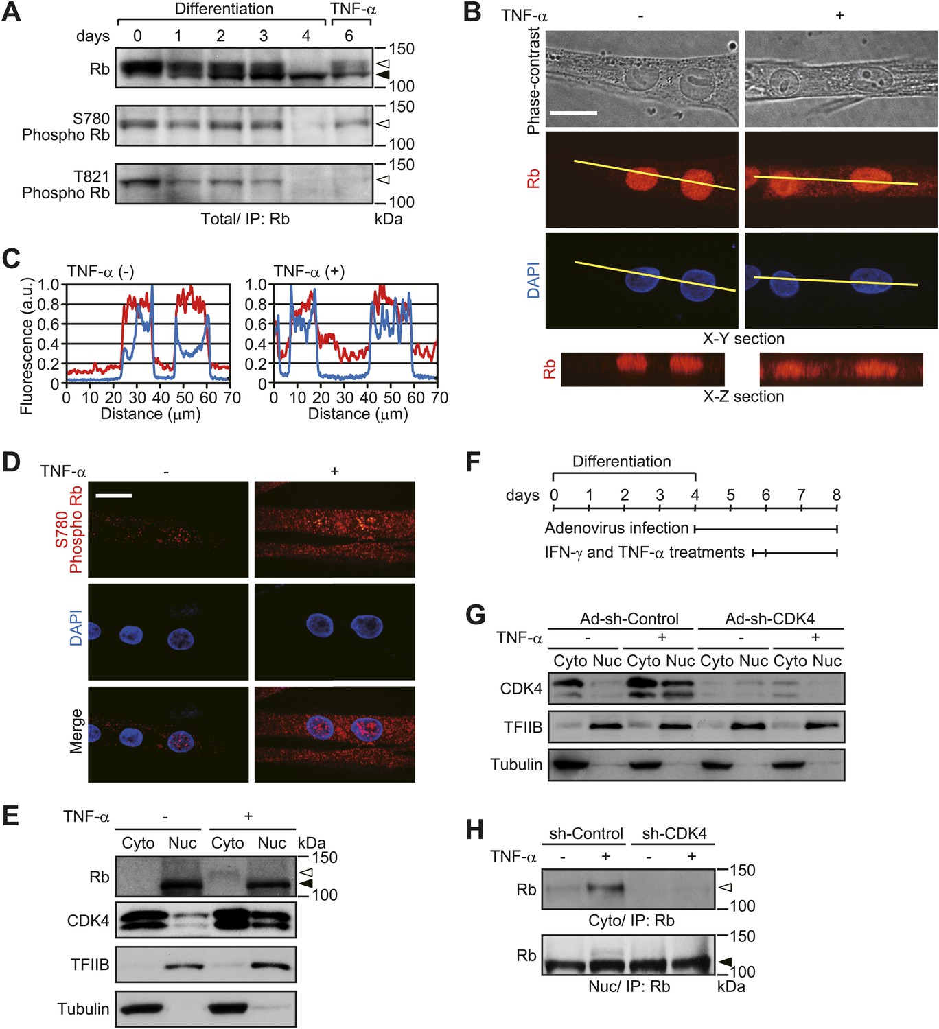
TNF-α induces cytoplasmic translocation of Rb.
(A–E) HSMMs were treated with TNF-α for 2 days. (A) CDK4-mediated phosphorylation of Rb is induced by TNF-α treatment. At the indicated time points after differentiation stimuli, total cell lysates were prepared and immunoprecipitated with anti-Rb antibody, followed by immunoblotting. TNF-α was added for the last 2 days of 6-day cultures. The open and solid arrowheads indicate the position of phosphorylated and unphosphorylated Rb, respectively. (B) Cytoplasmic translocation of Rb is caused by TNF-α treatment. Z-stack confocal images of Rb and 4′,6-diamidino-2-phenylindole (DAPI)-stained nuclei were obtained (50 slices at 0.3-μm intervals). X-Y section images for Rb and DAPI-stained nuclei and X-Z section images for Rb along yellow lines in the X-Y section images are shown. Scale bar, 20 μm. (C) Line plots denote the fluorescence intensities of Rb (red lines) and DAPI (blue lines) along the yellow lines in B. Intensity values were normalized by the maximum value of each plot. a.u., arbitrary units. (D) Phosphorylated Rb is localized in the cytoplasm. Confocal images for Rb phosphorylated at S780 (red) and DAPI-stained nuclei (blue). Scale bar, 20 μm. (E) Nuclear CDK4 expression is increased after TNF-α treatment. Cytoplasmic (Cyto) and nuclear (Nuc) lysates were analyzed by immunoblotting with the antibodies indicated. Tubulin and TFIIB were used as loading controls for cytoplasmic and nuclear lysates, respectively. The open and solid arrowheads indicate the position of phosphorylated and unphosphorylated Rb. (F–H) HSMMs were infected with adenoviruses expressing control non-target shRNA or shRNA against CDK4 at a MOI of 10 pfu/nucleus and then treated with TNF-α for 2 days. (F) Experimental design and reference time frame. (G) The cytoplasmic (Cyto) and nuclear (Nuc) lysates were analyzed by immunoblotting. (H) TNF-α-induced cytoplasmic translocation of Rb is prevented by CDK4 depletion. The cytoplasmic and nuclear lysates were subjected to immunoprecipitation and probed by immunoblotting. The open and solid arrowheads indicate the position of phosphorylated and unphosphorylated Rb, respectively.
Figure 2
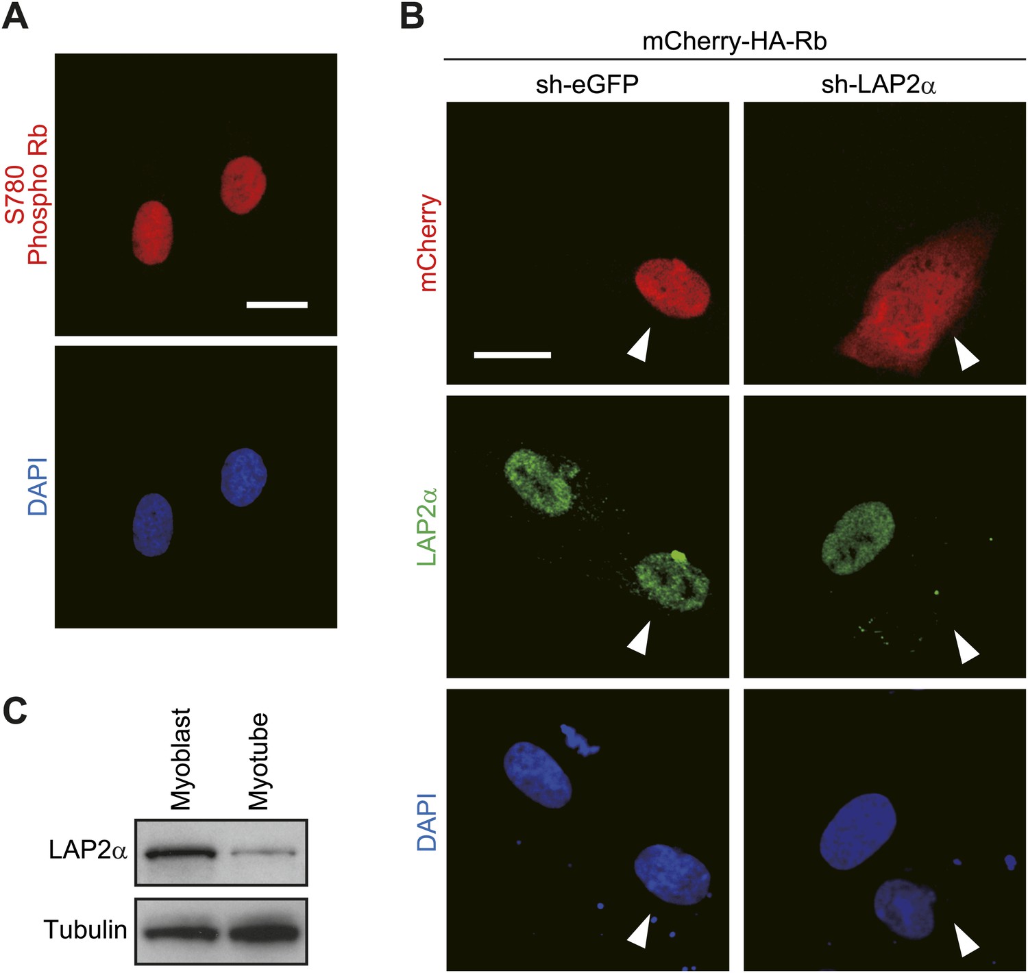
Loss of LAP2α affects the cytoplasmic translocation of Rb.
(A) Phosphorylated Rb is localized in the nucleus in human skeletal muscle myoblasts. Confocal images for Rb phosphorylated at S780 and DAPI-stained nuclei. Scale bar, 20 μm. (B) Rb is translocated to the cytoplasm by LAP2α depletion. Human skeletal muscle myoblasts were transfected with an mCherry-HA-Rb expression plasmid together with an shRNA expression plasmid against eGFP or LAP2α. Confocal images for mCherry, LAP2α and DAPI-stained nuclei. The arrowheads indicate transfected cells. Scale bar, 20 μm. (C) LAP2α expression is decreased in HSMMs. Total cell lysates from human skeletal muscle myoblasts and HSMMs were subjected to immunoblotting.
Figure 3

TNF-α disrupts sarcomeric organization.
(A–E) HSMMs were treated with TNF-α for 2 days. (A) Contractile activity of HSMMs is impaired by TNF-α treatment. EPS was applied to HSMMs. The percentage of beating cells from a total of 100 HSMMs is shown. Results are presented as mean ± SD from three independent experiments. *p<0.002, determined by the Student’s t-test. (B–E) Sarcomeric organization of HSMMs. Confocal images for α-actinin (B) and merged images of F-actin (green) and α-actinin (red) (C) are shown. Scale bar, 10 μm in B and 5 μm in C. (D) Line plots of α-actinin fluorescence intensity along individual myofibrils (denoted by yellow lines in C). Intensity values were normalized by the maximum value for each fibril. a.u., arbitrary units. (E) Autocorrelation analyses of the α-actinin distribution. Scale bar, 5 μm.
Figure 4
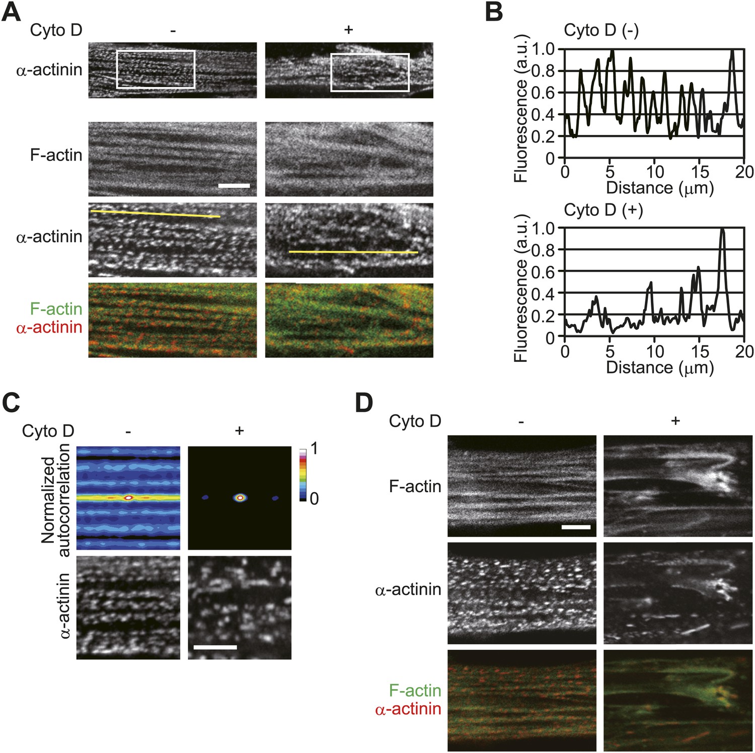
Inhibition of actin polymerization disorganizes sarcomeric assembly.
(A–D) HSMMs were treated with 2 μM cytochalasin D (Cyto D) for 30 min (A–C) or 60 min (D) at room temperature. (A) The periodic arrangement of α-actinin is not well ordered after 30 min of Cyto D-treatment. Merged images of F-actin (green) and α-actinin (red). Scale bar, 5 μm. (B) Line plots of α-actinin fluorescence intensity along individual myofibrils (denoted by yellow lines in A). Intensity values were normalized by the maximum value for each fibril. a.u., arbitrary units. (C) Autocorrelation analyses of the α-actinin distribution. Scale bar, 5 μm. (D) The lateral periodicity in the α-actinin distribution is strongly disordered by 60 min of treatment. Merged images of F-actin (green) and α-actinin (red). Scale bar, 5 μm.
Figure 5
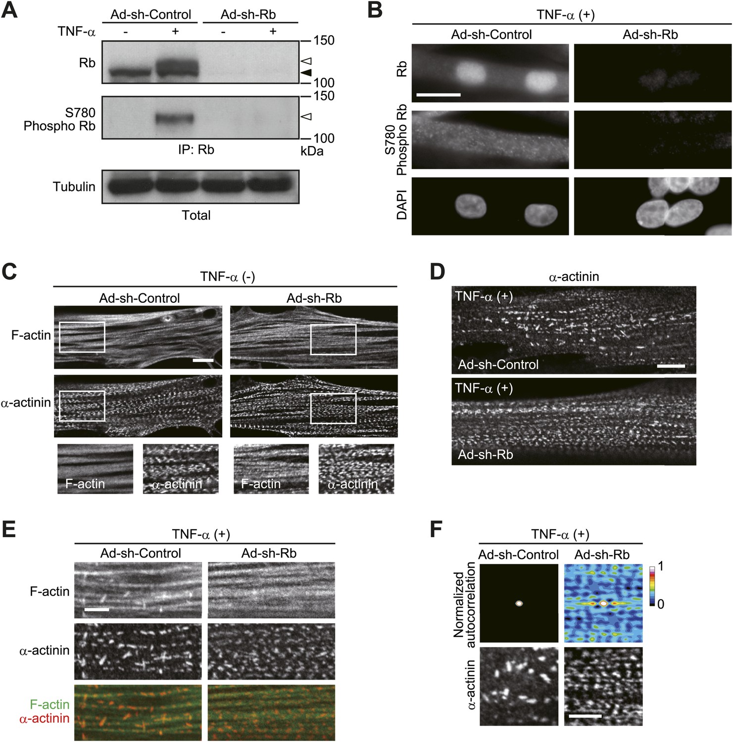
Rb contributes to TNF-α-induced sarcomeric disorganization.
(A–F) HSMMs were infected with adenoviruses expressing control non-target shRNA or shRNA against Rb at a MOI of 10 pfu/nucleus and then treated with TNF-α for 2 days. (A and B) Depletion of Rb protein was verified by immunoblotting (A) and epifluorescence microscopy (B). The open and solid arrowheads indicate the position of phosphorylated and unphosphorylated Rb, respectively. Scale bar, 20 μm. (C) Confocal images for F-actin and α-actinin. Scale bar, 10 μm. (D and E) TNF-α-induced sarcomeric disorganization is attenuated by Rb depletion. Confocal images for α-actinin (D) and merged images of F-actin (green) and α-actinin (red) (E). Scale bar, 10 μm in D and 5 μm in E. (F) Autocorrelation analyses of the α-actinin distribution. Scale bar, 5 μm.
Figure 6
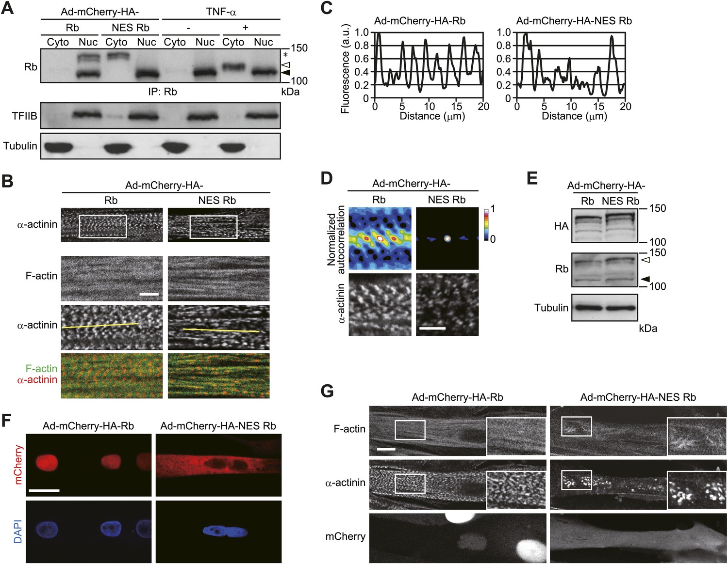
Sarcomeric organization is impaired by cytoplasmic Rb.
(A–G) HSMMs were infected with adenoviruses expressing mCherry-HA-Rb or mCherry-HA-NES Rb at a MOI of 10 pfu/nucleus (A–D) or 50 pfu/nucleus (E–G) for 4 days. (A) The expression of exogenous Rb proteins. Cytoplasmic (Cyto) and nuclear (Nuc) lysates were prepared from infected HSMMs in parallel with TNF-α-treated HSMMs. The lysates were subjected to immunoprecipitation and probed by immunoblotting. The open and solid arrowheads indicate the position of phosphorylated and unphosphorylated Rb. Asterisk indicates the position of exogenous Rb. (B) Sarcomeric structure is not well ordered in NES Rb-expressing HSMMs. Merged images of F-actin (green) and α-actinin (red). Scale bar, 5 μm. (C) Line plots of α-actinin fluorescence intensity along individual myofibrils (denoted by yellow lines in B). Intensity values were normalized by the maximum value for each fibril. a.u., arbitrary units. (D) Autocorrelation analyses of the α-actinin distribution. Scale bar, 5 μm. (E and F) The expression and distribution of exogenous Rb proteins were analyzed by immunoblotting (E) and confocal microscopy (F). The open and solid arrowheads indicate the position of exogenous and endogenous Rb, respectively. Scale bar, 20 μm. (G) Sarcomeric structure is strongly disordered in NES Rb-expressing HSMMs. Confocal images for F-actin and α-actinin. Scale bar, 10 μm.
Figure 7 with 1 supplement
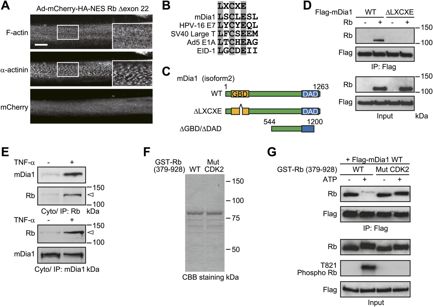
The function of cytoplasmic Rb is rendered through its interaction with LXCXE motif-containing proteins.
(A) Sarcomeric structure is not disordered in NES Rb Δexon 22-expressing HSMMs. HSMMs were infected with an adenovirus expressing mCherry-HA-NES Rb Δexon 22 at a MOI of 50 pfu/nucleus for 4 days. Confocal images for F-actin and α-actinin. Scale bar, 10 μm. (B) mDia1 contains the LXCXE motif. Sequence alignment of the LXCXE motif of mDia1 and other known Rb-binding proteins. (C) The primary structure of mDia1 and its mutants. Scheme represents location of GBD and DAD of mDia1. Numbers denote amino acid positions in mDia1 isform2. The LXCXE motif is present in GBD (amino acid positions 153 to 157). ΔGBD/ΔDAD, doubly deleted mDia1 lacking both GBD and DAD. (D) The LXCXE motif is required for the in vitro interaction between Rb and mDia1. Purified Flag-tagged mDia1 proteins were mixed with full-length recombinant Rb protein and immunoprecipitates were analyzed by immunoblotting. (E) Rb interacts with mDia1 after TNF-α treatment. HSMMs were treated with TNF-α for 2 days. The cytoplasmic lysates were subjected to immunoprecipitation and probed by immunoblotting. The open arrowheads indicate the position of phosphorylated Rb. (F) Expression and purification of GST fusion Rb proteins. The purity of bacterially expressed GST-Rb proteins was evaluated by SDS-PAGE, followed by CBB staining. GST-Rb wild-type protein encompasses amino acids 379–928. GST-Rb Mut CDK2 contains serine/threonine to alanine substitutions at CDK2-specific phosphorylation sites (S612 and T821). (G) GST-Rb proteins were preincubated with CDK4/Cyclin D1 and CDK2/Cyclin E proteins in the presence or absence of ATP and then mixed with purified Flag-mDia1 protein. The interaction between mDia1 and Rb proteins was analyzed by immunoprecipitation with anti-Flag antibody-agarose beads.
Figure 7—figure supplement 1
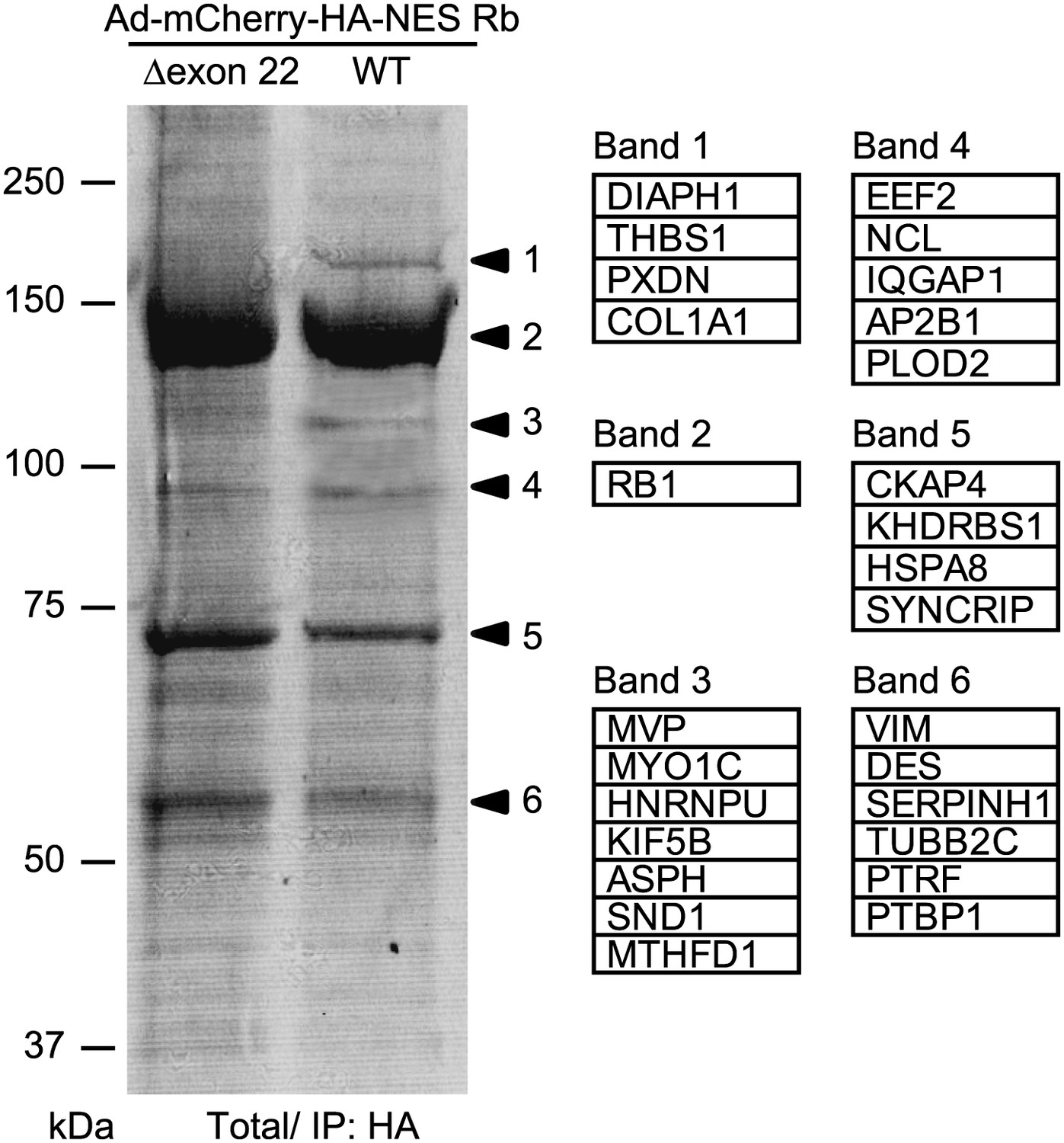
Identification of NES Rb-binding protein.
HSMMs were infected with adenoviruses expressing mCherry-HA-NES Rb WT or mCherry-HA-NES Rb Δexon 22 at a MOI of 50 pfu/nucleus for 4 days. Total cell lysates from adenovirus-infected HSMMs were immunoprecipitated with anti-HA antibody-conjugated agarose beads. The bound proteins were analyzed by SDS-PAGE and visualized by CBB staining. Individual protein bands were excised from SDS-PAGE gel and protein samples were subjected to electrospray ionization mass spectrometric analysis. The lists of proteins from the bands are shown.
Figure 8

Cytoplasmic Rb colocalizes with mDia1 in atrophied skeletal muscle.
(A–C′) Normal and atrophied tibialis anterior muscles were surgically excised from cancer patients. Cross sections (A) and longitudinal sections (B–C′) were stained. (A) Hematoxylin and eosin-stained cryosections. Scale bar, 400 μm. (B–C′) Localization of Rb and mDia1 in normal and atrophied tibialis anterior muscles. Confocal images of cryosections stained with anti-mDia1 and anti-α-actinin antibodies (B) or anti-Rb and anti-mDia1 antibodies (C, magnified in C′). Scale bar, 10 μm.
Figure 9

TNF-α-induced sarcomeric disorganization is prevented by constitutively active mDia1.
(A–F) HSMMs were infected with adenoviruses expressing GFP or GFP-mDia1 ΔGBD/ΔDAD at a MOI of 1 pfu/nucleus and then treated with TNF-α for 2 days. (A) The expression was analyzed by immunoblotting. (B) Confocal images for α-actinin. Scale bar, 5 μm. (C) TNF-α-induced contractile dysfunction is diminished by constitutively active mDia1. EPS was applied to HSMMs. The percentage of beating cells from a total of 100 HSMMs is shown. Results are presented as mean ± SD from three independent experiments. *p<0.002; **p<0.02, determined by the Student’s t-test. (D and E) Constitutively active mDia1 recovers TNF-α-induced sarcomeric disorganization. Confocal images for α-actinin (D) and merged images of F-actin (green) and α-actinin (red) (E). Scale bar, 10 μm in D and 5 μm in E. (F) Autocorrelation analyses of the α-actinin distribution. Scale bar, 5 μm.
Figure 10
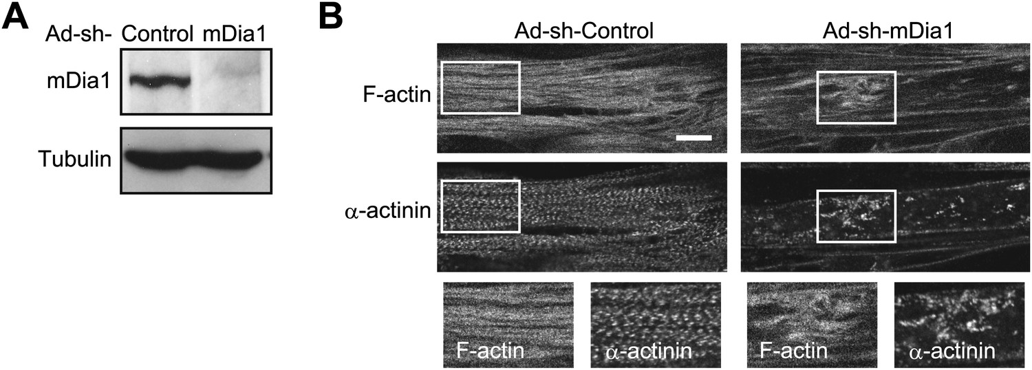
mDia1 is critical for sarcomeric organization.
(A and B) HSMMs were infected with adenoviruses expressing control non-target shRNA or shRNA against mDia1 at a MOI of 5 pfu/nucleus for 4 days. (A) Depletion of mDia1 protein was verified by immunoblotting. (B) Confocal images for F-actin and α-actinin. Scale bar, 10 μm.
Figure 11

The expression of cell-cycle regulators and mitochondrial biogenesis factors after TNF-α treatment.
HSMMs were treated with TNF-α for 2 days. Quantification of the expression of cell-cycle regulators and mitochondrial biogenesis factors. Results are presented as mean ± SD from three independent experiments. *p<0.02, determined by the Student’s t-test.
Author response image 1
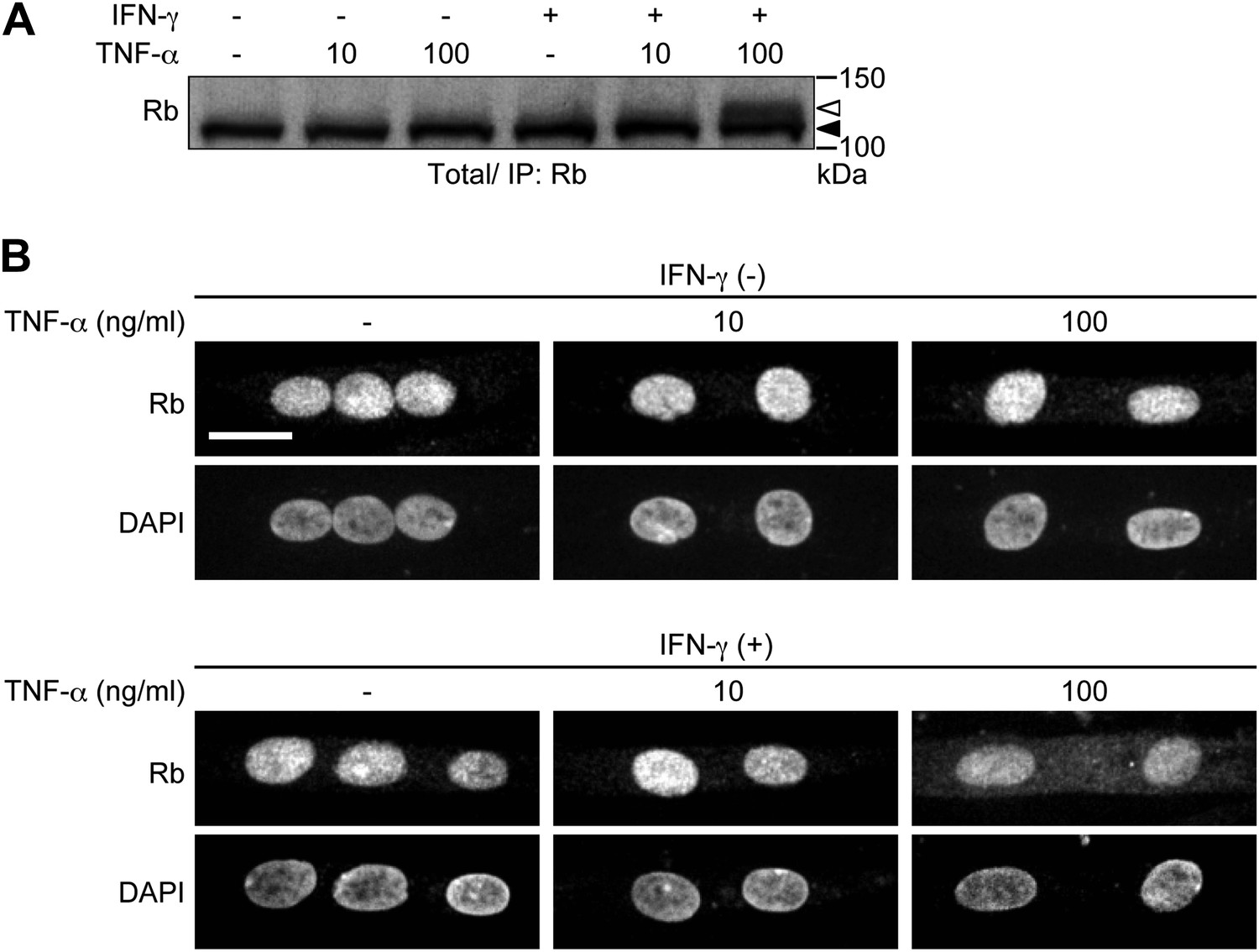
HSMMs were treated with TNF-α (10 or 100 ng/ml) for 2 days with or without IFN-γ pretreatment (100 ng/ml, 8 hr). TNF-α was repeatedly added every 24 hr. A. Total cell lysates were prepared and immunoprecipitated with anti-Rb antibody, followed by immunoblotting. The open and solid arrowheads indicate the position of phosphorylated and unphosphorylated Rb, respectively. B. Confocal images for Rb and DAPI-stained nuclei. Scale bar, 20 μm.
Videos
Video 1
Image of live beating HSMMs in response to EPS.
https://doi.org/10.7554/eLife.01228.006
Video 2
Image of live beating TNF-α-treated HSMMs in response to EPS.
https://doi.org/10.7554/eLife.01228.007Tables
Table 1
Subject characteristics
| Biosample diagnosis | Normal | Atrophy |
| Gender and age, years | Female, 15 | Female, 14 |
| Cancer diagnosis | Osteosarcoma | Synovial sarcoma |
| Cancer location | Shin bone | Soft tissues of shin |
| Height, cm | 166 | 159 |
| Weight, kg | 50 | 50 |
| BMI, kg/m2 | 18.14 | 19.78 |
-
BMI, body mass index
Download links
A two-part list of links to download the article, or parts of the article, in various formats.
Downloads (link to download the article as PDF)
Open citations (links to open the citations from this article in various online reference manager services)
Cite this article (links to download the citations from this article in formats compatible with various reference manager tools)
Cytoplasmic translocation of the retinoblastoma protein disrupts sarcomeric organization
eLife 2:e01228.
https://doi.org/10.7554/eLife.01228


