Cell type-specific and time-dependent light exposure contribute to silencing in neurons expressing Channelrhodopsin-2
- Baylor College of Medicine, United States
- Jan and Dan Duncan Neurological Research Institute at Texas Children’s Hospital, United States
Figures
Figure 1
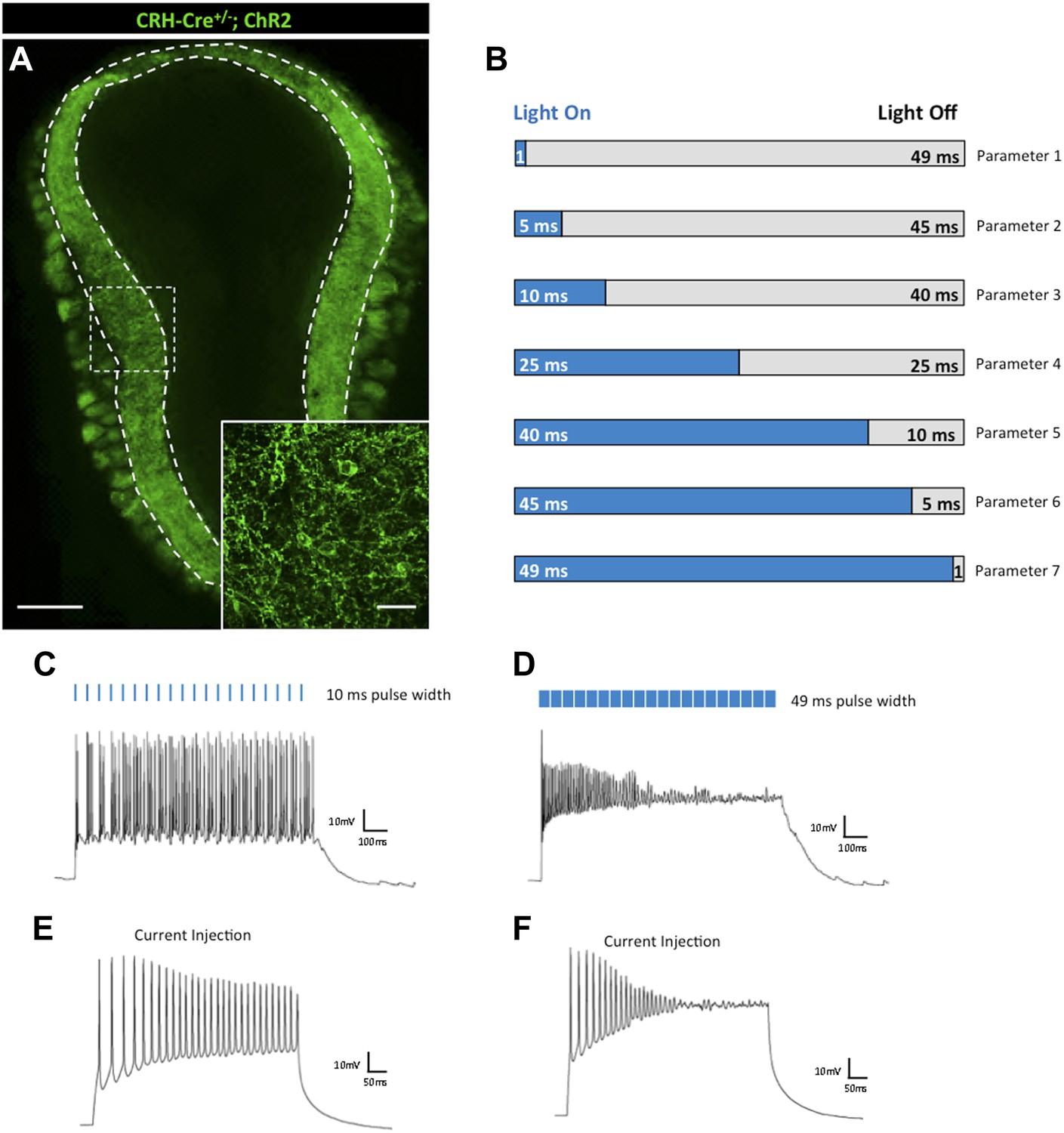
Effects of light pulse duration on CRH-expressing interneurons of the olfactory bulb.
(A) Crh-Cre+/−; ROSALSL-ChR2-EYFP mice express the ChR2-EYFP fusion protein in CRH-expressing interneurons of the external plexiform layer (EPL) of the main olfactory bulb (scale bar, 0.5 mm). Inlay represents zoomed image of ChR2-expressing interneurons of the EPL (scale bar, 100 μM). (B) Firing responses of ChR2-expressing neurons were recorded for seven different stimulation parameters. Each light stimulation parameter consists of a single train comprised of 20 light pulses (≈40 mW/mm2) at 20 Hz. Pulse width is the only condition that varies among the seven parameters. Parameter 1–1 ms pulse width/49 ms intervals, Parameter 2–5 ms pulse width/45 ms intervals, Parameter 3–10 ms pulse width/40 ms intervals, Parameter 4–25 ms pulse width/25 ms intervals, Parameter 5–40 ms pulse width/10 ms intervals, Parameter 6–45 ms pulse width/5 ms intervals, Parameter 7–49 ms pulse width/1 ms intervals. (C) Robust firing of a CRH-expressing EPL interneuron in response to brief light pulses (20 Hz, 10 ms pulse width). (D) Prolonged light pulse duration (20 Hz, 49 ms pulse width) leads to depolarization block in CRH interneurons. (E) Moderate current injection (60 pA) elicits regular firing of ChR2-expressing CRH interneurons. (F) High current injection (160 pA) results in depolarization block of ChR2-expressing CRH interneurons.
Figure 2
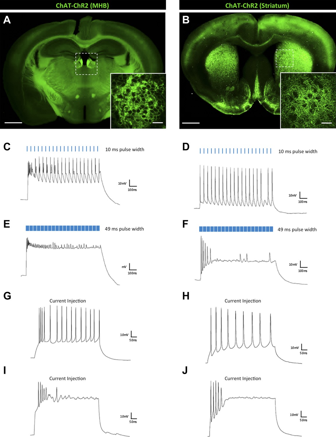
Effects of light pulse duration on ChAT-expressing interneurons of the MHB and striatum.
(A) Chat-ChR2 transgenic mice display ChR2-EYFP expression in the medial habenula (MHB) (scale bar, 1 mm) and (B) striatum (scale bar, 1 mm). Inlays show zoomed images of respective ChR2-expressing MHB or striatal interneurons (scale bars, 100 μM). Regular firing of ChR2-expressing ChAT interneurons of the (C) MHB and (D) striatum in response to brief light pulses (20 Hz, 10 ms pulse width). Prolonged light pulse duration (20 Hz, 49 ms pulse width) leads to depolarization block in (E) MHB and (F) striatal ChAT interneurons. Moderate current injection results in steady firing of ChR2-expressing (G) MHB (50 pA) and (H) striatal (80 pA) ChAT interneurons. High current injection results in depolarization block of ChR2-expressing (I) MHB (150 pA) and (J) striatal (300 pA) ChAT interneurons.
Figure 3

Effects of light pulse duration on a heterogeneous SST interneuron population.
(A–E) Sst-Cre+/−; ROSA26LSL-tdTomato animals express the tdTomato reporter in SST-positive interneurons, which display heterogeneous morphologies (scale bars, 50 μM). (F) Sst-Cre+/−; ROSALSL-ChR2-EYFP mice display diffuse expression of ChR2-EYFP throughout the cortex (scale bar, 1 mm). Inlay shows zoomed image of ChR2-expressing SST cortical interneurons (scale bar, 100 μM). (G) Steady firing of a regular-spiking ChR2-expressing SST cortical interneuron in response to brief light pulses (20 Hz, 10 ms pulse width). (H) Prolonged light pulse duration (20 Hz, 49 ms pulse width) leads to depolarization block in regular-spiking SST cortical interneurons. (I) Moderate current injection (30 pA) leads to steady firing of regular-spiking SST cortical interneurons expressing ChR2. (J) High current injection (100 pA) results in depolarization block of regular-spiking ChR2-expressing SST cortical interneurons. (K) Steady firing of a fast-spiking SST cortical interneuron in response to brief light pulse stimulation (20 Hz, 10 ms pulse width). (L) Prolonged light pulse duration (20 Hz, 49 ms pulse width) leads to robust firing in fast-spiking SST cortical interneurons. (M) Current injection (120 pA) leads to steady firing of fast-spiking SST interneurons. (N) High current injection (500 pA) results in robust firing of fast-spiking SST cortical interneurons.
Figure 4
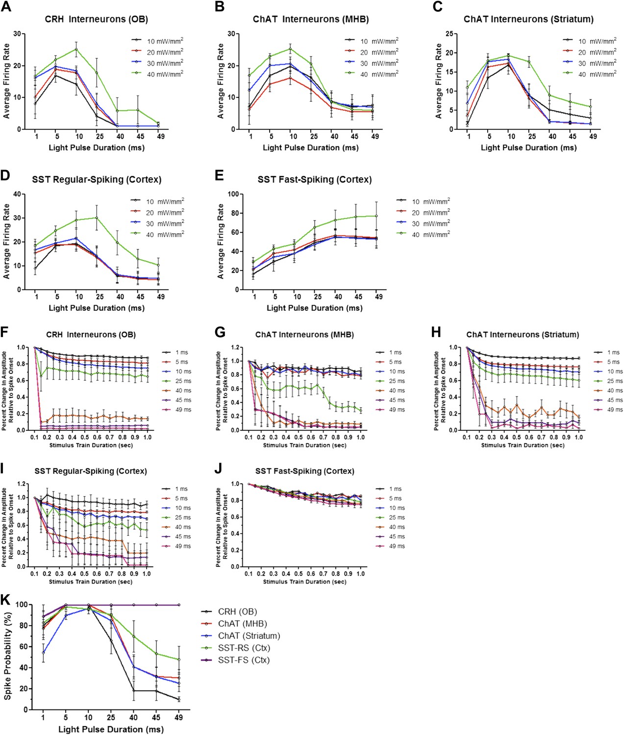
Firing dynamics of diverse interneurons in response to varying light pulse duration.
Average firing rates of ChR2-expressing (A) CRH (N = 5–10 cells/intensity from 4 animals), (B) ChAT MHB (N = 4–9 cells/intensity from 5 animals), (C) ChAT striatal (N = 6–15 cells/intensity from 5 animals), (D) regular-spiking SST (N = 5–9 cells/intensity from 4 animals), and (E) fast-spiking SST interneurons (N = 4-9 cells/intensity from 4 animals) in response to variable light intensity and pulse duration (20 Hz). (F–J) Interneuron amplitudes, normalized to spike onset, in response to increasing pulse width. (K) Spike probabilities of various interneuron cell types in response to increasing pulse widths. Data points represent averages ± SEM. OB = Olfactory Bulb, MHB = Medial Habenula.
Figure 5

Principal excitatory cell types are less susceptible to light-induced depolarization block.
Thy1-ChR2 transgenic mice display ChR2-EYFP expression in (A) excitatory mitral cells of the main olfactory bulb (scale bar, 0.5 mm)—note that ChR2 expression is also observed throughout the granule cell layer of the olfactory bulb due to axon collaterals from mitral/tufted cells and ChR2-expressing centrifugal inputs from the piriform cortex–and (B) layer V cortical pyramidal neurons (scale bar, 1 mm). Inlays display zoomed images of ChR2-expressing olfactory bulb mitral cells (scale bar, 100 μM) or cortical pyramidal neurons (scale bar, 200 μM). (C) Mitral cells display steady firing in response to brief light pulses (20 Hz, 10 ms pulse width) and (D) enhanced firing in response to prolonged light pulse duration (20 Hz, 49 ms pulse width). (E) Steady firing of ChR2-expressing pyramidal cells in response to brief light pulse stimulation (20 Hz, 10 ms pulse width) and (F) prolonged light pulse duration (20 Hz, 40 ms pulse width). OB = Olfactory Bulb.
Figure 6

Firing dynamics of principal cell types in response to varying light pulse duration.
Average firing rates of (A) mitral cells (N = 8–14 cells/intensity from 4 animals) and (B) cortical pyramidal cells (N = 6 cells/intensity from 3 animals) in response to variable light intensity and increasing pulse width (20 Hz). (C) Mitral cell and (D) pyramidal cell amplitudes, normalized to spike onset, in response to increasing pulse width. (E) Spike probabilities of mitral cells and cortical pyramidal neurons in response to increasing pulse widths. (F) Minimal pulse widths required to elicit single action potentials in various neuron populations. Data points represent averages ± SEM.
Figure 7
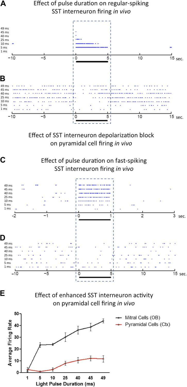
Effects of light pulse duration on ChR2-expressing neurons in vivo.
Increasing light pulse duration onto (A) a presumptive regular-spiking SST cortical interneuron (median latency to spike = 1.8 ms, N = 7 cells from 5 animals) leads to depolarization block and (B) disinhibition of a presumptive cortical pyramidal cell. Increasing light pulse duration onto (C) a presumptive fast-spiking SST cortical interneuron (median latency to spike = 6 ms, N = 3 cells from 5 animals) results in enhanced interneuron firing and (D) subsequent inhibition of a presumptive cortical pyramidal cell. In contrast to regular-spiking interneurons, increasing light pulse duration onto excitatory (E) mitral cells and cortical pyramidal cells enhance average firing rate with increasing pulse width. OB = Olfactory Bulb.
Download links
A two-part list of links to download the article, or parts of the article, in various formats.
Downloads (link to download the article as PDF)
Open citations (links to open the citations from this article in various online reference manager services)
Cite this article (links to download the citations from this article in formats compatible with various reference manager tools)
Cell type-specific and time-dependent light exposure contribute to silencing in neurons expressing Channelrhodopsin-2
eLife 3:e01481.
https://doi.org/10.7554/eLife.01481


