dPob/EMC is essential for biosynthesis of rhodopsin and other multi-pass membrane proteins in Drosophila photoreceptors
- Hiroshima University, Japan
- Heilongjiang Academy of Agricultural Sciences, China
Figures
Figure 1

Identification of CG6750 as an essential gene for rhodopsin 1 (Rh1) biosynthesis.
(A) Observation of fluorescent protein localizations in CG6750e02662 mosaic retinas by the water immersion technique. RFP (red) indicates wild-type photoreceptors (R1–R8). Arrestin2::GFP (green) shows endogenous Rh1 localization in R1–R6 peripheral photoreceptors. (B) Schematic drawing of CG6750 and insertion/deletion mutants. The dPob-null mutant allele, dPob∆4, was created by the recombination of two FRTs on dPobf07762 and dPobCB−0279−3 using an FRT/FLP-based deletion method. (C, D) Immunostaining of dPobe02662 (C) and dPob∆4 (D) retinas expressing RFP as a wild-type cell marker (magenta) by anti-Rh1 antibody (green). Asterisks show mutant cells. Scale bar: 5 μm (A, C, D).
Figure 2
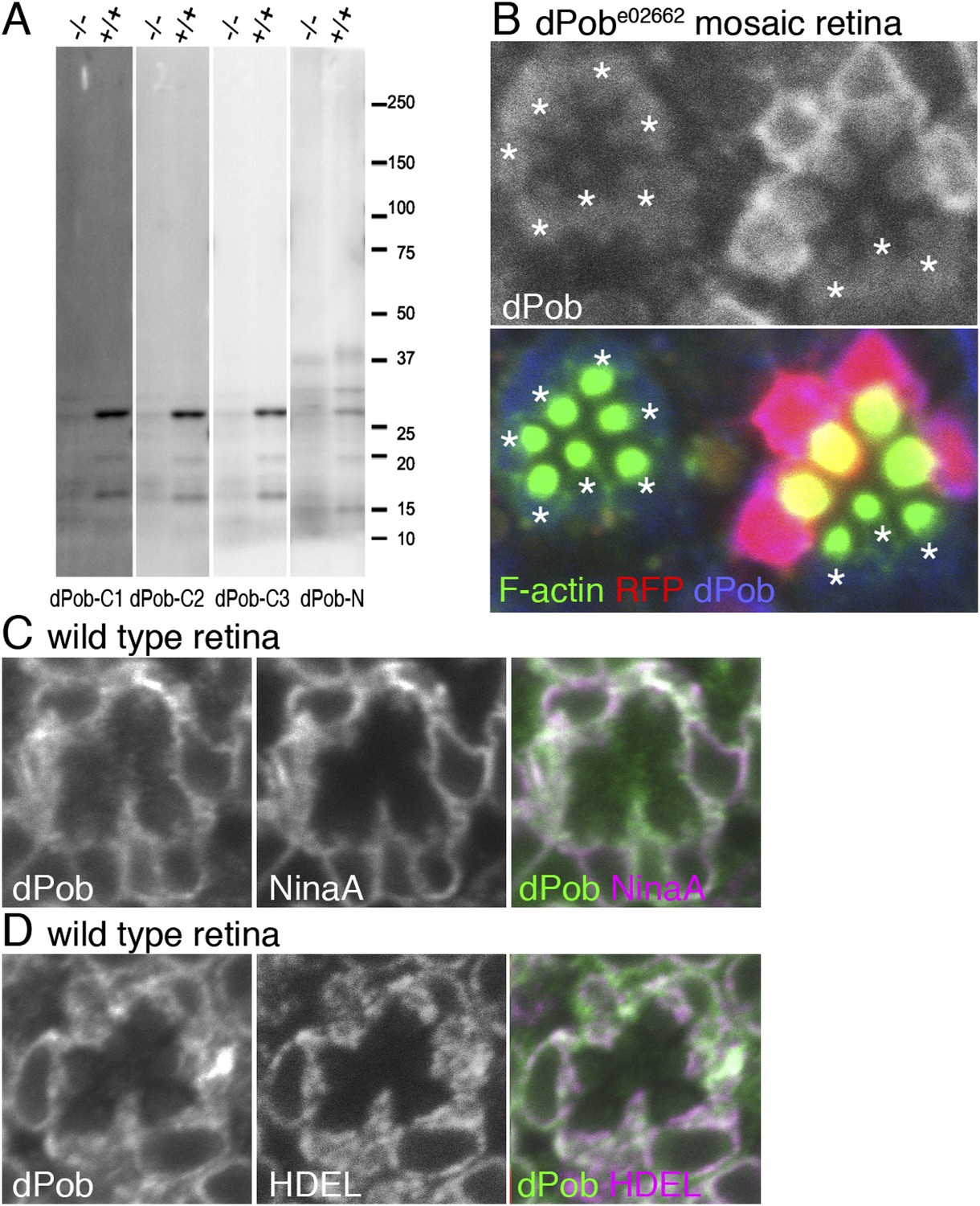
Construction of antisera against dPob.
(A) Immunoblotting of wild-type (+/+) and dPobe02662 homozygous (−/−) extracts from whole larvae using antiserum against dPob N- and C-terminal polypeptides. (B) Immunostaining of a dPobe02662 mosaic retina expressing RFP (red) as a wild-type cell marker (not shown) by rat anti-dPob-C1 antiserum (blue) and phalloidin (green). Asterisks show dPobe02662 homozygous photoreceptors. (C, D) Immunostaining of wild-type retinas by anti-dPob (green) and anti-NinaA (C) or anti-HDEL (D) antisera. Scale bar: 5 μm (B–D).
Figure 3
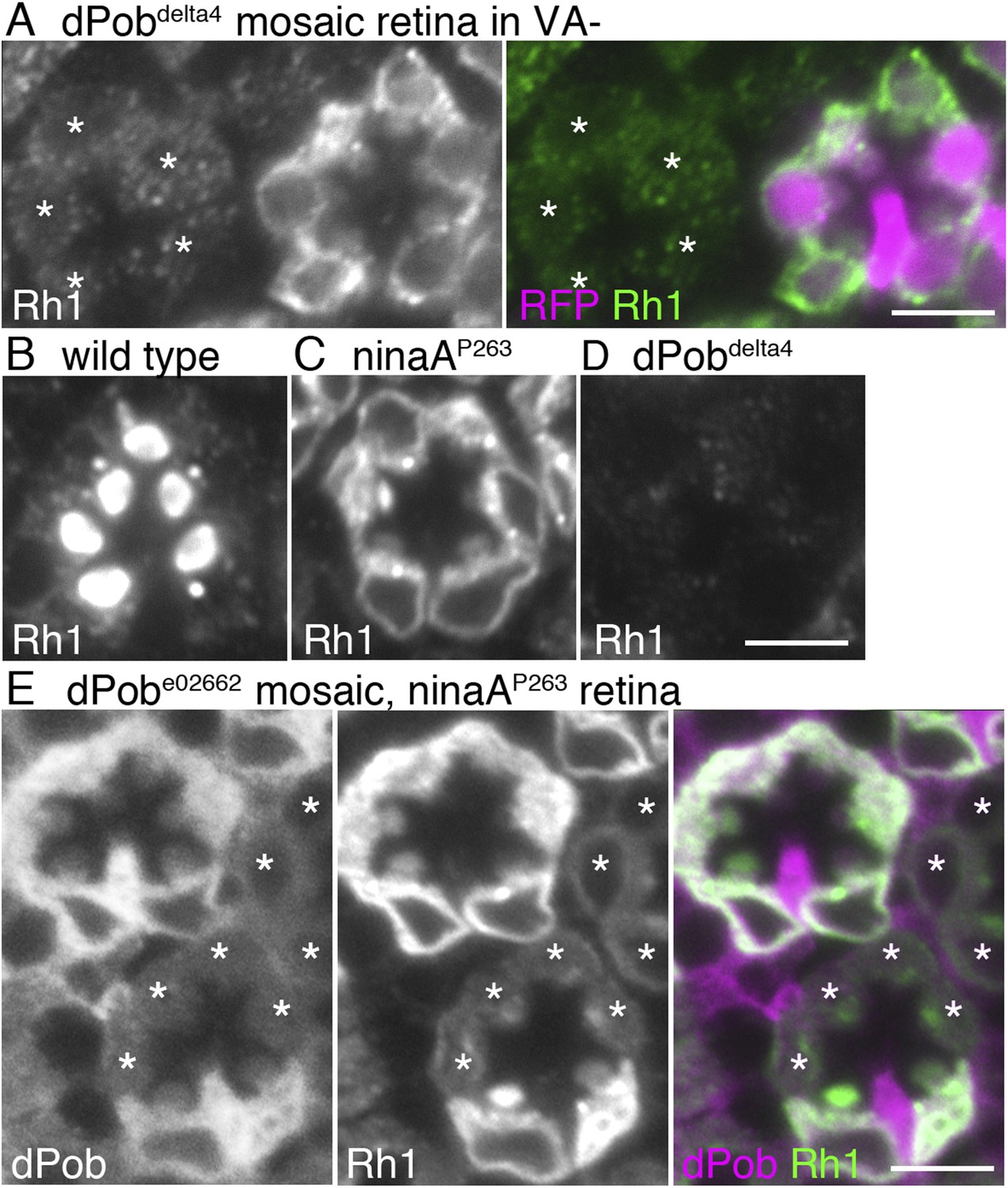
dPob stabilizes rhodopsin 1 (Rh1) apoprotein.
(A) Immunostaining of a dPob∆4 mosaic retina from a fly reared in vitamin A (VA)-deficient medium by anti-Rh1 antibody. Asterisks show dPob∆4 homozygous photoreceptors. (B–D) Immunostaining of a wild-type (B), ninaAp263(C), or dPob∆4 (D) ommatidium of flies reared in normal vitamin A-containing medium. (E) Immunostaining of a dPobe02662 mosaic retina in ninaAp263 homozygous mutant background from a fly reared in normal medium. Asterisks show dPob∆4 homozygous photoreceptors. Scale bar: 5 μm (A–E).
Figure 4
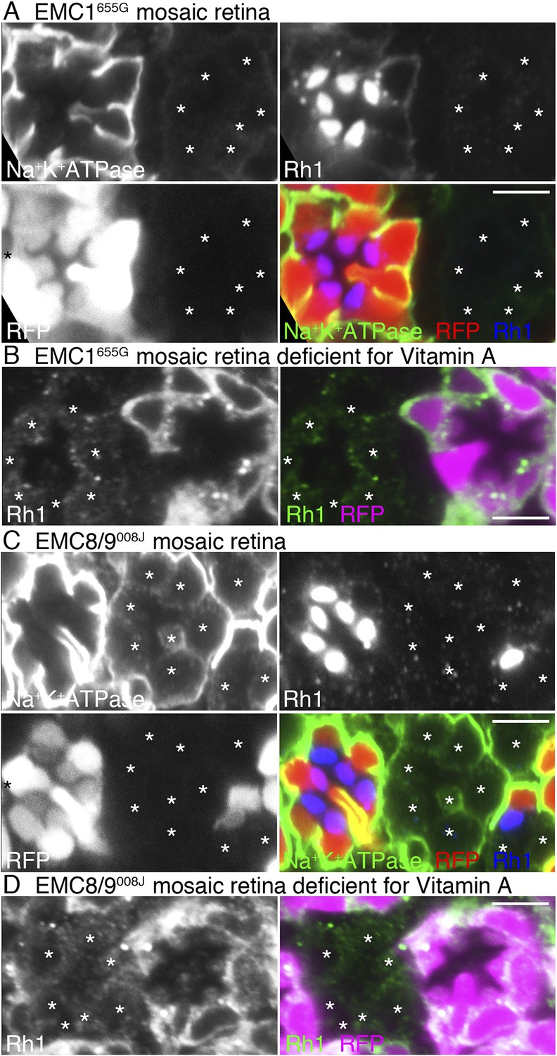
Loss of rhodopsin 1 (Rh1) apoprotein in EMC1 and EMC8/9 deficiency.
Immunostaining of a EMC1655G mosaic retina (A, B) or a EMC8/9008J mosaic retina (C, D) reared in normal (A, C) and vitamin A-deficient media (B, D). Asterisks show EMC1655G or EMC8/9008J homozygous photoreceptors. RFP (red) indicates wild-type photoreceptors (R1–R8). (A, C) Na+K+-ATPase, green; Rh1, blue; RFP, red. (B, D) Rh1, green; RFP, magenta. Scale bar: 5 μm (A–D).
Figure 5
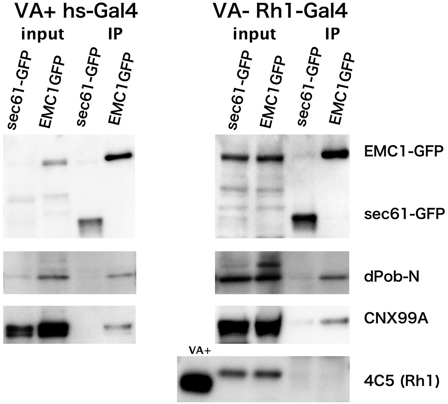
Co-immunoprecipitation of EMC1::GFP with dPob and calnexin (Cnx).
Immunoblotting of precipitates with anti-GFP antibody from the head extract was prepared from Rh1-Gal4/UAS-EMC1::GFP or sec61::GFP flies reared in a vitamin A (VA)-deficient medium (left) or heat shock (hs)-Gal4/UAS-EMC1::GFP or sec61::GFP flies reared in a vitamin A-containing normal medium (right). The mature form of rhodopsin 1 (Rh1) is accumulated in the rhabdomeres in normal medium but not in vitamin A-deficient medium. Instead of the mature form, an N-glycosylated immature form of Rh1 with a larger molecular weight accumulated in the endoplasmic reticulum of flies reared in the vitamin A-deficient medium. In both input extracts prepared from Rh1-Gal4/UAS-EMC1::GFP or sec61::GFP flies there is a band with the same position as EMC1GFP; this band will be the protein cross-reacting to anti-GFP antibody.
Figure 6
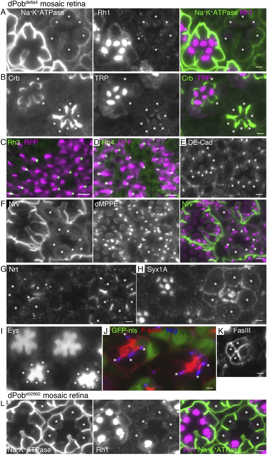
Essential role of dPob in the biosynthesis of multi-pass transmembrane proteins.
Immunostaining of a dPob∆4 mosaic retina (A–H) or a dPobe02662 mosaic retina (I). Asterisks show dPob homozygous photoreceptors. (A) Na+K+-ATPase, green; Rh1, magenta. (B) Crb, green; TRP1, magenta. (C, D) Rh3 (C) and Rh4 (D), green; RFP (wild-type cell marker), magenta. Although the boundary between dPob∆4 and wild-type cells is unclear, all green signals are attached to RFP-expressing cell bodies, indicating that mutant R7 cells do not express Rh3 (C) or Rh4 (D). (E) DE-Cad staining. (F) Nrv, the beta subunit of Na+K+-ATPase, green; dMPPE, magenta. (G) Nrt staining. (H) Syx1A staining. (I) Eys staining. (J) Nrg, blue; F-actin, red; GFP-nls (wild-type cell marker), green. (K) FasIII staining. (L) Na+K+-ATPase, green; Rh1, magenta. Scale bar: 2 μm (A, B), 10 μm (C, D), 2 μm (E–I).
Figure 7
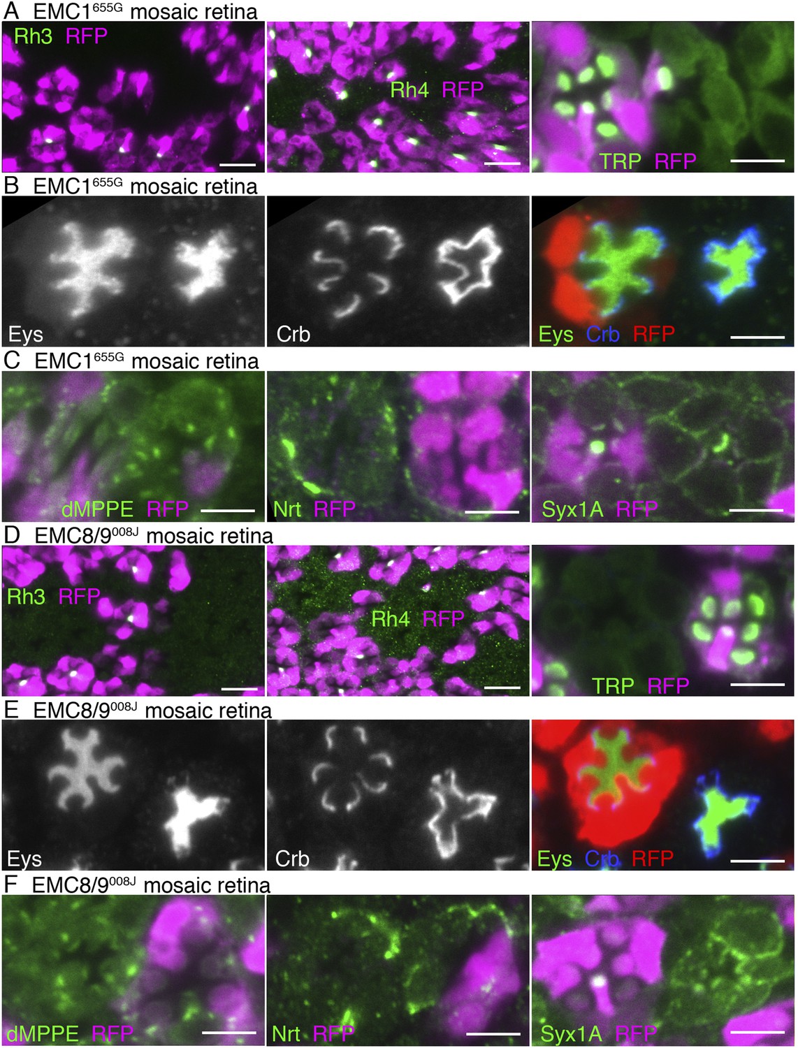
Essential role of EMC1 and EMC8/9 in the biosynthesis of multi-pass transmembrane proteins.
Immunostaining of a EMC1655G mosaic retina (A, B, C) or a EMC8/9008J mosaic retina (D, E, F). (A, D) Left: Rh3, middle: Rh4, right: TRP in green, RFP in magenda. (B, E) Eys in green, Crb in blue, and RFP, wild-type cell marker in red. (C, F) Left: dMPPE, middle: Nrt, right: Syx1A in green, RFP in magenda. Scale bar: 10 μm (left and middle in A, D), 5 μm (right in A, D), 5 μm (B, C, E, F).
Figure 8
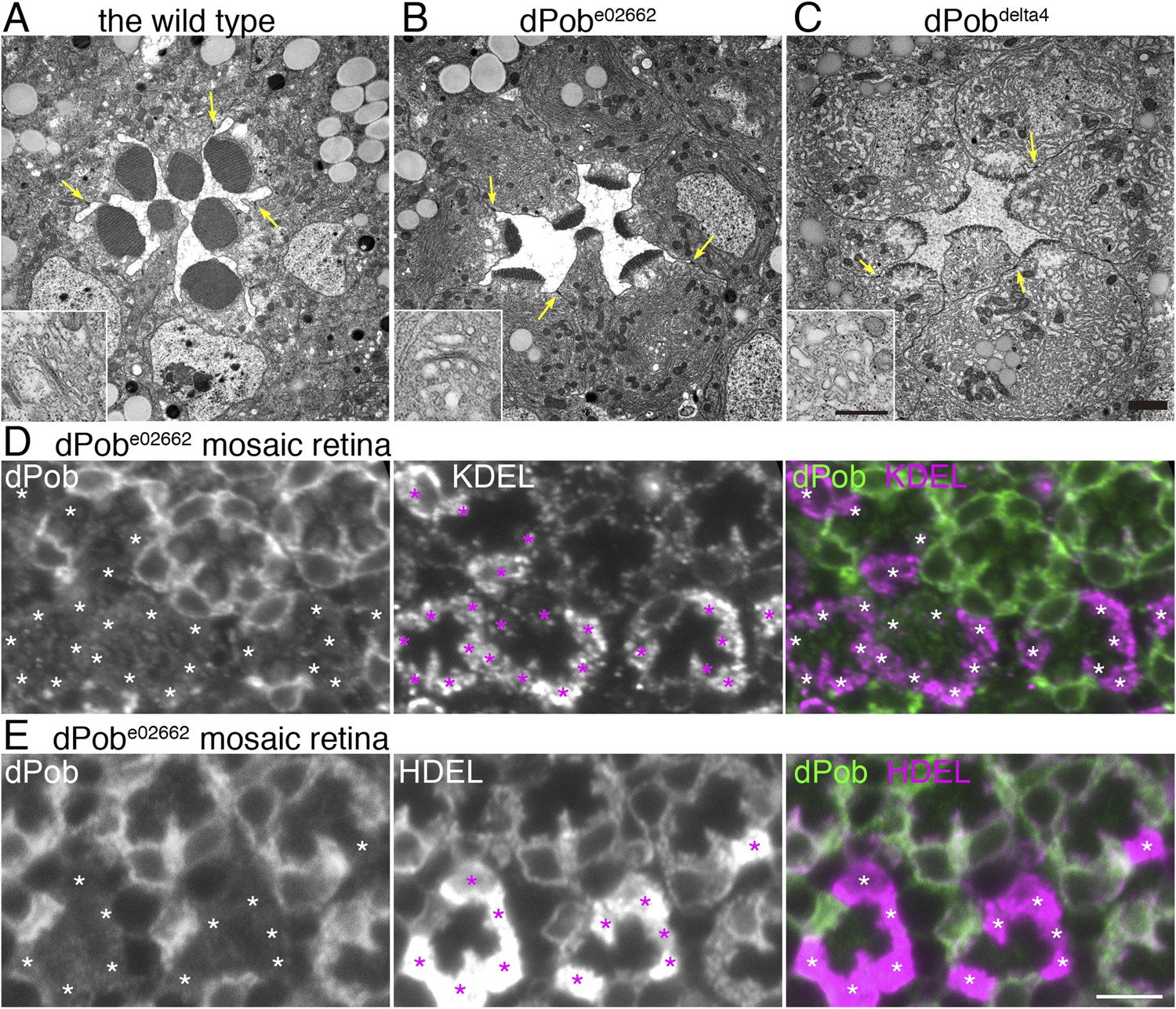
Endoplasmic reticulum membrane amplification and unfolded protein response (UPR) induced in dPob∆4 photoreceptor.
(A–C) Electron microscopy of late pupal photoreceptors: wild-type (A), dPobe02662 (B), and dPob∆4 photoreceptors (C). Arrow indicate adherens junctions. Insets show Golgi bodies. (D, E) Immunostaining of a dPobe02662 mosaic retina. dPob is shown in green and KDEL (D) or HDEL (E) are shown in magenta. Asterisks show dPob∆4 homozygous photoreceptors. Scale bar: 1 μm (A–C), 5 μm (D, E).
Figure 9
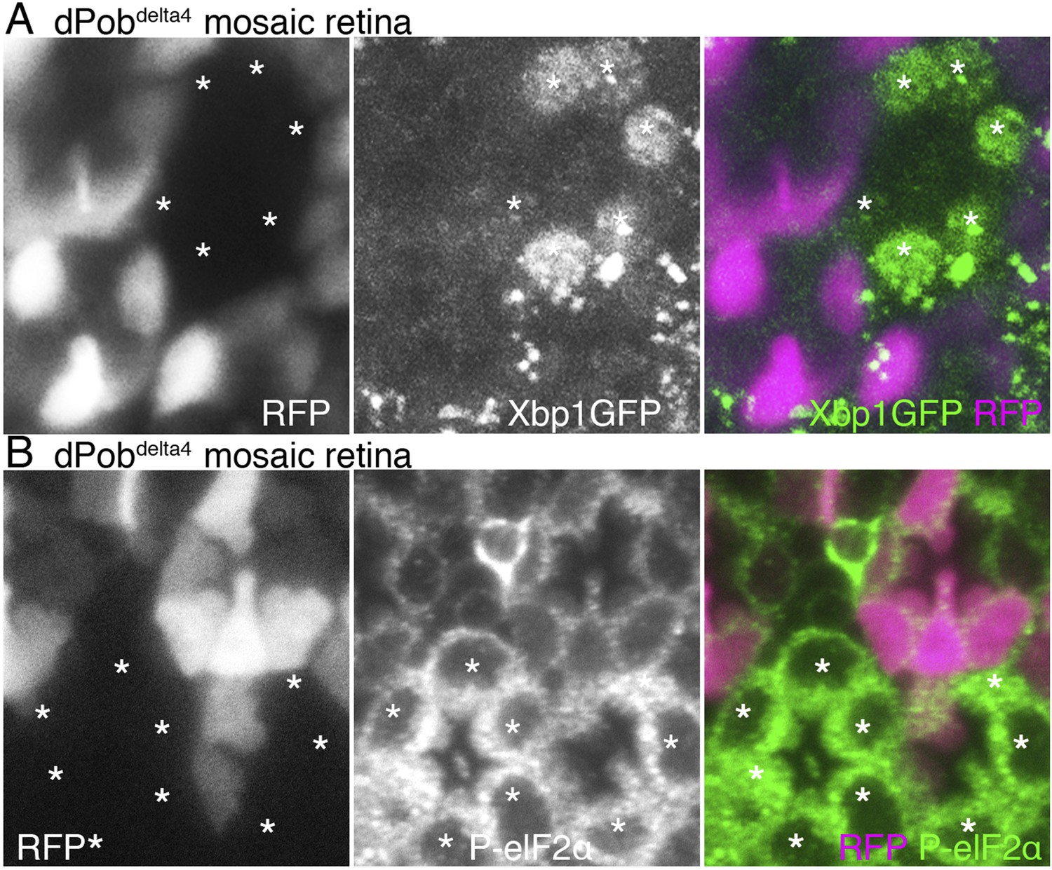
Unfolded protein response (UPR) induced in dPob∆4 photoreceptor.
(A) Projection image from the Z-series section with a 1 μm interval of dPob∆4 mosaic retina expressing RFP (magenta) as a wild-type cell marker and Xbp1:GFP as a UPR sensor. The Xbp1:GFP signal (green) is enhanced by immunostaining using anti-GFP antibody. Asterisks show dPob∆4 homozygous photoreceptors. (B) Immunostaining of a dPob∆4 mosaic retina expressing RFP (magenta) as a wild-type cell marker. Phosphorylated eukaryotic translation Initiation Factor 2α is shown in green. Asterisks show dPob∆4 homozygous photoreceptors.
Figure 10
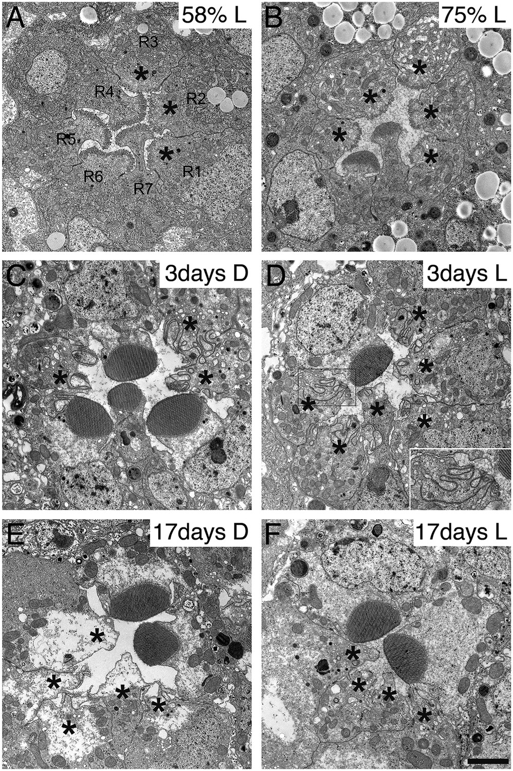
Development and degeneration of dPob∆4 photoreceptor rhabdomeres.
Electron microscopy of pupal and adult dPob∆4 mosaic retinas. Asterisks show dPob∆4 homozygous photoreceptors. Scale bar: 1 μm. (A, B) dPob∆4 mosaic ommatidia from 58% pupal development (A) and 73% pupal development (B) under constant light (L) condition. (C–F) dPob∆4 mosaic ommatidia from flies reared in complete darkness (D) (C, E) or under 12 hr light/12 hr dark conditions (D, F). Ommatidia from 3-day-old (C, D) and 17-day-old (E, F) flies. (D, inset) dPob∆4 R5 photoreceptor rhabdomere at higher magnification.
Download links
A two-part list of links to download the article, or parts of the article, in various formats.
Downloads (link to download the article as PDF)
Open citations (links to open the citations from this article in various online reference manager services)
Cite this article (links to download the citations from this article in formats compatible with various reference manager tools)
dPob/EMC is essential for biosynthesis of rhodopsin and other multi-pass membrane proteins in Drosophila photoreceptors
eLife 4:e06306.
https://doi.org/10.7554/eLife.06306


