Homo naledi, a new species of the genus Homo from the Dinaledi Chamber, South Africa
- University of the Witwatersrand, South Africa
- University of Wisconsin-Madison, United States
- Texas A&M University, United States
- Duke University, United States
- University of Zurich, Switzerland
- University of Arkansas, United States
- University of Kent, United Kingdom
- Max Planck Institute for Evolutionary Anthropology, Germany
- Mercyhurst University, United States
- New York University, United States
- New York Consortium in Evolutionary Primatology, United States
- Dartmouth College, United States
- University of Colorado Denver, United States
- Loughborough University, United Kingdom
- Tulane University, United States
- Lehman College, United States
- American Museum of Natural History, United States
- University of Cape Town, South Africa
- Museo Nacional de Ciencias Naturales, Spain
- Modesto Junior College, United States
- Louisiana State University, United States
- Nazarbayev University, Kazakhstan
- University of Missouri, United States
- University of Kentucky College of Medicine, United States
- Simon Fraser University, Canada
- Université de Montréal, Canada
- Australian National University, Australia
- Biology Department, Universidad Autònoma de Madrid, Spain
- Midwestern University, United States
- Liverpool John Moores University, United Kingdom
- University of Pisa, Italy
- Chaffey College, United States
- University of Johannesburg, South Africa
- George Washington University, United States
- University of Colorado School of Medicine, United States
- Croatian Natural History Museum, Croatia
- University of Iowa, United States
- Lincoln Memorial University, United States
- Smithsonian Institution, United States
- Department of Anthropology, Lakehead University, Canada
- Institute of Vertebrate Paleontology and Paleoanthropology, China
Figures
Figure 1
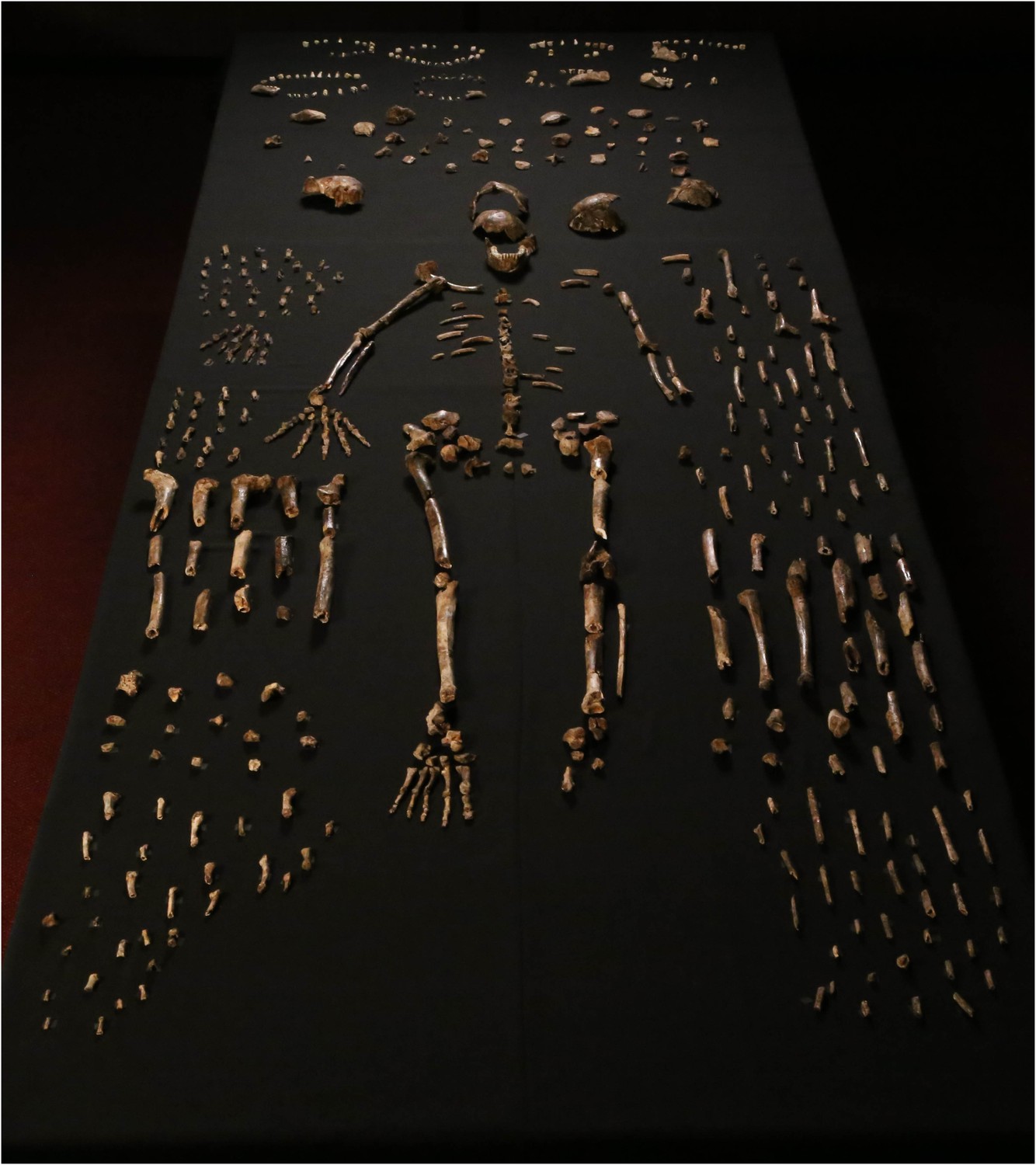
Dinaledi skeletal specimens.
The figure includes approximately all of the material incorporated in this diagnosis, including the holotype specimen, paratypes and referred material. These make up 737 partial or complete anatomical elements, many of which consist of several refitted specimens. Specimens not identified to element, such as non-diagnostic long bone or cranial fragments, and a subset of fragile specimens are not shown here. The ‘skeleton’ layout in the center of the photo is a composite of elements that represent multiple individuals. This view is foreshortened; the table upon which the bones are arranged is 120-cm wide for scale.
Figure 2
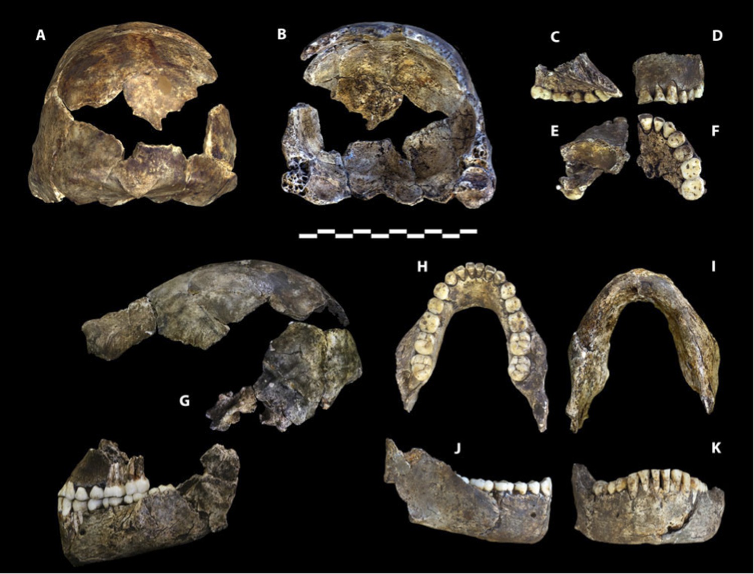
Holotype specimen of Homo naledi, Dinaledi Hominin 1 (DH1).
U.W. 101-1473 cranium in (A) posterior and (B) frontal views (frontal view minus the frontal fragment to show calvaria interior). U.W. 101-1277 maxilla in (C) medial, (D) frontal, (E) superior, and (F) occlusal views. (G) U.W. 101-1473 cranium in anatomical alignment with occluded U.W. 101-1277 maxilla and U.W. 101-1261 mandible in left lateral view. U.W. 101-1277 mandible in (H) occlusal, (I) basal, (J) right lateral, and (K) anterior views. Scale bar = 10 cm.
Figure 3
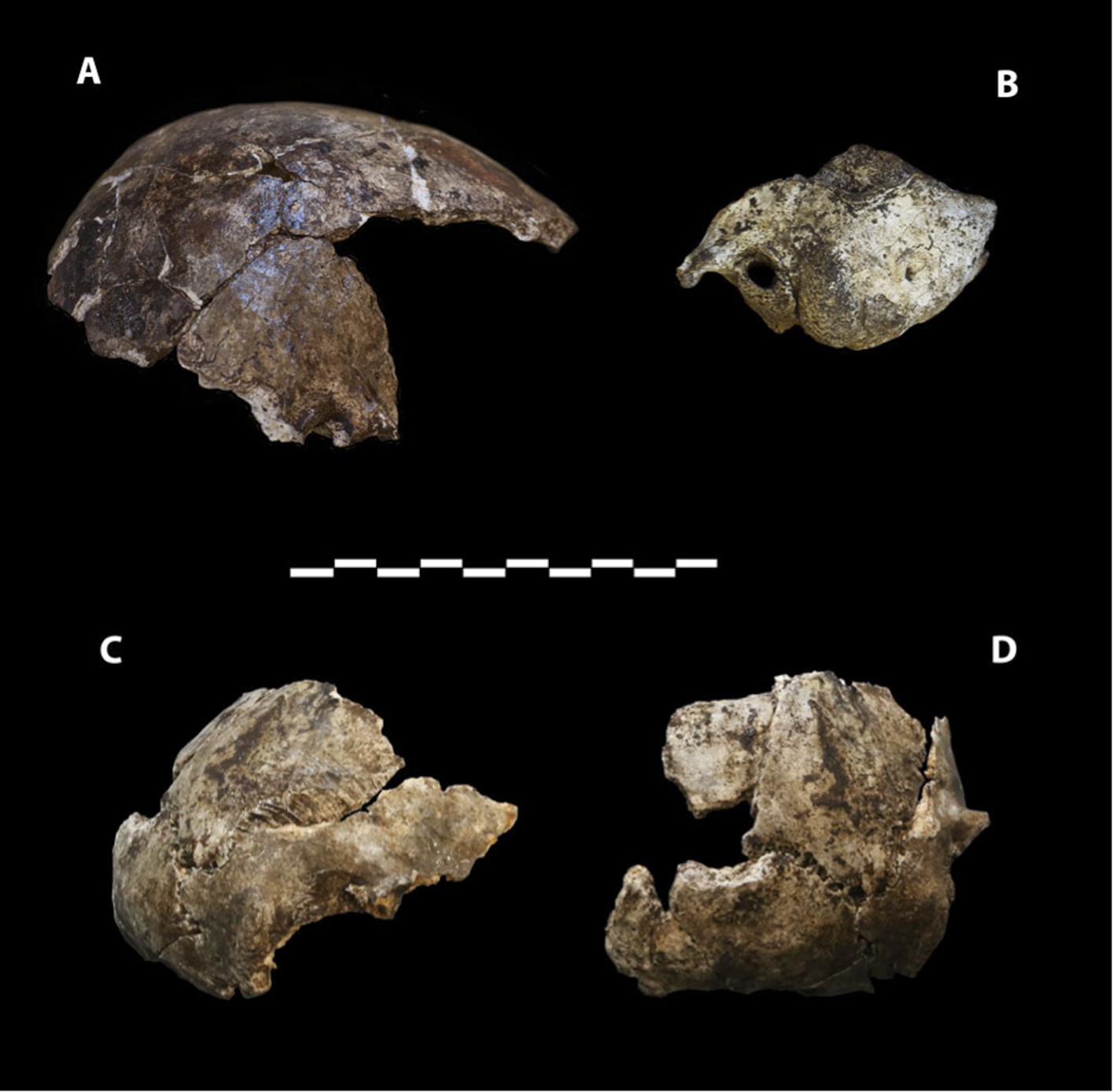
Cranial paratypes.
(A) DH2, right lateral view. (B) DH5, left lateral view. (C) DH4, right lateral view. (D) DH4, posterior view. Scale bar = 10 cm.
Figure 4

Paratype DH3.
(A) Frontal view. (B) Left lateral view, with calvaria in articulation with the mandible (U.W. 101-361). (C) Basal view. Mandible in (D) medial view; (E) occlusal view; (F) basal view. DH3 was a relatively old individual at time of death, with extreme tooth wear. Scale bar = 10 cm.
Figure 5
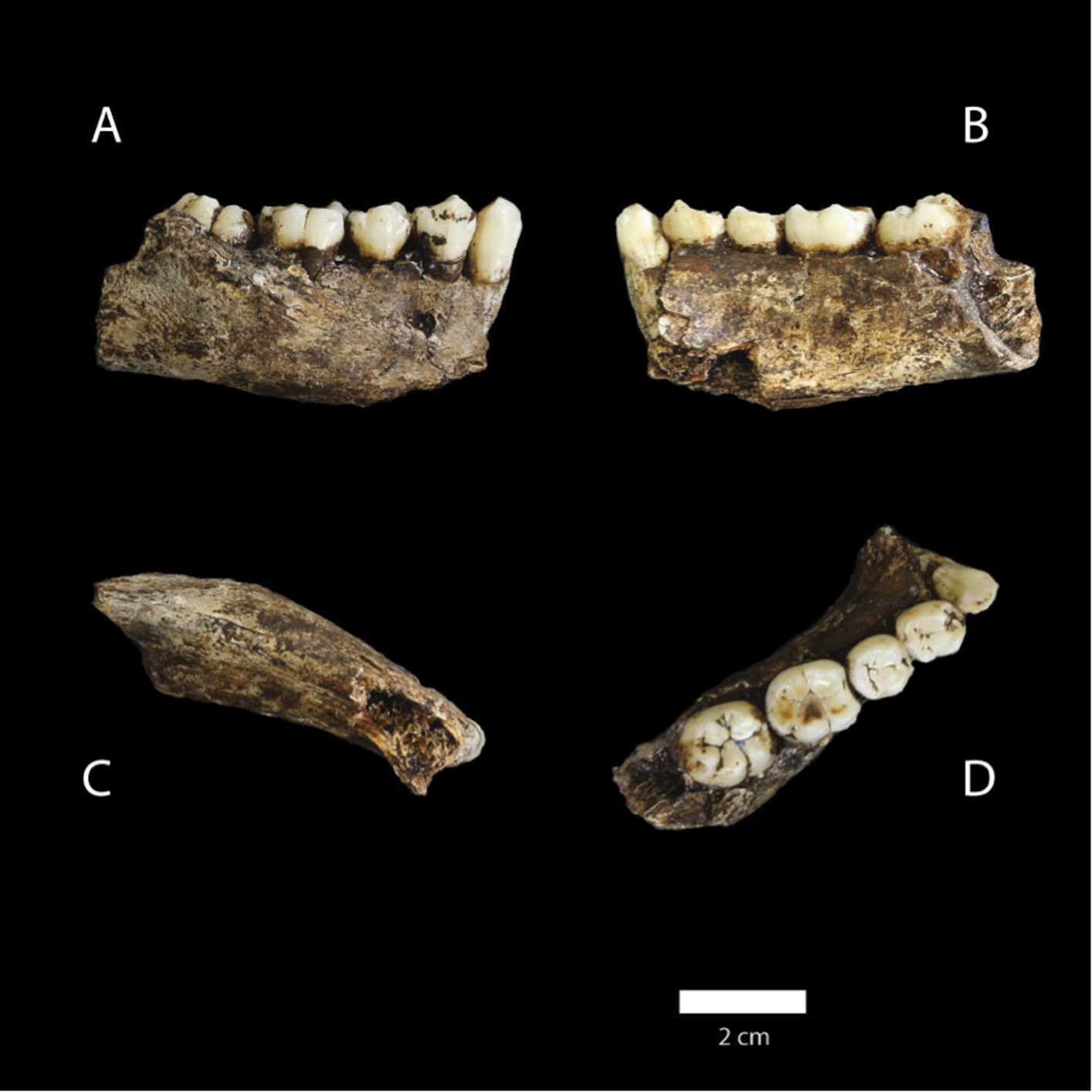
U.W. 101-377 mandible.
(A) Lateral view; (B) medial view; (C) basal view; (D) occlusal view. (D) The distinctive mandibular premolar morphology with elongated talonids in unworn state. Scale bar = 2 cm.
Figure 6
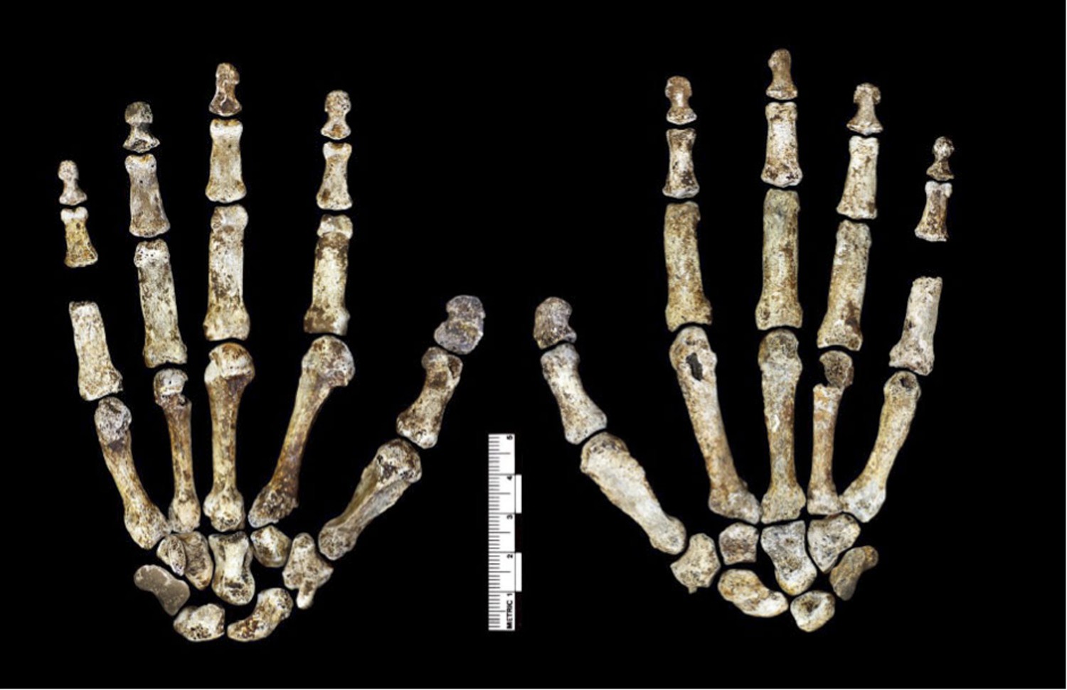
Hand 1.
Palmar view on left; dorsal view on right. This hand was discovered in articulation and all bones are represented except for the pisiform. The proportions of digits are humanlike and visually apparent, as are the expanded distal apical tufts on all digits, the robust pollical ray, and the unique first metacarpal morphology.
Figure 7
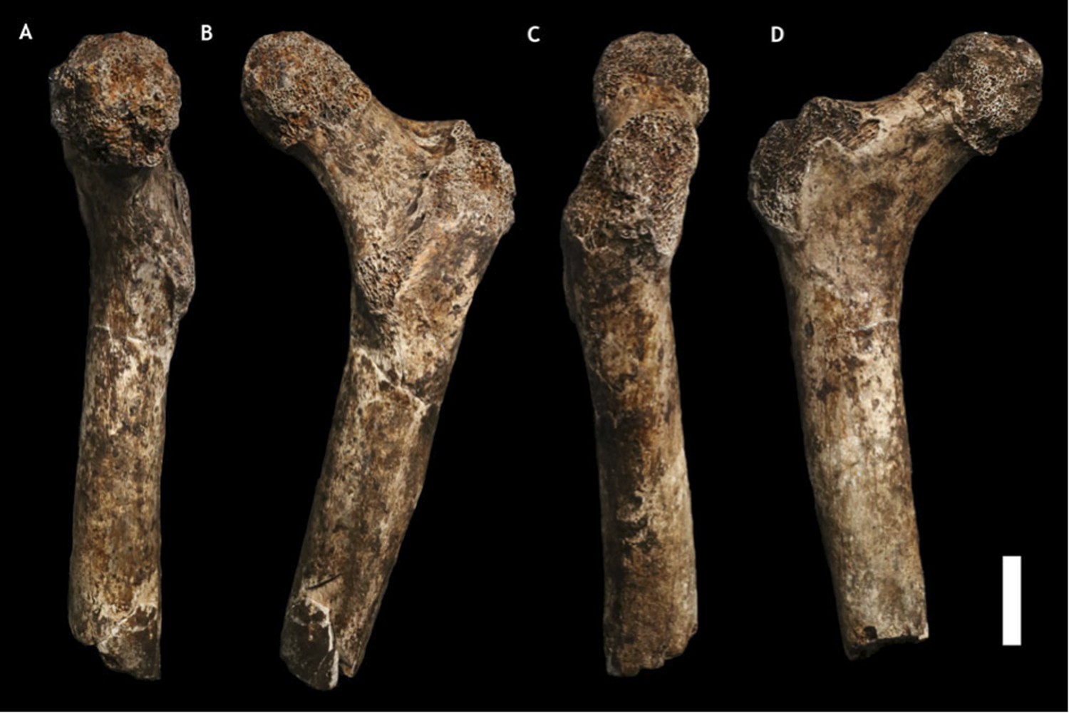
U.W. 101-1391 paratype femur.
(A) Medial view; (B) posterior view; (C) lateral view; (D) anterior view. The femur neck is relatively long and anteroposteriorly compressed. The anteversion of the neck is evident in medial view. Scale bar = 2 cm.
Figure 8
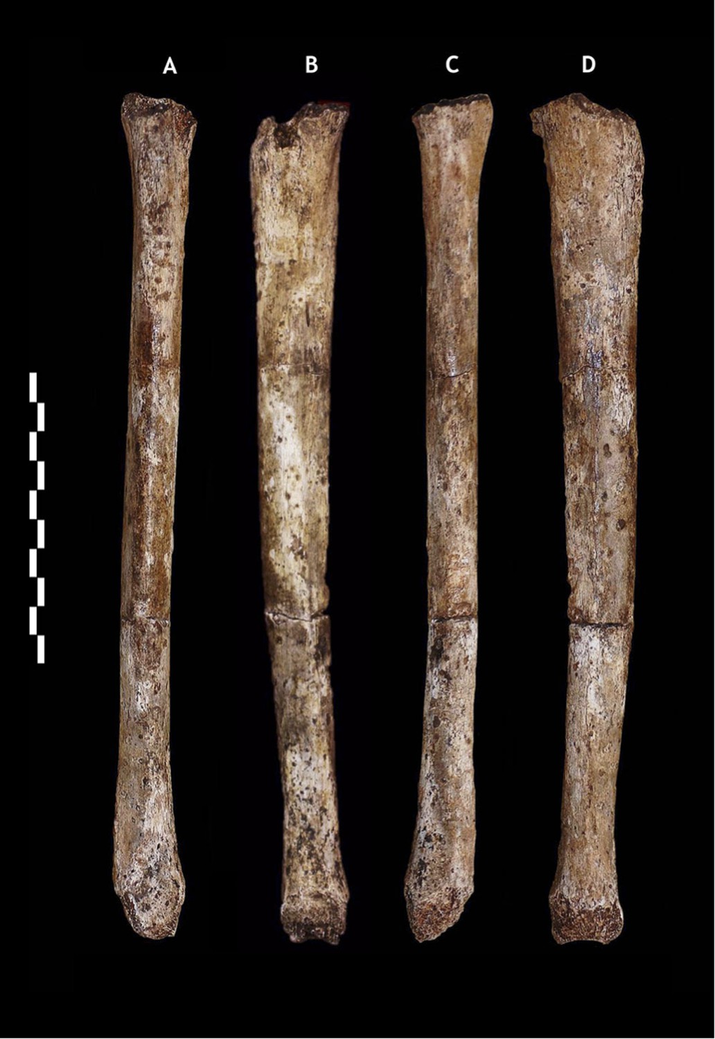
U.W. 101-484 paratype tibia.
(A) Anterior view; (B) medial view; (C) posterior view; (D) lateral view. The tibiae are notably slender for their length. Scale bar = 10 cm.
Figure 9

Foot 1 in (A) dorsal view; and (B) medial view.
(C) Proximal articular surfaces of the metatarsals of Foot 1, shown in articulation to illustrate transverse arch structure. Scale bar = 10 cm.
Figure 10

Maximum tibia length in H. naledi and other hominins.
Maximum tibia length for U.W. 101-484, compared to other nearly complete hominin tibia specimens. Australopithecus afarensis represented by A.L. 288-1 and KSD-VP-1/1 (Haile-Selassie et al., 2010); Homo erectus represented by D3901 from Dmanisi and KNM-WT 15000; Homo habilis by OH 35; Homo floresiensis by LB1 and LB8 (Brown et al., 2004; Morwood et al., 2005). Chimpanzee and contemporary European ancestry humans from Cleveland Museum of Natural History (Lee, 2001); Andaman Islanders from Stock (2013). Vertical lines represent sample ranges; bars represent 1 standard deviation.
Figure 11

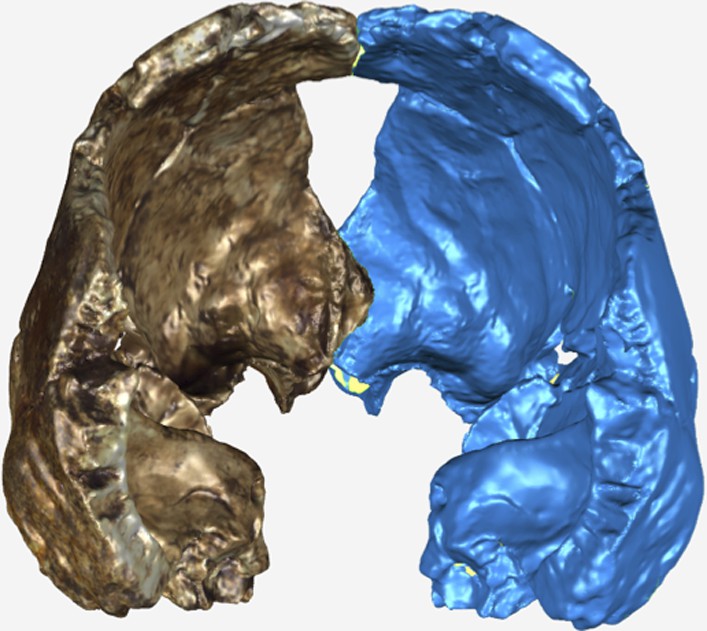
Virtual reconstruction of the endocranium of the larger composite cranium from DH1 and DH2 overlaid with the ectocranial surfaces.
(A) Lateral view. (B) Superior view. The resulting estimate of endocranial volume is 560cc. Scale bar = 10 cm.
Figure 12
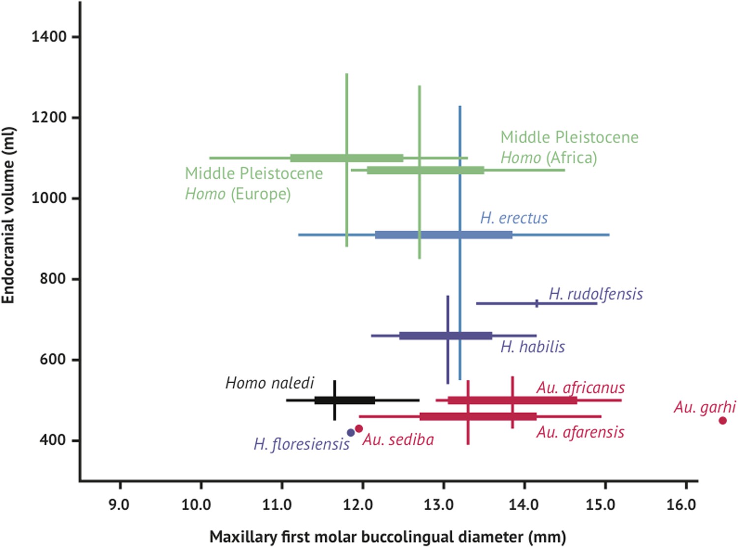
Brain size and tooth size in hominins.
The buccolingual breadth of the first maxillary molar is shown here in comparison to endocranial volume for many hominin species. H. naledi occupies a position with relatively small molar size (comparable to later Homo) and relatively small endocranial volume (comparable to australopiths). The range of variation within the Dinaledi sample is also fairly small, in particular in comparison to the extensive range of variation within the H. erectus sensu lato. Vertical lines represent the range of endocranial volume estimates known for each taxon; each vertical line meets the horizontal line representing M1 BL diameter at the mean for each taxon. Ranges are illustrated here instead of data points because the ranges of endocranial volume in several species are established by specimens that do not preserve first maxillary molars.
Figure 13
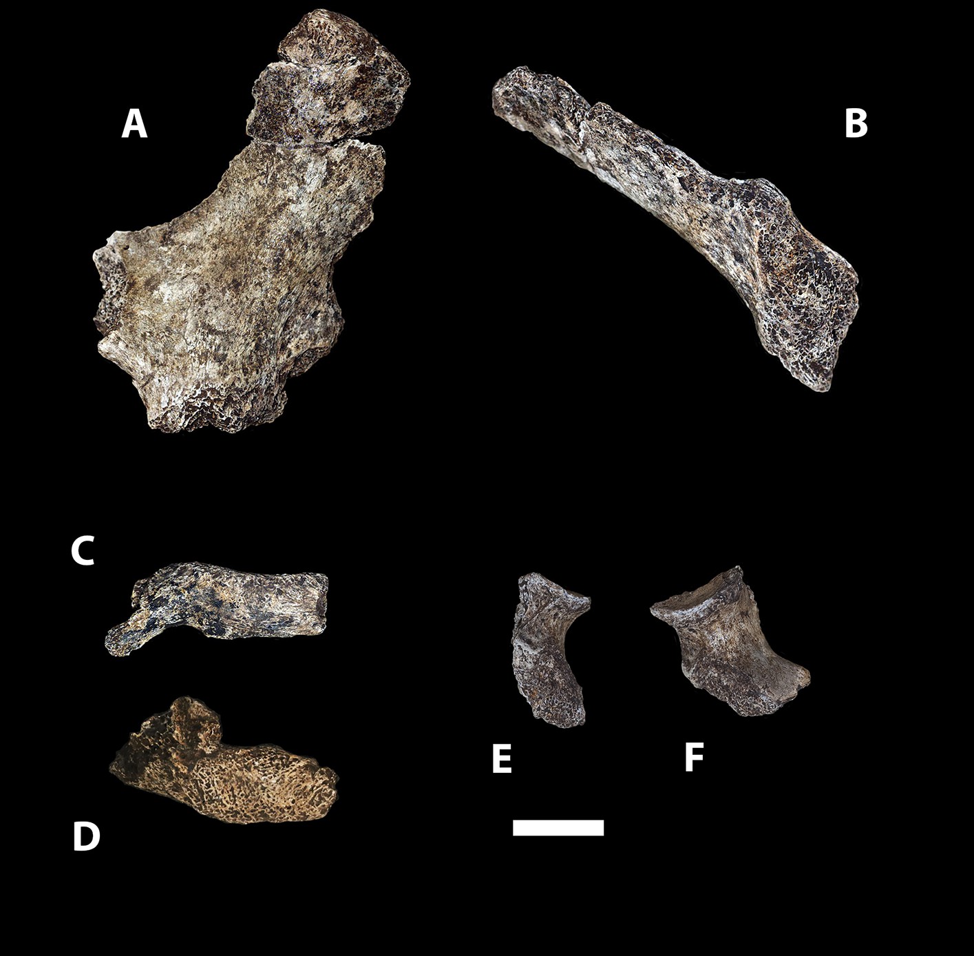
Selected pelvic specimens of H. naledi.
U.W. 101-1100 ilium in (A) lateral view showing a weak iliac pillar relatively near the anterior edge of the ilium, with no cristal tubercle development; (B) anterior view, angled to demonstrate the degree of flare, which is clear in comparison to the subarcuate surface. U.W. 101-723 immature sacrum in (C) anterior view; and (D) superior view. U.W. 101-1112 ischium in (E) lateral view; and (F) anterior view, demonstrating relatively short tuberacetabular diameter. Scale bar = 2 cm.
Figure 14
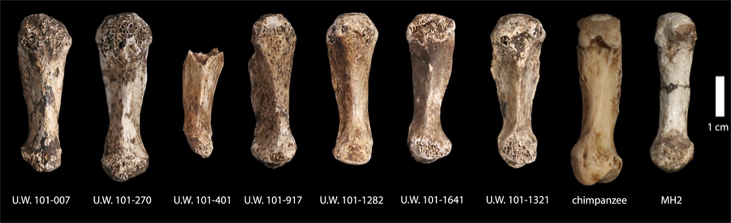
First metacarpals of H. naledi.
Seven first metacarpals have been recovered from the Dinaledi Chamber. U.W. 101-1321 is the right first metacarpal of the associated Hand 1 found in articulation. U.W. 101-1282 and U.W. 101-1641 are anatomically similar left and right first metacarpals, which we hypothesize as antimeres, both were recovered from excavation. U.W. 101-007 was collected from the surface of the chamber, and exhibits the same distinctive morphological characteristics as all the first metacarpals in the assemblage. All of these show a marked robusticity of the distal half of the bone, a very narrow, ‘waisted’ appearance to the proximal shaft and proximal articular surface, prominent crests for attachment of M. opponens pollicis and M. first dorsal interosseous, and a prominent ridge running down the palmar aspect of the bone. The heads of these metacarpals are dorsopalmarly flat and strongly asymmetric, with an enlarged palmar-radial protuberance. These distinctive features are present among all the first metacarpals in the Dinaledi collection, and are absent from any other hominin sample. Their derived nature is evident in comparison to apes and other early hominins, here illustrated with a chimpanzee first metacarpal and the MH2 first metacarpal of Australopithecus sediba.
Figure 15
Posterior view of the virtual reconstruction of DH3.
The resultant mirror image is displayed in blue. The antimeres were aligned by the frontal crest and sagittal suture using the Manual Registration function in GeoMagic Studio 14.0.
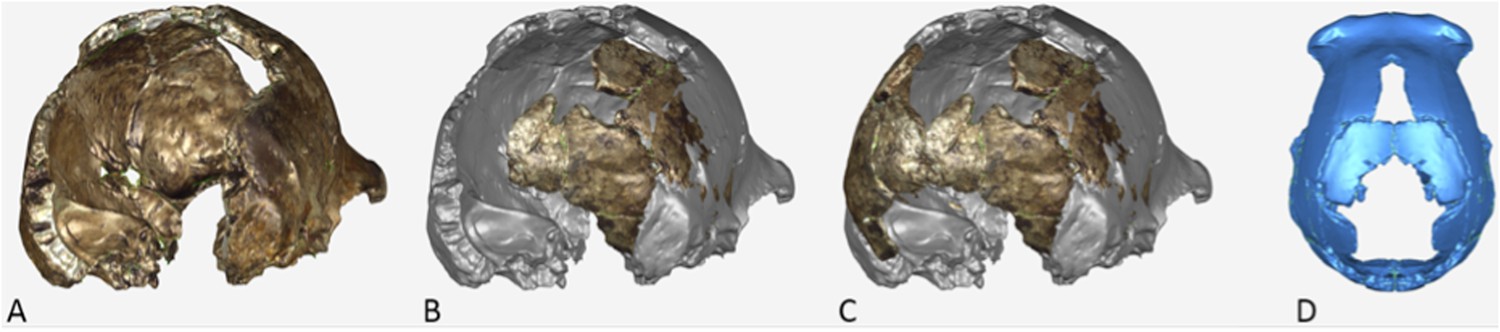
Figure 16
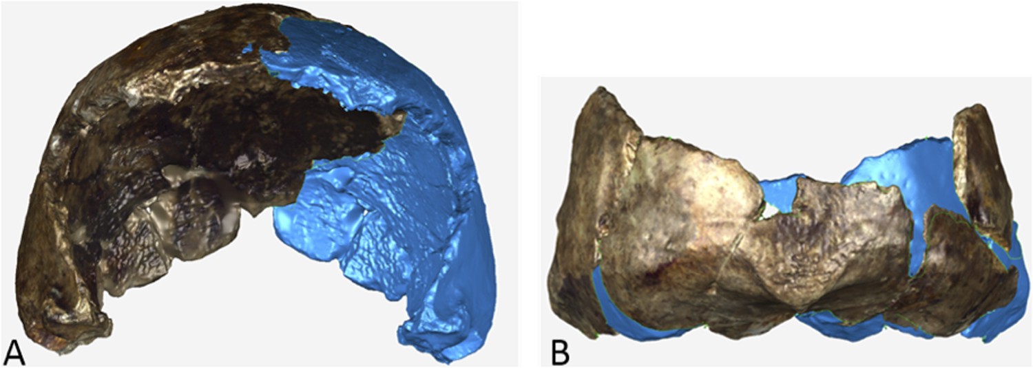
Virtual reconstruction of (A) DH2 and (B) occipital portion of DH1.
The actual specimen displays its original coloration and the mirror imaged portion is illustrated in blue.
Figure 17
Postero-lateral view of the virtual reconstruction of a composite cranium from DH3 and DH4.
(A) The surface scan of DH3 was mirror imaged and merged as described in Supplementary Note 8. (B) The scan of DH4 was aligned to the DH3 model. (C) DH4 was then mirror imaged to complete the occipital contour (D).
Figure 18
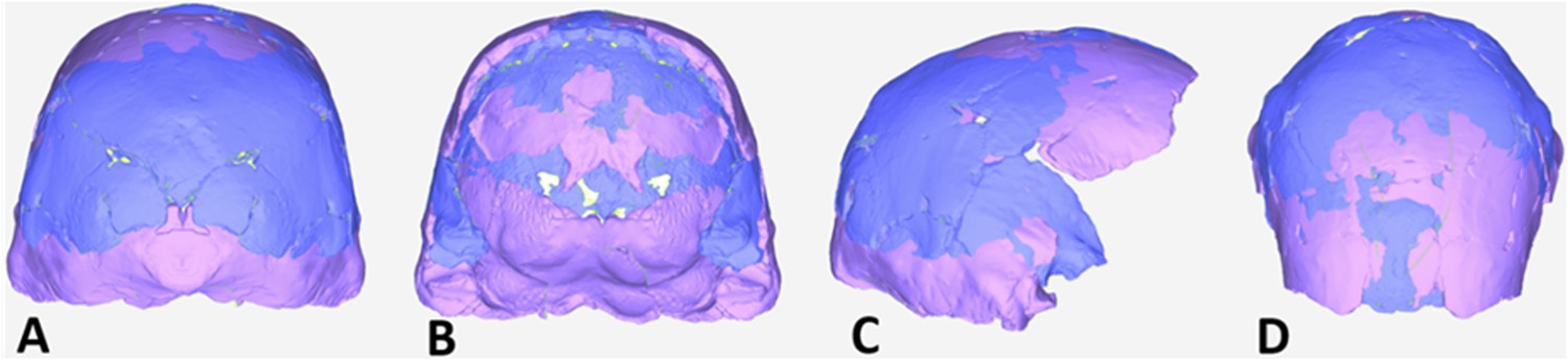
Virtual reconstruction of a composite cranium from DH1 and DH2.
The surface model of DH2 (blue), consisting of the original scan merged with the mirror image, was then uploaded and aligned with the mirror-imaged DH1 model (pink). Note the similarity in size and shape between DH1 and DH2 observed in the posterior (A) anterior (B) lateral (C) and superior (D) views.
Figure 19
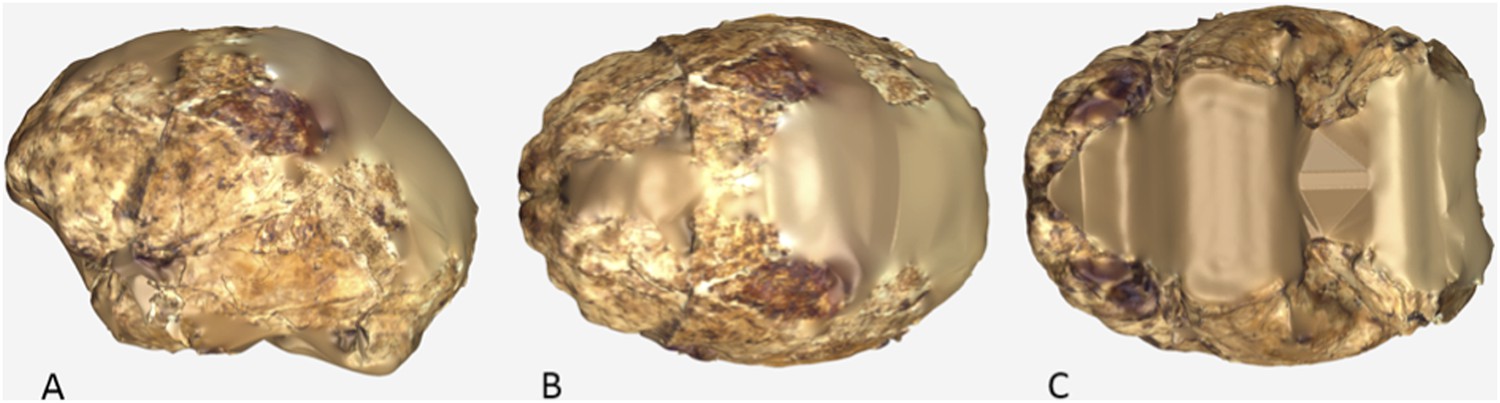
Virtual reconstruction of the endocranium of the composite cranium from DH3 and DH4.
(A) Lateral view. (B) Superior view. (C) Inferior view. In all views, anterior is to towards the left.
Figure 20

Virtual reconstruction of the endocranium of the composite cranium from DH3 and DH4 overlaid with the ectocranial surfaces.
(A) Lateral view. (B) Superior view.
Figure 21
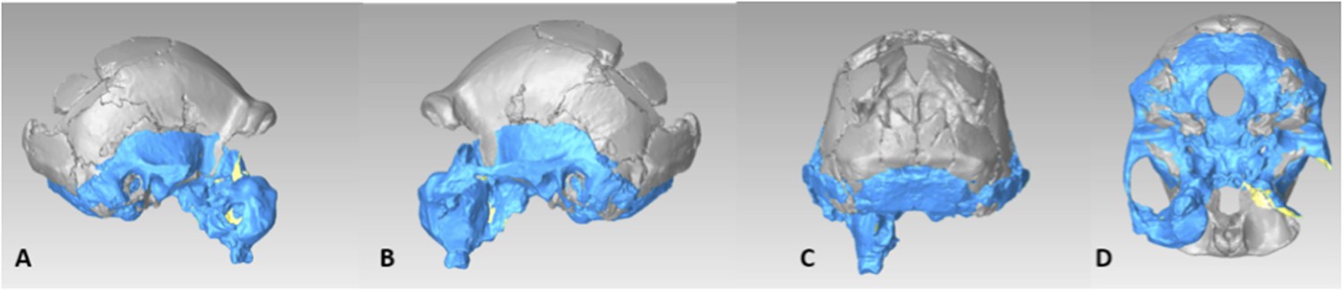
Virtual reconstruction the DH3/DH4 cranial base using a model of Sts 19.
(A) Right lateral view. (B) Left lateral view. (C) Posterior view. (D) Inferior view.
Figure 22
Virtual reconstruction the DH3/DH4 endocranial volume using a cranial base model of Sts 19.
Right lateral view.
Tables
Table 1
Cranial and mandibular measurements for H. naledi, early hominins, and modern humans
| Measurement definitions as in Wood (1991) | P. aethiopicus | P. boisei | P. robustus | Au. afarensis | Au. africanus | Au. sediba | H. naledi | H. habilis | H. rudolfensis | H. erectus | MP Homo | H. sapiens | |
|---|---|---|---|---|---|---|---|---|---|---|---|---|---|
| Cranium | |||||||||||||
| Cranial capacity | – | 410 | 485 | 493 | 457 | 467 | 420 | 513 | 610 | 776 | 865 | 1266 | 1330 |
| Porion height | 6 | 72 | 74 | – | 86 | 70 | 67 | 81 | 77 | 90 | 94 | 101 | 112 |
| Posterior cranial length | 3 | 58 | 47 | 54 | 60 | 44 | – | 65 | 60 | 70 | 79 | 99 | 81 |
| Bi-parietal breadth | 9 | 94 | 98 | – | 90 | 99 | 100 | 103 | 107 | 118 | 129 | 142 | 132 |
| Bi-temporal breadth | 10 | 110 | 109 | 108 | 115 | 104 | 101 | 107 | 112 | 126 | 131 | 146 | 127 |
| Closest approach of temporal lines | – | crest* | crest* | crest* | crest* | 21 | 56 | 52 | 35 | 51 | 72 | 101 | 96 |
| Supraorbital height index | – | 46 | 53 | 50 | 51 | 60 | 56 | 56 | 64 | 59 | 56 | 62 | 71 |
| Minimum post-orbital breadth | – | 62 | 66 | 70 | 77 | 67 | 70 | 68 | 75 | 78 | 89 | 96 | 97 |
| Superior facial breadth | 49 | 100 | 107 | 109 | – | 95 | 86 | 86 | 97 | 113 | 110 | 124 | 107 |
| Post-orbital constriction index† | – | 62 | 61 | 64 | – | 69 | 81 | 79 | 72 | 74 | 81 | 80 | 91 |
| EAM area (as an ellipse)‡ | – | 77 | 80 | 103 | 70 | 96 | – | 38 | 76 | – | 95 | 85 | 61 |
| Root of zygomatic process origin | – | P4 | P4 | P3 to M1 | P4 to M1 | P4 to M1 | P4 | P3 to P4 | P4 to M1 | P4 to M1 | P4 to M1 | M1 | M1 |
| Petromedian angle | 137 | 50 | 45 | 50 | 31 | 33 | – | 55 | 48 | – | 52 | 55 | 46 |
| Maxilloalveolar process | |||||||||||||
| Maxilloalveolar length | 87 | 94 | 78 | 69 | 67 | 71 | 63 | 57 | 65 | 68 | 66 | 69 | 55 |
| Maxilloalveolar breadth | 88 | 83 | 76 | 69 | 68 | 66 | 63 | 71 | 68 | 72 | 70 | 72 | 62 |
| Palate breadth | 91 | 32 | 40 | 35 | 30 | 36 | 29 | 44 | 38 | 40 | 38 | 56 | 40 |
| Palate depth at incisive fossa | – | 3 | 11 | 10 | 10 | 9 | 10 | 5 | 10 | 13 | 11 | 10 | 9 |
| Palate depth at M1 | 103 | 7 | 18 | 11 | 11 | 13 | 10 | 10 | 12 | 16 | 15 | 18 | 13 |
| Mandible | |||||||||||||
| Symphysis height | 141 | 37 | 49 | 42 | 39 | 37 | 32 | 33 | 31 | 37 | 35 | 34 | 34 |
| Symphysis width | 142 | 26 | 28 | 25 | 20 | 21 | 18 | 18 | 20 | 24 | 18 | 17 | 14 |
| Symphysis area at M1 (as an ellipse)‡ | 146 | 757 | 1114 | 835 | 623 | 606 | 452 | 467 | 393 | 723 | 519 | 474 | 365 |
| Corpus height at M1 | 150 | 38 | 42 | 36 | 34 | 32 | 30 | 26 | 29 | 36 | 31 | 31 | 28 |
| Corpus breadth at M1 | 151 | 25 | 29 | 26 | 20 | 21 | 18 | 16 | 20 | 22 | 19 | 19 | 13 |
| Corpus area at M1 (as an ellipse)‡ | 152 | 742 | 955 | 736 | 540 | 539 | 405 | 326 | 425 | 631 | 458 | 469 | 296 |
| Mental foramen height index§ | – | 51 | 50 | 54 | 58 | 53 | 50 | 40 | 46 | 49 | 48 | 48 | 50 |
-
*
At least in presumed males.
-
†
Post-orbital breadth/superior facial breadth × 100.
-
‡
Following the formula (π × (corpus height/2) × (corpus breadth/2)).
-
§
Height of mental foramen from alveolar border relative to corpus height at the mental foramen.
-
MP, Middle Pleistocene.
-
Unless otherwise indicated measurements are defined as in Wood (1991). Chord distances are in mm. Data for H. naledi collected from original fossils or laser scans by DJdeR and HMG; comparative data collected by DJdeR on original fossils and casts and supplemented by data from Wood (1991).
Table 2
Dental measures for H. naledi and comparative hominin species
| Maxillary | |||||||||||||||||
|---|---|---|---|---|---|---|---|---|---|---|---|---|---|---|---|---|---|
| I1 | I2 | C | P3 | P4 | M1 | M2 | M3 | ||||||||||
| MD | LL | MD | LL | MD | LL | MD | BL | MD | BL | MD | BL | MD | BL | MD | BL | ||
| Au. anamensis | n | 3 | 5 | – | 2 | 6 | 7 | 7 | 6 | 5 | 3 | 12 | 10 | 10 | 8 | 9 | 8 |
| mean | 10.8 | 8.7 | – | 7.3 | 11.0 | 10.6 | 9.9 | 12.6 | 8.9 | 13.6 | 11.5 | 12.9 | 13.0 | 14.4 | 12.5 | 14.2 | |
| range | 9.1–12.4 | 8.2–9.3 | – | 7.0–7.5 | 9.9–12.3 | 9.1–11.8 | 8.2–11.8 | 10.1–14.3 | 7.2–12.1 | 12.6–14.2 | 7.8–14.3 | 9.0–16.7 | 10.9–16.3 | 12.9–16.1 | 11.1–15.7 | 13.0–15.7 | |
| Au. afarensis | n | 7 | 8 | 9 | 9 | 15 | 15 | 12 | 10 | 18 | 12 | 16 | 13 | 10 | 11 | 11 | 11 |
| mean | 10.7 | 8.4 | 7.5 | 7.2 | 9.9 | 10.8 | 8.8 | 12.4 | 9.1 | 12.4 | 12.0 | 13.4 | 12.9 | 14.6 | 12.7 | 14.5 | |
| range | 9.9–11.8 | 7.1–9.7 | 6.6–8.2 | 6.2–8.1 | 8.8–11.6 | 9.3–12.5 | 7.7–9.7 | 11.3–13.4 | 7.6–10.8 | 11.1–14.5 | 10.5–13.8 | 12.0–15.0 | 12.1–13.6 | 13.4–15.2 | 10.9–14.8 | 13.1–16.3 | |
| Au. africanus | n | 15 | 15 | 11 | 10 | 16 | 13 | 26 | 25 | 20 | 20 | 21 | 20 | 23 | 24 | 27 | 28 |
| mean | 10.7 | 8.3 | 6.9 | 6.8 | 9.9 | 10.3 | 9.2 | 12.7 | 9.5 | 13.4 | 12.9 | 13.9 | 14.1 | 15.7 | 14.2 | 16.0 | |
| range | 9.4–12.5 | 7.4–9.1 | 5.8–8.0 | 5.6–7.9 | 8.8–11.0 | 8.7–12.0 | 8.5–10.2 | 10.7–14.5 | 7.2–11.0 | 12.4–15.3 | 11.7–14.4 | 12.9–15.3 | 12.1–16.3 | 12.8–17.9 | 11.2–16.9 | 13.1–18.6 | |
| Au. sediba | n | 1 | 1 | 1 | 1 | 1 | 1 | 1 | 1 | 1 | 1 | 1 | 1 | 1 | 1 | 2 | 2 |
| mean | 10.1 | 6.9 | 7.2 | 6.6 | 9.0 | 8.8 | 9.0 | 11.2 | 9.3 | 12.1 | 12.9 | 12.0 | 12.9 | 13.7 | 13.0 | 13.5 | |
| range | – | – | – | – | – | – | – | – | – | – | – | – | – | – | 12.6–13.3 | 12.9–14.1 | |
| H. naledi | n | – | 5 | 4 | 8 | 10 | 9 | 10 | 10 | 7 | 7 | 12 | 13 | 11 | 9 | 7 | 7 |
| mean | 9.4 | 6.5 | 6.6 | 6.2 | 8.1 | 8.6 | 8.0 | 10.5 | 8.1 | 11.0 | 11.6 | 11.7 | 12.2 | 12.8 | 11.6 | 12.4 | |
| range | 8.8–9.8 | 6.3–7.0 | 6.3–7.0 | 5.8–6.6 | 7.3–8.9 | 8.0–9.6 | 7.7–8.4 | 9.8–11.0 | 7.7–8.7 | 10.5–11.2 | 10.5–12.4 | 11.2–12.4 | 11.0–13.0 | 11.9–13.6 | 11.0–12.7 | 11.4–13.4 | |
| H. habilis | n | 2 | 2 | 4 | 4 | 2 | 3 | 7 | 7 | 8 | 8 | 13 | 13 | 7 | 7 | 7 | 7 |
| mean | 10.6 | 8.0 | 7.4 | 6.6 | 9.0 | 9.8 | 9.0 | 11.9 | 9.2 | 12.1 | 12.7 | 13.0 | 12.7 | 14.3 | 12.3 | 14.7 | |
| range | 10.1–11.1 | 7.3–8.7 | 6.7–8.1 | 6.0–7.9 | 8.5–9.4 | 8.5–11.6 | 8.1–9.6 | 11.0–12.7 | 8.5–9.9 | 11.0–13.1 | 11.6–13.9 | 12.1–14.1 | 11.8–13.5 | 13.5–16.2 | 11.3–13.9 | 13.2–16.6 | |
| H. rudolfensis | n | 1 | 1 | – | – | 1 | 1 | 1 | 1 | 2 | 2 | 2 | 2 | 2 | 2 | 1 | 1 |
| mean | 12.3 | 7.7 | – | – | 11.5 | 12.5 | 10.5 | 13.6 | 10.2 | 12.5 | 14.0 | 14.0 | 14.3 | 15.8 | 13.3 | 13.5 | |
| range | – | – | – | – | – | – | – | – | 9.7–10.7 | 11.1–13.8 | 13.9–14.2 | 13.3–14.8 | 14.1–14.6 | 14.1–17.6 | – | – | |
| H. erectus | n | 11 | 12 | 6 | 6 | 12 | 12 | 27 | 27 | 30 | 29 | 34 | 32 | 22 | 22 | 16 | 16 |
| mean | 10.3 | 8.1 | 7.7 | 8.0 | 9.5 | 10.0 | 8.5 | 11.8 | 8.1 | 11.6 | 12.2 | 13.2 | 12.0 | 13.3 | 10.5 | 12.8 | |
| range | 8.1–12.6 | 7.0–11.7 | 6.0–8.3 | 6.9–8.5 | 8.5–11.1 | 9.0–11.8 | 7.1–10.1 | 9.5–13.8 | 7.0–9.4 | 9.9–13.4 | 10.1–14.6 | 11.0–15.9 | 10.3–13.6 | 10.9–15.5 | 8.7–14.7 | 10.4–15.8 | |
| H. neanderthalensis | n | 28 | 37 | 35 | 41 | 28 | 29 | 16 | 17 | 21 | 19 | 23 | 24 | 27 | 28 | 22 | 21 |
| mean | 9.7 | 8.5 | 8.0 | 8.4 | 8.8 | 10.1 | 8.0 | 10.6 | 7.8 | 10.6 | 11.6 | 12.3 | 10.9 | 12.5 | 9.9 | 12.3 | |
| range | 8.2–11.8 | 7.3–9.9 | 5.8–9.3 | 5.8–9.9 | 7.2–10.0 | 7.6–11.4 | 6.6–9.3 | 8.4–11.8 | 5.9–11.5 | 8.3–11.7 | 9.5–13.5 | 11.0–14.2 | 8.9–15.9 | 10.8–14.6 | 8.2–11.4 | 9.8–14.6 | |
| H. heidelbergensis | n | 21 | 23 | 19 | 21 | 27 | 29 | 25 | 25 | 22 | 23 | 25 | 24 | 24 | 23 | 26 | 27 |
| mean | 9.6 | 7.8 | 7.7 | 7.8 | 8.8 | 9.8 | 7.9 | 10.6 | 7.6 | 10.3 | 11.2 | 11.9 | 10.2 | 12.3 | 8.9 | 11.6 | |
| range | 8.7–10.7 | 7.1–9.9 | 7.2–8.4 | 7.3–8.6 | 8.1–11.0 | 8.8–11.8 | 7.1–9.0 | 9.2–12.2 | 7.0–8.8 | 9.1–11.5 | 9.9–12.3 | 10.3–13.2 | 8.1–12.1 | 11.1–13.8 | 7.6–11.0 | 10.0–13.2 | |
| MP/LP African Homo | n | 6 | 6 | 7 | 8 | 4 | 4 | 6 | 6 | 10 | 10 | 14 | 14 | 20 | 20 | 9 | 9 |
| mean | 9.0 | 7.8 | 7.4 | 7.2 | 8.9 | 9.7 | 8.4 | 10.8 | 8.1 | 10.8 | 12.3 | 13.2 | 11.0 | 12.9 | 9.2 | 11.7 | |
| range | 6.3–10.9 | 6.6–8.7 | 6.0–9.3 | 6.1–8.5 | 8.2–9.5 | 8.8–10.0 | 8.1–8.7 | 9.9–11.8 | 7.5–9.3 | 9.4–12.8 | 10.4–14.0 | 12.0–15.0 | 7.8–13.0 | 11.0–15.0 | 7.6–10.2 | 10.0–13.2 | |
| Mandibular | |||||||||||||||||
|---|---|---|---|---|---|---|---|---|---|---|---|---|---|---|---|---|---|
| I1 | I2 | C | P3 | P4 | M1 | M2 | M3 | ||||||||||
| MD | LL | MD | LL | MD | LL | MD | BL | MD | BL | MD | BL | MD | BL | MD | BL | ||
| Au. anamensis | n | 2 | 1 | 4 | 3 | 7 | 7 | 8 | 8 | 8 | 8 | 9 | 10 | 7 | 7 | 8 | 8 |
| mean | 6.9 | 7.4 | 7.8 | 8.3 | 10.0 | 10.4 | 12.4 | 9.2 | 9.1 | 11.3 | 12.9 | 12.3 | 14.0 | 13.4 | 15.3 | 13.4 | |
| range | 6.8–6.9 | – | 6.6–8.7 | 7.9–8.6 | 6.6–13.9 | 9.2–11.4 | 11.3–13.4 | 8.6–10.0 | 7.4–9.8 | 9.6–13.2 | 11.6–13.8 | 10.2–14.8 | 13.0–15.9 | 12.3–14.9 | 13.7–17.0 | 12.1–15.2 | |
| Au. afarensis | n | 7 | 8 | 7 | 6 | 13 | 16 | 27 | 26 | 24 | 21 | 32 | 26 | 31 | 27 | 26 | 23 |
| mean | 6.7 | 7.1 | 6.7 | 8.0 | 8.8 | 10.4 | 9.6 | 10.6 | 9.8 | 11.0 | 13.1 | 12.6 | 14.3 | 13.4 | 15.3 | 13.5 | |
| range | 5.6–7.7 | 5.6–8.0 | 5.0–8.0 | 6.7–8.8 | 7.5–11.7 | 8.0–12.4 | 7.9–12.6 | 8.9–13.8 | 7.7–11.4 | 9.8–12.8 | 10.1–14.8 | 11.0–14.0 | 12.1–16.5 | 11.1–15.2 | 13.4–18.1 | 11.3–15.3 | |
| Au. africanus | n | 11 | 12 | 12 | 13 | 23 | 25 | 20 | 21 | 25 | 23 | 29 | 32 | 38 | 38 | 34 | 35 |
| mean | 6.2 | 6.7 | 7.2 | 7.9 | 9.4 | 10.1 | 9.7 | 11.5 | 10.4 | 11.6 | 14.0 | 13.0 | 15.7 | 14.5 | 16.3 | 14.6 | |
| range | 4.8–6.9 | 5.7–7.9 | 5.6–8.1 | 6.6–9.2 | 8.5–10.7 | 8.2–12.1 | 8.8–11.0 | 9.9–13.9 | 8.7–12.3 | 9.3–13.2 | 12.4–15.8 | 11.2–15.1 | 14.2–17.7 | 12.8–16.8 | 13.5–18.5 | 12.2–16.8 | |
| Au. sediba | n | – | 1 | – | 1 | 2 | 2 | 1 | 1 | 1 | 1 | 2 | 2 | 2 | 2 | 2 | 2 |
| mean | – | 5.9 | – | 6.6 | 7.7 | 8.0 | 8.1 | 9.2 | 8.8 | 9.7 | 13.1 | 11.4 | 14.5 | 12.8 | 14.9 | 13.2 | |
| range | – | – | – | – | 7.3–8.0 | 7.4–8.6 | – | – | – | – | 13.1–13.1 | 11.3–11.5 | 14.4–14.5 | 12.3–13.2 | 14.9–14.9 | 12.5–13.6 | |
| H. naledi | n | 7 | 7 | 5 | 6 | 7 | 7 | 9 | 10 | 6 | 6 | 11 | 11 | 9 | 9 | 6 | 5 |
| mean | 6.1 | 5.4 | 6.9 | 5.9 | 7.1 | 7.1 | 9.0 | 8.8 | 8.7 | 9.1 | 12.2 | 10.7 | 13.3 | 11.2 | 13.4 | 12.1 | |
| range | 5.7–7.0 | 5.3–5.9 | 6.6–7.4 | 5.9–6.0 | 6.4–7.5 | 6.9–7.4 | 8.2–9.4 | 8.2–9.7 | 8.3–9.0 | 8.5–10.2 | 11.3–12.7 | 10.3–11.4 | 12.3–14.0 | 10.7–12.2 | 12.9–13.7 | 11.7–12.8 | |
| H. habilis | n | 2 | 2 | 2 | 2 | 3 | 2 | 4 | 4 | 3 | 3 | 5 | 5 | 4 | 4 | 4 | 4 |
| mean | 6.4 | 6.8 | 7.4 | 7.6 | 8.7 | 9.0 | 9.6 | 9.6 | 9.9 | 10.5 | 13.7 | 11.9 | 15.0 | 13.5 | 15.4 | 13.3 | |
| range | 6.4–6.5 | 6.7–7.0 | 7.2–7.7 | 7.6–7.6 | 7.6–9.6 | 7.9–10.0 | 9.0–10.6 | 8.6–11.1 | 9.0–10.5 | 9.9–11.0 | 13.0–14.8 | 10.9–12.8 | 14.2–15.7 | 12.0–15.1 | 14.8–15.9 | 12.4–14.4 | |
| H. rudolfensis | n | – | 1 | – | 1 | – | 1 | 3 | 3 | 6 | 6 | 5 | 5 | 6 | 5 | 3 | 3 |
| mean | – | 5.4 | – | 6.7 | – | 8.3 | 9.9 | 11.1 | 10.1 | 11.4 | 14.0 | 12.7 | 16.0 | 13.7 | 16.4 | 14.1 | |
| range | – | – | – | – | – | – | 9.0–10.7 | 9.5–12.3 | 8.8–11.8 | 9.8–12.2 | 12.8–15.2 | 11.4–13.2 | 14.0–18.3 | 12.7–14.9 | 15.6–17.0 | 13.1–14.6 | |
| H. erectus | n | 11 | 12 | 14 | 16 | 14 | 16 | 30 | 30 | 25 | 26 | 43 | 43 | 41 | 40 | 26 | 27 |
| mean | 6.2 | 6.4 | 7 | 7.2 | 8.7 | 9 | 9 | 10.1 | 8.7 | 10.1 | 12.7 | 11.9 | 13.3 | 12.5 | 12.7 | 11.7 | |
| range | 4.8–7.4 | 5.8–7.1 | 5.3–8.1 | 6.4–8.5 | 7.0–10.3 | 8.0–10.4 | 7.0–12.0 | 8.2–12.0 | 7.2–10.3 | 8.0–12.5 | 9.9–14.8 | 10.1–13.3 | 11.3–15.3 | 10.8–14.3 | 10.0–15.2 | 10.0–14.2 | |
| H. neanderthalensis | n | 9 | 16 | 23 | 31 | 36 | 41 | 20 | 21 | 23 | 25 | 38 | 40 | 26 | 27 | 18 | 20 |
| mean | 5.6 | 7.2 | 6.8 | 7.8 | 7.8 | 8.8 | 7.9 | 9.1 | 7.8 | 9.4 | 11.8 | 11.1 | 12.1 | 11.3 | 12.0 | 11.0 | |
| range | 4.2–6.4 | 5.2–8.8 | 5.9–7.5 | 6.8–9.0 | 6.7–8.8 | 6.8–10.3 | 6.6–9.1 | 8.0–10.3 | 6.5–9.4 | 8.5–10.5 | 10.1–13.6 | 10.2–12.9 | 9.3–14.0 | 8.8–12.4 | 11.2–13.9 | 9.9–12.2 | |
| H. heidelbergensis | n | 21 | 22 | 19 | 20 | 23 | 24 | 22 | 22 | 26 | 26 | 29 | 29 | 29 | 29 | 32 | 32 |
| mean | 5.6 | 6.7 | 6.5 | 7.3 | 7.6 | 8.7 | 7.9 | 8.9 | 7.2 | 8.7 | 11.3 | 10.6 | 11.2 | 10.5 | 11.5 | 10.0 | |
| range | 4.8–6.5 | 6.0–7.5 | 6.0–7.2 | 6.6–8.0 | 6.9–9.0 | 7.3–10.0 | 7.2–9.0 | 7.6–11.6 | 6.6–8.8 | 7.2–11.7 | 10.4–13.8 | 9.6–13.0 | 9.7–14.6 | 8.5–13.9 | 9.7–13.2 | 8.6–12.5 | |
| MP/LP African Homo | n | 5 | 5 | 8 | 8 | 8 | 8 | 8 | 8 | 12 | 9 | 16 | 16 | 20 | 20 | 13 | 13 |
| mean | 6.0 | 6.8 | 6.8 | 7.2 | 8.8 | 9.6 | 8.6 | 9.8 | 8.6 | 10.3 | 13.1 | 11.8 | 12.5 | 11.7 | 12.4 | 11.5 | |
| range | 5.7–6.4 | 6.1–7.2 | 5.6–8.3 | 6.4–8.0 | 7.8–10.0 | 8.8–10.3 | 7.7–9.0 | 8.6–11.2 | 6.9–9.6 | 9.3–11.4 | 10.7–14.2 | 10.0–13.0 | 10.8–15.0 | 9.2–13.6 | 10.6–13.5 | 9.9–12.7 | |
-
MP, Middle Pleistocene and LP, Late Pleistocene.
Table 3
Dinaledi body mass estimates from femur specimens preserving subtrochanteric diameters
| Specimen ID | Side | AP subtrochanteric breadth | ML subtrochanteric breadth | Mass (a) | Mass (b) |
|---|---|---|---|---|---|
| U.W. 101-002 | R | 18.5 | 23.6 | 40.0 | 44.7 |
| U.W. 101-003 | R | 21.6 | 31.4 | 54.5 | 55.8 |
| U.W. 101-018 | R | 18.1 | 23.8 | 39.7 | 44.4 |
| U.W. 101-226 | L | 19.1 | 24.0 | 41.3 | 45.7 |
| U.W. 101-1136 | R | 16.9 | 25.5 | 39.7 | 44.4 |
| U.W. 101-1391 | R | 18.8 | 23.9 | 40.8 | 45.3 |
| U.W. 101-1475 | L | 18.8 | 29.0 | 46.5 | 49.7 |
| U.W. 101-1482 | L | 20.7 | 28.9 | 49.7 | 52.1 |
-
Regression equations described in ‘Materials and methods’. Mass (a) based on forensic statures from European individuals. Mass (b) based on multiple population sample. The two estimates diverge somewhat for smaller femora.
Additional files
-
Supplementary file 1
Holotype and paratype specimens and referred materials.
- https://doi.org/10.7554/eLife.09560.028
-
Supplementary file 2
Traits of H. naledi and comparative species.
- https://doi.org/10.7554/eLife.09560.029
Download links
A two-part list of links to download the article, or parts of the article, in various formats.
Downloads (link to download the article as PDF)
Open citations (links to open the citations from this article in various online reference manager services)
Cite this article (links to download the citations from this article in formats compatible with various reference manager tools)
Homo naledi, a new species of the genus Homo from the Dinaledi Chamber, South Africa
eLife 4:e09560.
https://doi.org/10.7554/eLife.09560


