Regulative synthesis of capsular polysaccharides in the pathogenesis of Streptococcus suis
- Key Laboratory of Pathogenic Microbiology and Immunology, Institute of Microbiology, Chinese Academy of Sciences, China
Figures
Figure 1
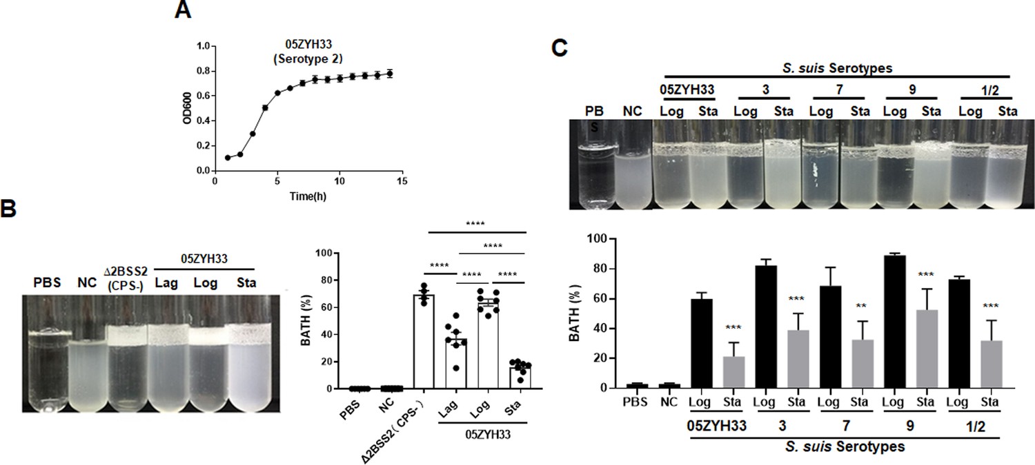
Monitoring the capsular polysaccharide (CPS) content of S. suis serotypes in culture.
(A) The growth curve 108 of 05ZYH33 (serotype 2) in tryptic soy broth (TSB)-FBS culture was assessed by measuring the optical density at 600 nm (OD600) at specified time points. Data present the mean ± SD of three independent experiments (n=10). (B) The hydrophobicity of 05ZYH33 during the lag, log, and stationary (Sta) phases and the CPS-deficient strain Δ2BSS2 (log phase) was measured by bacterial adhesion to hydrocarbon (BATH) assays (n=4–7). (C) The hydrophobicity of indicated serotype strains in the log and stationary phases was measured by BATH assays. A representative photo of the BATH assay (upper panel) and BATH values (lower panel) (n=3–6). Δ2BSS2 refers to an isogenic strain lacking CPS; nasal cavity (NC) represents a 05ZYH33 suspension without hydrocarbon. A one-way analysis of variance (ANOVA) with Tukey’s post hoc test was utilized in B, and an unpaired, two-tailed Student’s t-test was employed while in C. **, p<0.01. ***, p<0.001. ****, p<0.0001.
Figure 2
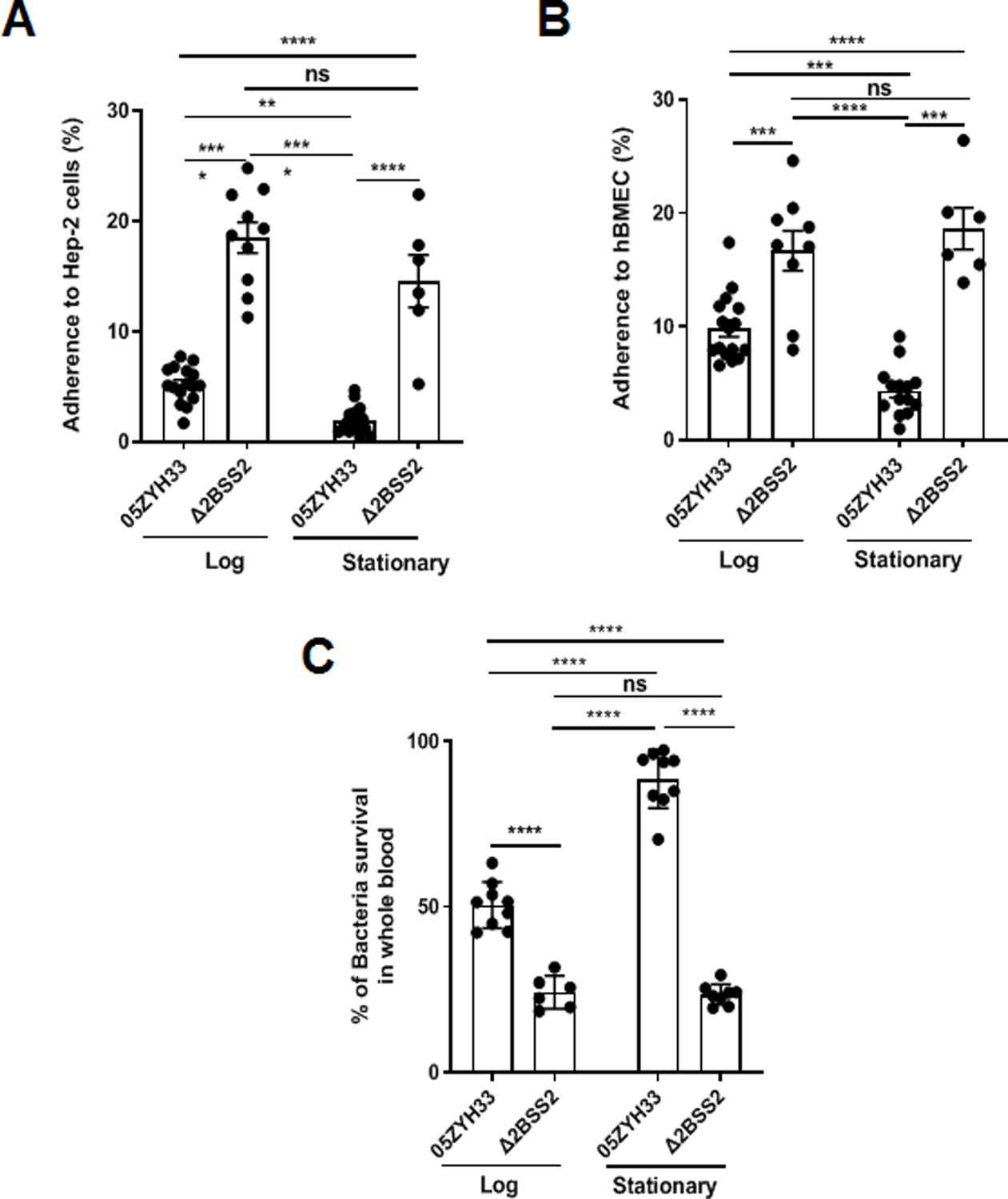
The adherence of 05ZYH33 to epithelial cells and its resistance to bacterial killing.
HEp-2 or human brain microvascular endothelial cells (HBMECs) cells were inoculated with 05ZYH33 or Δ2BSS2 in either the log or stationary phase at an infection MOI of 10. (A) Adherence rate of S. suis to HEp-2 cells. (B) Adherence rate of S. suis to HBMECs. (C) S. suis strains in log and stationary phases were incubated with whole blood from naive mice for a duration of 3 hr, and the viability of S. suis was determined. Data are from three independent experiments and presented as means ± SEM (n=6–16). **p<0.01, ***p<0.001, ****p<0.0001; ns, not significant.
Figure 3
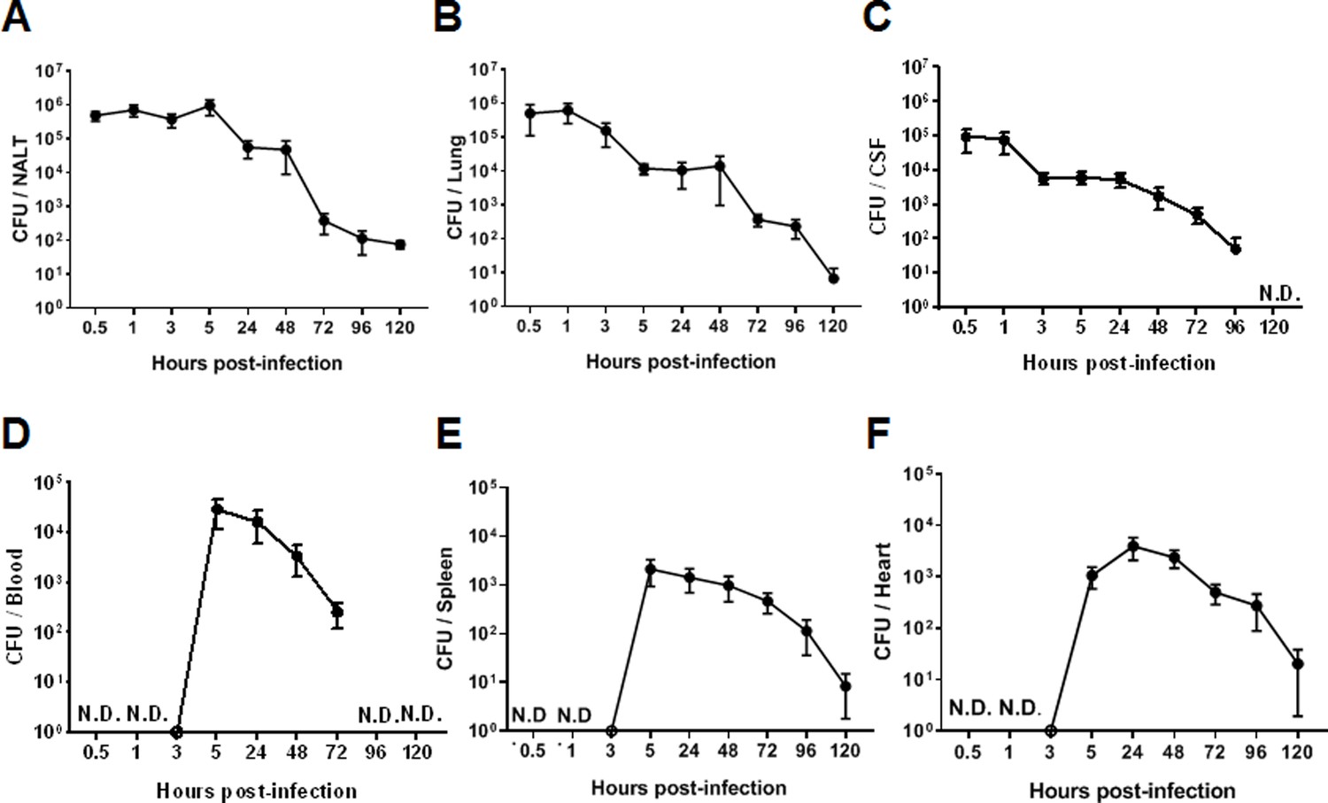
The intranasal infection course in mice.
Mice were intranasally inoculated with 05ZYH33. Total colony-forming units (CFUs) in various bodily compartments were assessed at the indicated time points after inoculation, (A) nasal-associated lymphoid tissue (NALT), (B) the cerebrospinal fluid (CSF), (C) the lung, (D) the blood, (E) the spleen, and (F) the heart. The data presented are derived from two independent experiments and presented as means ± SEM (n=6). ND, not detected.
Figure 4
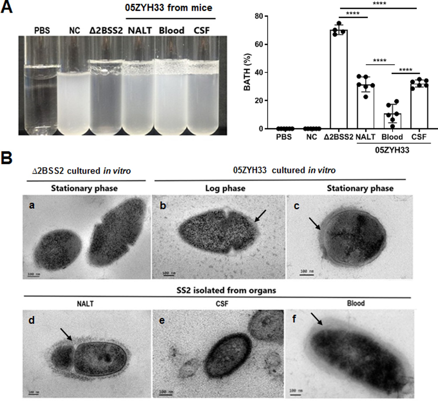
The capsular polysaccharide (CPS) of 05ZYH33 isolated from various bodily compartments was evaluated following infection.
05ZYH33 was intranasally inoculated in mice. Twelve hours after inoculation, colony-forming units (CFUs) in nasal-associated lymphoid tissue (NALT), the cerebrospinal fluid (CSF), and the blood were assessed for (A) hydrophobicity of bacterial cells by bacterial adhesion to hydrocarbon (BATH) assay and (B) CPS by transmission electron microscopy (TEM). Arrows indicate the CPS layers. Scale bar, 100 nm. Data in (A) are from two independent experiments and presented as means ± SEM (n=4–6). Nasal cavity (NC), 05ZYH33 suspension without hydrocarbon. ****p<0.0001.
Figure 5
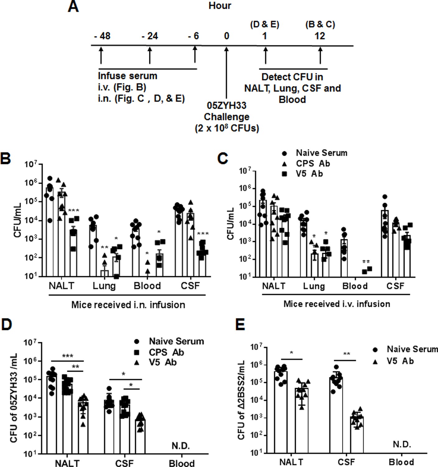
The role of anti-capsular polysaccharide (CPS) or anti-V5 serum in 05ZYH33 infection.
(A) Schematic illustration of passive transfer of antisera and infection in mice. (B, C) Anti-V5 or anti-CPS antiserum was transferred through the intranasally (i.n.) or intravenous (i.v.) route. Six hours later, mice were i.n. challenged with 05ZYH33, then euthanized 12 hr post-challenge. Colony-forming units (CFUs) of 05ZYH33 in nasal-associated lymphoid tissue (NALT), the lungs, the blood, and the cerebrospinal fluid (CSF) were determined. (D, E) Mice were infused with anti-V5 or anti-CPS serum through the i.n. route and inoculated i.n. with 05ZYH33 or Δ2BSS2 6 hr later. One hour later, mice were euthanized, and CFUs in NALT, the blood, and the CSF were determined. Data are the mean ± SEM of three independent experiments (n=10). *p<0.05, **p<0.01, ***p<0.001.
Figure 6
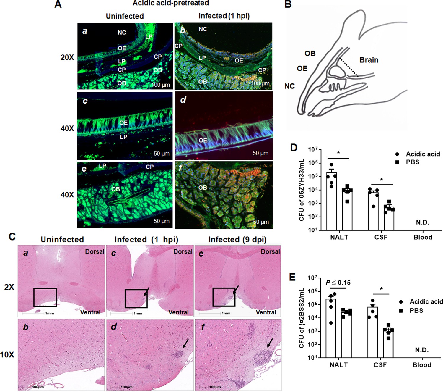
Detection of the presence of 05ZYH33 in the tissue of olfactory nerve area.
Mice were inoculated with acetic acid through the nostril, and 1 hr later, infected intranasally (i.n.) with 05ZYH33. (A) Sagittal views of the olfactory system. Sections of the distal nasal cavity and olfactory epithelium from uninfected (a, c, and e) and 05ZYH33-infected (b, d, and f) mice. Red or orange, 05ZYH33; green, the neuronal marker β-tubulin III; blue, DNA. NC, nasal cavity; OE, olfactory epithelium; LP, lamina propria; CP, cribriform plate; OB, olfactory bulb. (B) A schematic drawing of the sagittal plane of the rodent nose elucidates the compartments of the olfactory bulb (OB), olfactory epithelium (OE), nasal cavity (NC), and brain. This panel is redrawn from ‘Mouse Olfactory System’ (inspiredpencil.com). The dotted line indicates the anteroposterior localization of the coronal sections in C. (C) One hour or nine days after 05ZYH33 infection, coronal brain sections were prepared and stained with hematoxylin and eosin (H&E). The sections display the regions of the ventral striatum and basal forebrain located behind the anterior olfactory nucleus. Arrows indicate infiltrated inflammatory cells in the lower areas of the ventral striatum or basal forebrain. (D, E) Mice were inoculated with acetic acid or phosphate-buffered saline (PBS), and 1 hr later infected i.n. with 05ZYH33 or ∆2BSS2. (D) Colony-forming units (CFUs) of 05ZYH33 or (E) ∆2BSS2 in nasal-associated lymphoid tissue (NALT), the cerebrospinal fluid (CSF), and blood were determined 1 hr after infection.
Additional files
-
Supplementary file 1
Information of the strains used in the study.
- https://cdn.elifesciences.org/articles/101760/elife-101760-supp1-v1.docx
-
MDAR checklist
- https://cdn.elifesciences.org/articles/101760/elife-101760-mdarchecklist1-v1.docx
Download links
A two-part list of links to download the article, or parts of the article, in various formats.
Downloads (link to download the article as PDF)
Open citations (links to open the citations from this article in various online reference manager services)
Cite this article (links to download the citations from this article in formats compatible with various reference manager tools)
Regulative synthesis of capsular polysaccharides in the pathogenesis of Streptococcus suis
eLife 13:RP101760.
https://doi.org/10.7554/eLife.101760.3


