Integration of sensory and fear memories in the rat medial temporal lobe
- School of Psychology, University of New South Wales, Australia
Figures
Figure 1
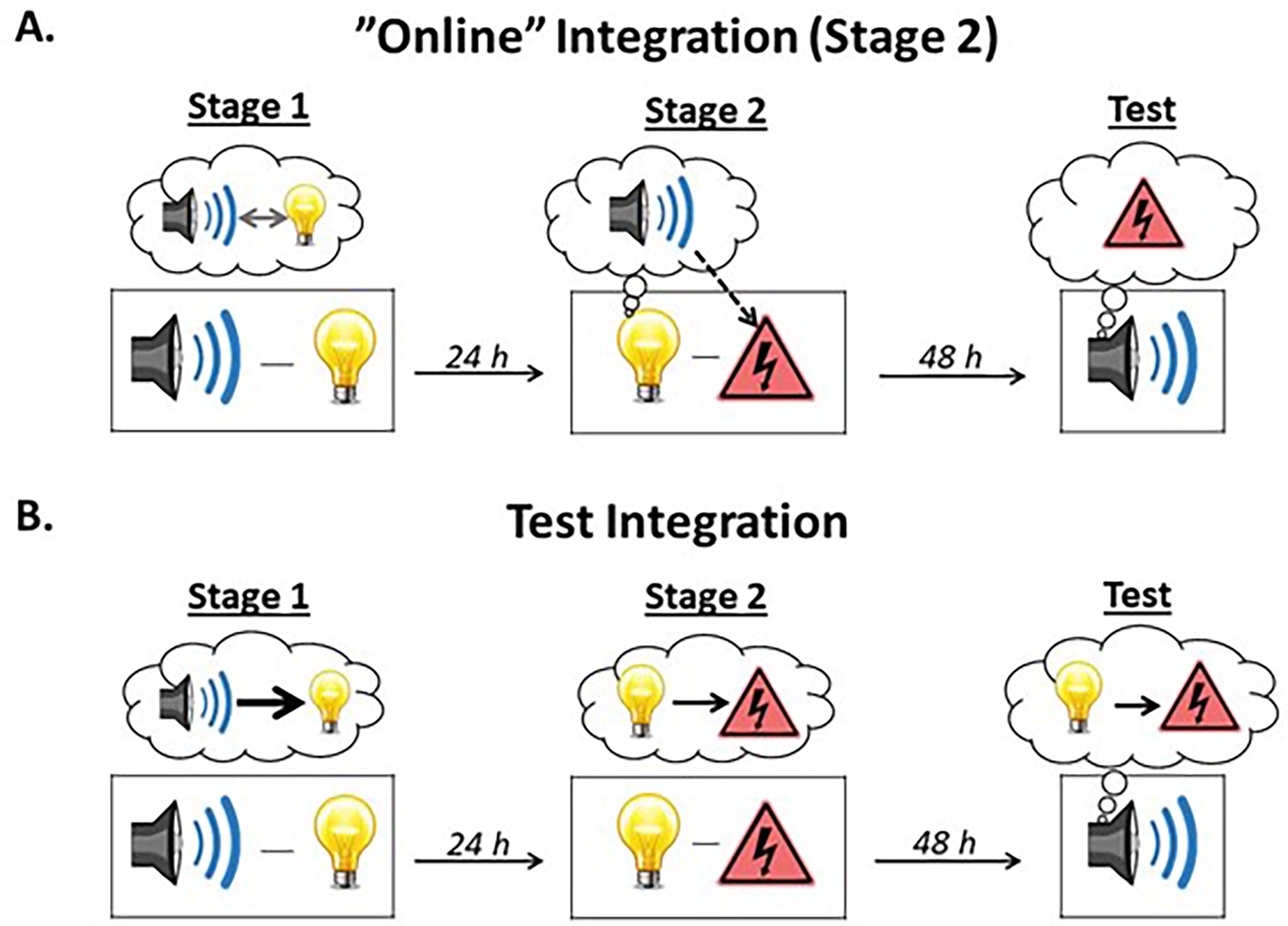
Illustration of the two types of integration.
(A) The sound-light and light-shock memories can be integrated ‘online’ during stage 2. When the subject is exposed to light-shock pairings in stage 2, the light activates the memory of its past associate, the sound, thereby allowing it to associate with the shock (i.e., subjects form a mediated sound-shock association). Test presentations of the sound then retrieve this mediated sound-shock association, resulting in the expression of fear. This type of integration is hypothesized to occur after few sound-light pairings in stage 1. Under these circumstances, subjects do not encode the order in which the events occur, resulting in mediated learning about the sound across direct conditioning of the light. (B) Alternatively, the sound-light and light-shock memories can be integrated at the time of testing. Here, when the sound is presented at test, the subject retrieves the sound-light memory formed in stage 1 and integrates (or chains) it with the light-shock memory formed in stage 2, resulting in the expression of fear. This type of integration is hypothesized to occur after many sound-light pairings in stage 1. Under these circumstances, subjects learn that the sound is followed by the light (sound →light) and that that light is followed by nothing (light→nothing). Hence, during the session of light-shock pairings in stage 2, the light does not activate the memory of the sound and the mediated sound-shock association does not form. Instead, during testing with the sound in stage 3, the light and its shock associate are strongly called to mind via the chain, sound→light→shock, resulting in the expression of fear responses.
Figure 2
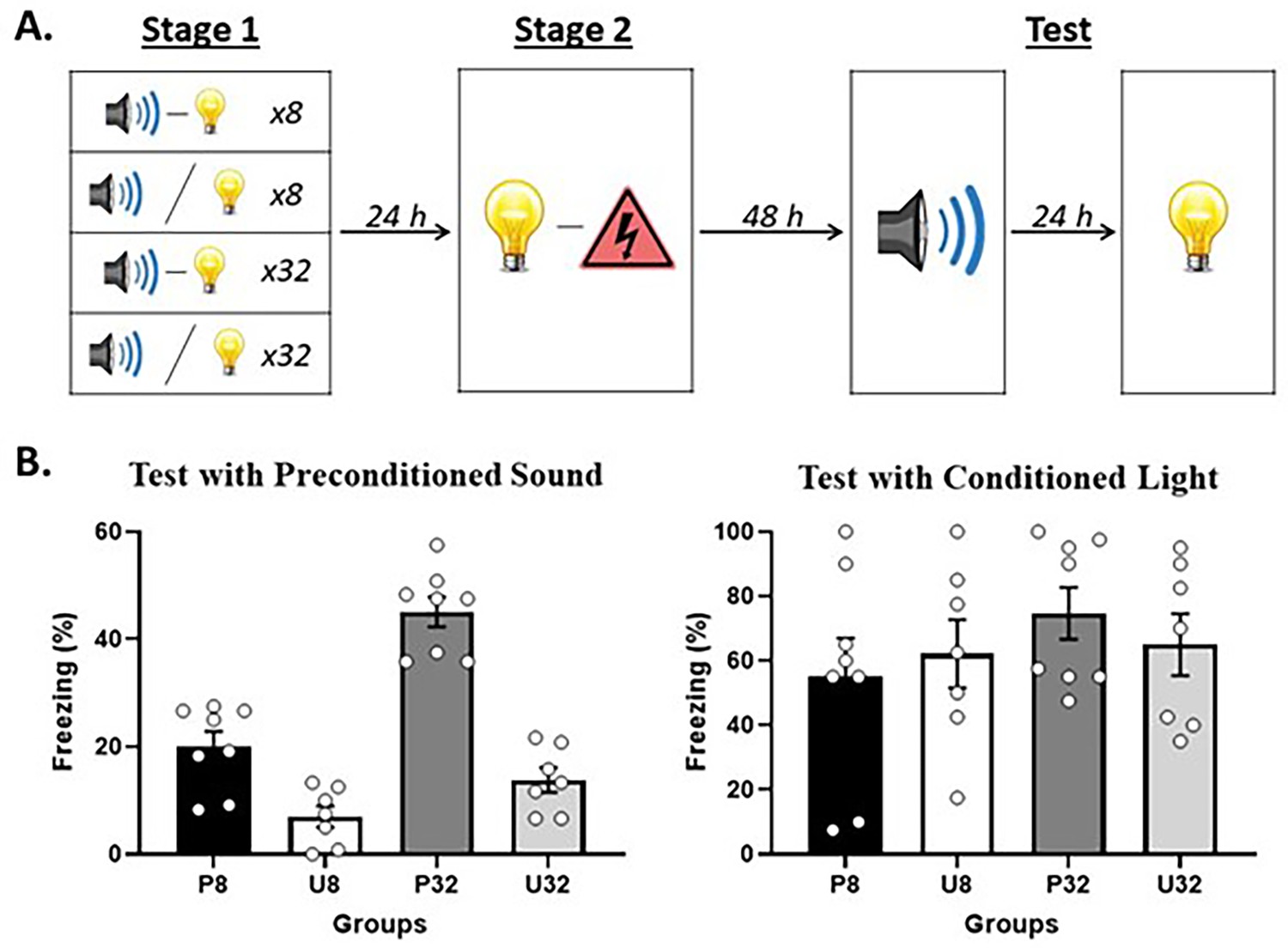
Demonstration of sensory preconditioned fear after few or many sound-light pairings in stage 1.
(A) Schematic of the behavioral protocol. In stage 1, rats were exposed to presentations of a sound and light. They either received 8 paired presentations in Group P8 (n = 8), 8 unpaired presentations in Group U8 (n = 7), 32 paired presentations in Group P32 (n = 8), or 32 unpaired presentations in Group U32 (n = 7). In stage 2, all rats received paired presentations of the light and shock. Finally, in stage 3 (Test), all rats were tested with the preconditioned sound alone and then with the conditioned light alone. (B) Percentage freezing to the preconditioned sound (left panel) and to the conditioned light (right panel), averaged across the eight trials of their respective tests. Data shown are means ± SEM.
Figure 3
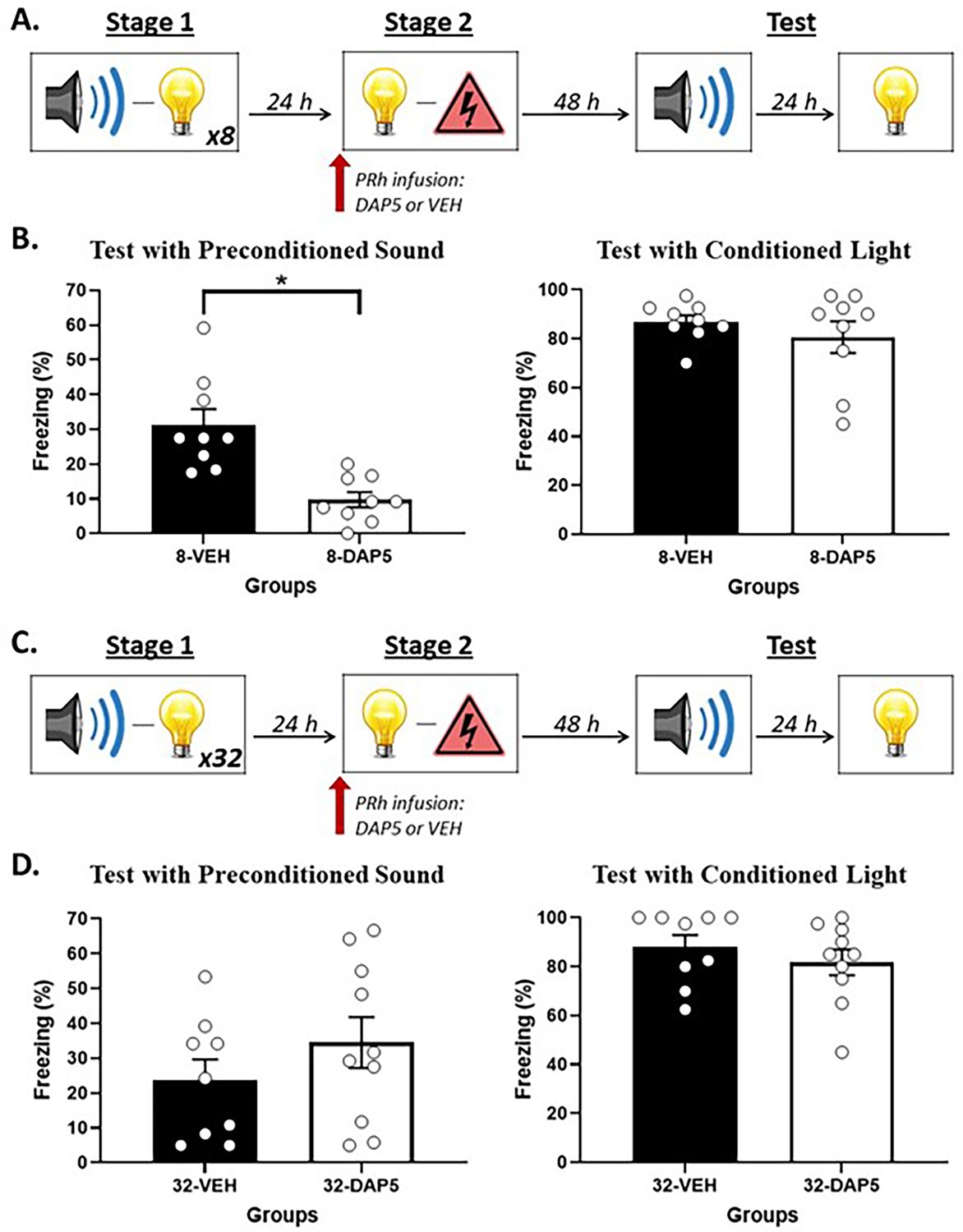
N-methyl-d-aspartate (NMDA) receptor activity in the perirhinal cortex (PRh) supports online integration in stage 2 after few sound-light pairings in stage 1 but NOT after many sound-light pairings.
(A) Schematic of the behavioral procedure for Experiment 2A (Group 8-VEH, n = 9; and Group 8-DAP5, n = 9). The red arrow indicates that the infusion of either DAP5 or vehicle (VEH) occurred before stage 2. (B) Percentage freezing to the preconditioned sound (left panel) and to the conditioned light (right panel), averaged across the eight trials of their respective tests. Data shown are means ± SEM. The asterisk (*) denotes a statistically significant difference (p<0.05). (C) Schematic of the behavioral procedure for Experiment 2B (Group 32-VEH, n = 9; and Group 32-DAP5, n = 10). The red arrow indicates that the infusion of either DAP5 or VEH occurred before stage 2. (D) Percentage freezing to the preconditioned sound (left panel) and to the conditioned light (right panel), averaged across the eight trials of their respective tests. Data shown are means ± SEM.
Figure 4
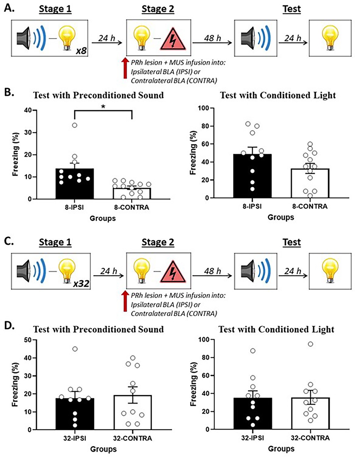
Perirhinal cortex-basolateral amygdala complex (PRh-BLA) communication is required for integration in stage 2 among rats that received just a few sound-light pairings in stage 1 but NOT among rats that received many sound-light pairings.
(A) Schematic of the behavioral procedure for Experiment 3A (Group 8-IPSI, n = 10; and Group 8-CONTRA, n = 12). The red arrow indicates that the infusion of muscimol occurred before stage 2. (B) Percentage freezing to the preconditioned sound (left panel) and to the conditioned light (right panel), averaged across the eight trials of their respective tests. Data shown are means ± SEM. The asterisk (*) denotes a statistically significant difference (p<0.05). (C) Schematic of the behavioral procedure for Experiment 3B (Group 32-IPSI, n = 10; and Group 8-CONTRA, n = 10). The red arrow indicates that the infusion of muscimol occurred before stage 2. (D) Percentage freezing to the preconditioned sound (left panel) and to the conditioned light (right panel), averaged across the eight trials of their respective tests. Data shown are means ± SEM.
Figure 5
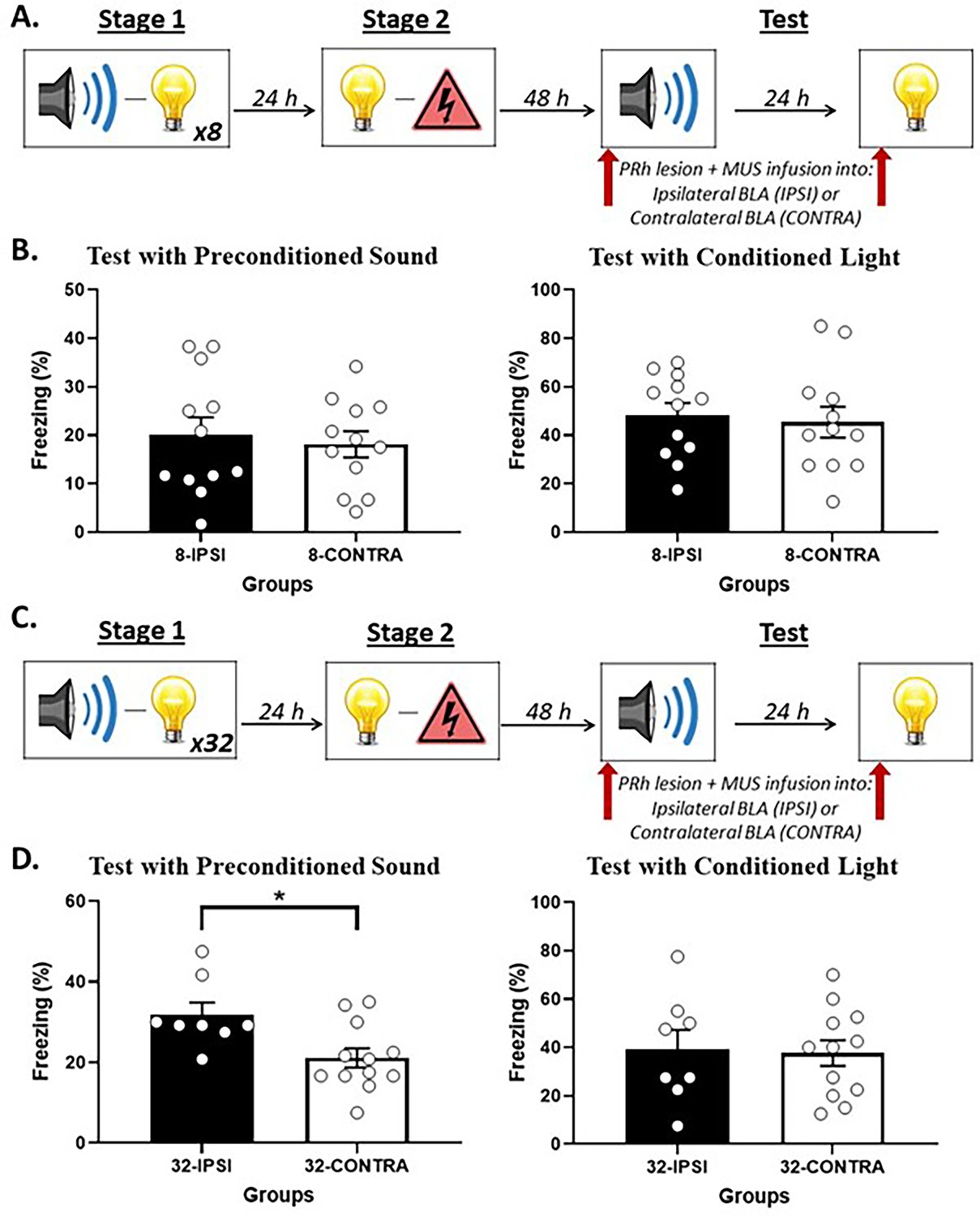
Perirhinal cortex-basolateral amygdala complex (PRh-BLA) communication is required for integration at test among rats that received many sound-light pairings in stage 1 but NOT among rats that received few sound-light pairings.
(A) Schematic of the behavioral procedure for Experiment 4A (Group 8-IPSI, n = 12; Group 8-CONTRA, n = 12). The red arrows indicate that the infusion of muscimol occurred before the test session of the preconditioned sound and before the test session of the conditioned light. (B) Percentage freezing to the preconditioned sound (left panel) and to the conditioned light (right panel), averaged across their eight respective test trials. Data shown are means ± SEM. (C) Schematic of the behavioral procedure for Experiment 4B (Group 32-IPSI, n = 8; Group 32-CONTRA, n = 12). The red arrows indicate that the infusion of muscimol occurred before the test session of the preconditioned sound and before the test session of the conditioned light. (D) Percentage freezing to the preconditioned sound (left panel) and to the conditioned light (right panel), averaged across the eight trials of their respective tests. Data shown are means ± SEM. The asterisks (*) denotes a statistically significant difference (p<0.05).
Figure 6
Cannula placements in the perirhinal cortex (PRh) taken from rats in Experiments 2A and 2B.
The most ventral portion of the cannulas are marked on coronal sections based on the atlas of Paxinos and Watson, 2006.
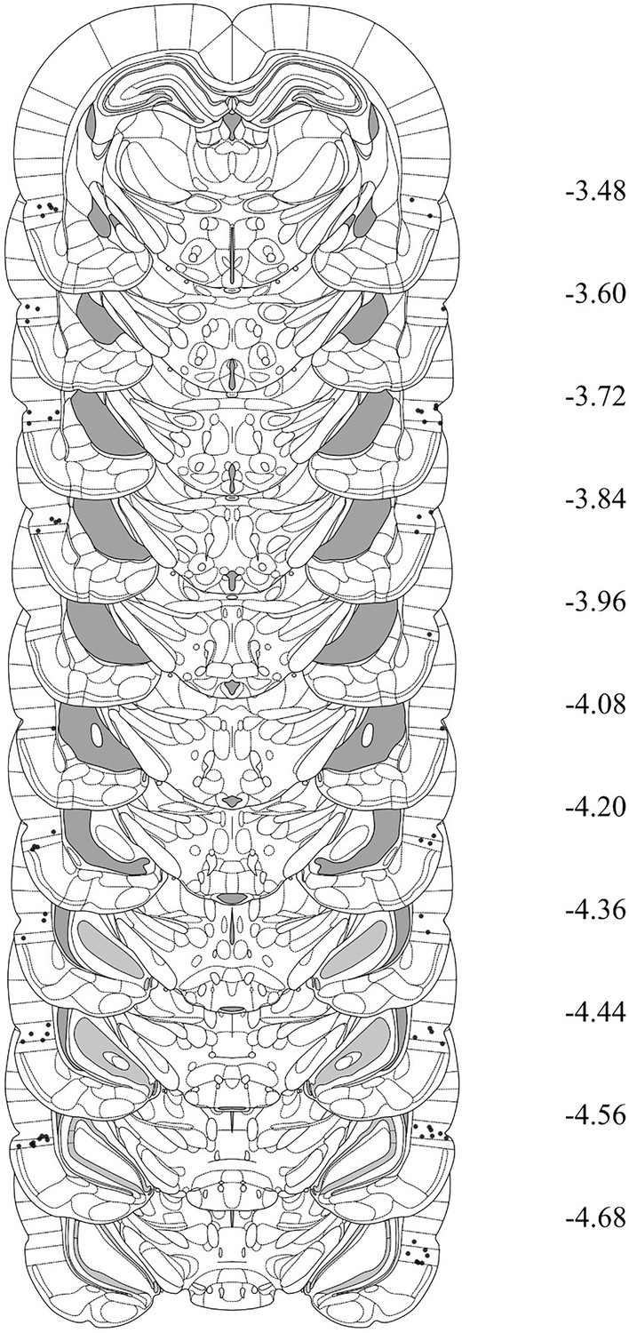
Figure 7
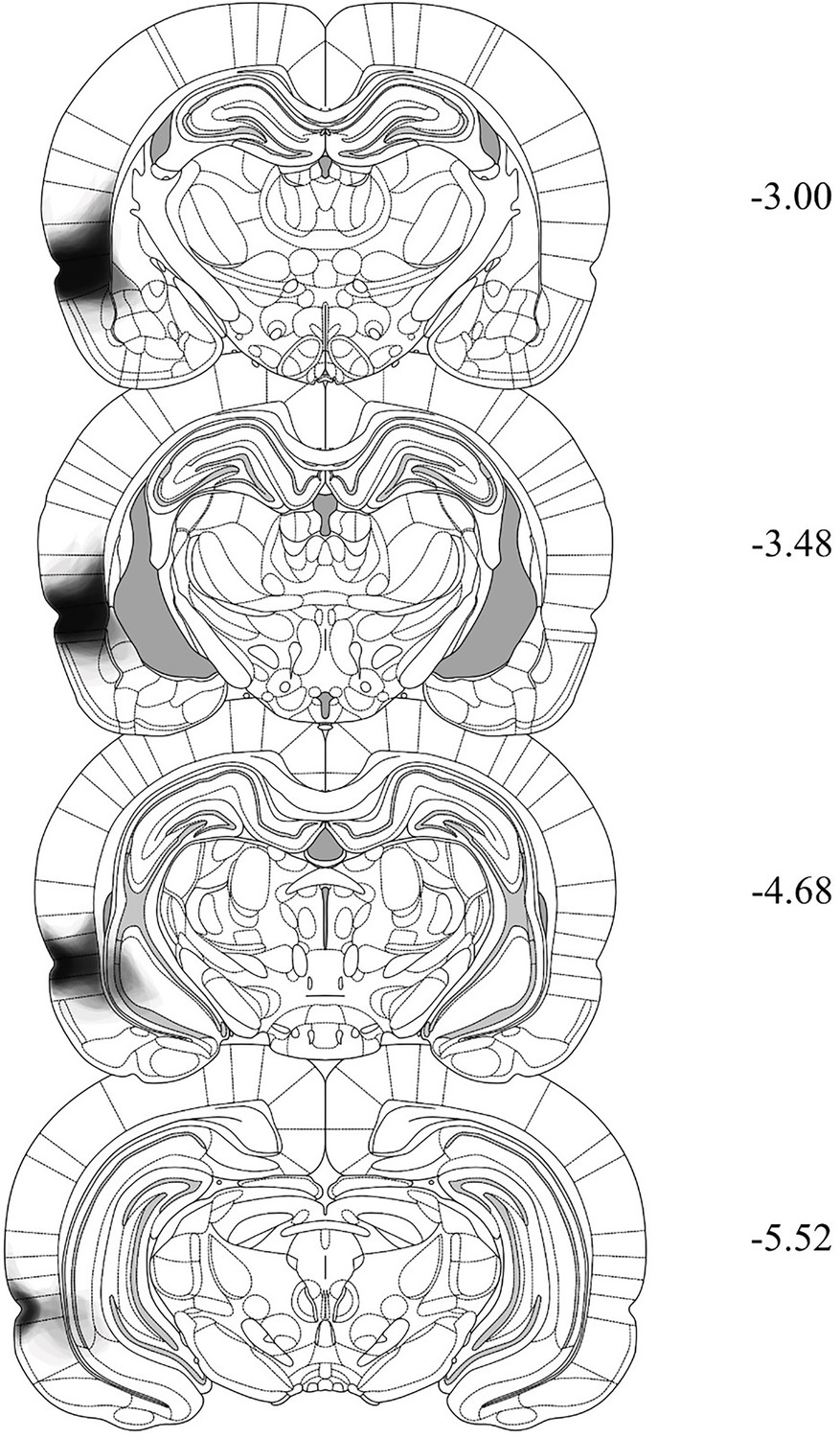
Histological reconstructions demonstrating the extent of N-methyl-d-aspartate (NMDA) lesions in the perirhinal cortex (PRh) taken from rats in Experiments 3A, 3B, 4A, and 4B.
The extent is shown across four coronal sections based on the atlas of Paxinos and Watson, 2006. The degree of shading indicates the number of cases exhibiting damage to those regions (e.g., darker areas result from a greater number of cases with damage to that region).
Figure 8

Cannula placements in the basolateral amygdala complex (BLA) taken from rats in Experiments 3A, 3B, 4A and 4B.
The most ventral portion of the cannulas are marked on coronal sections based on the atlas of Paxinos and Watson, 2006.
Additional files
Download links
A two-part list of links to download the article, or parts of the article, in various formats.
Downloads (link to download the article as PDF)
Open citations (links to open the citations from this article in various online reference manager services)
Cite this article (links to download the citations from this article in formats compatible with various reference manager tools)
Integration of sensory and fear memories in the rat medial temporal lobe
eLife 13:RP101965.
https://doi.org/10.7554/eLife.101965.3


