ORMDL3 restrains type I interferon signaling and anti-tumor immunity by promoting RIG-I degradation
- State Key Laboratory of Oncology in South China, Guangdong Provincial Clinical Research Center for Cancer, Sun Yat-sen University Cancer Center, China
- Center for Translational Medicine, The First Affiliated Hospital, Sun Yat-sen University, China
Figures
Figure 1 with 1 supplement
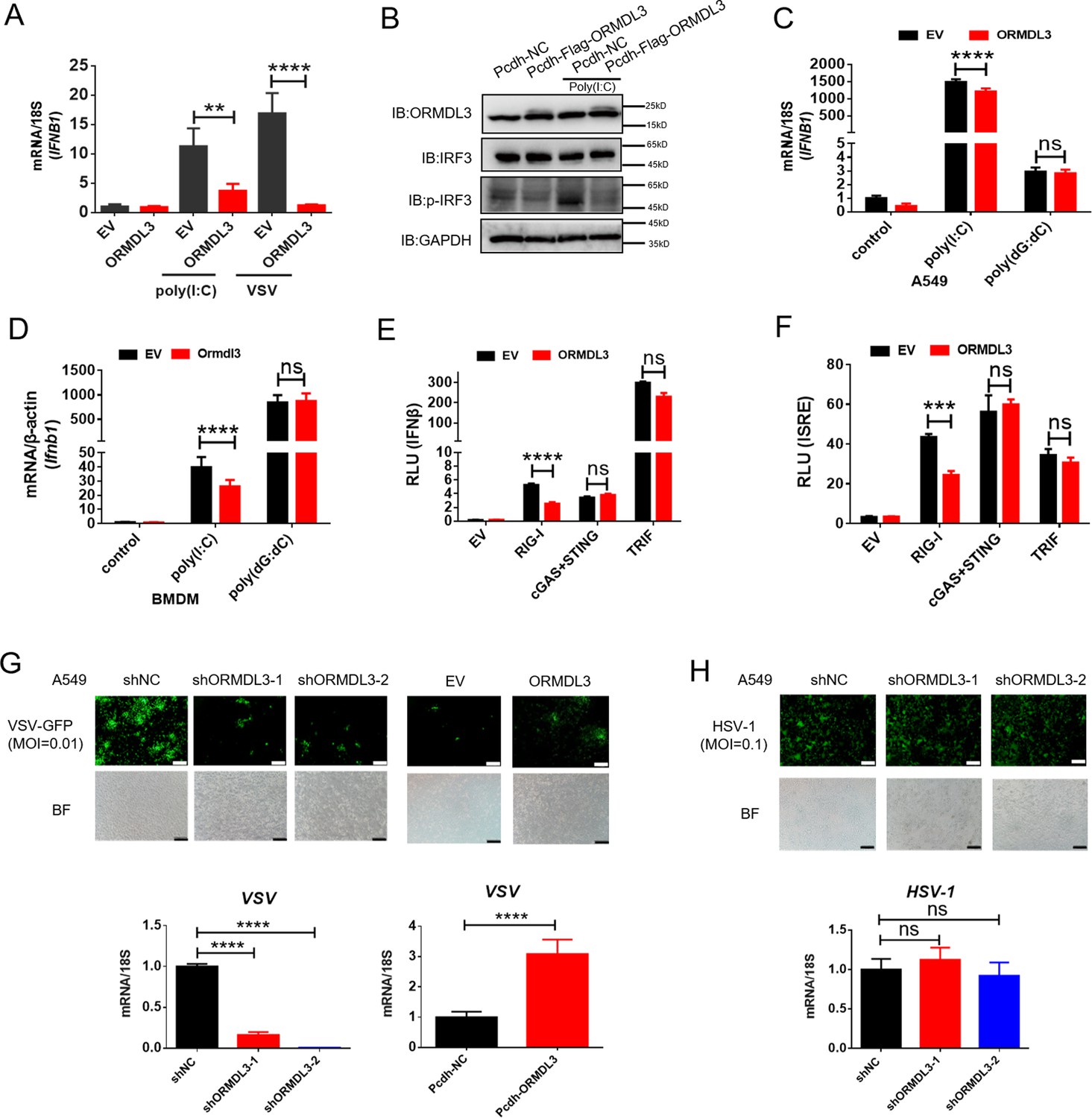
ORMDL3 negatively regulates RIG-I-like receptor (RLR)-induced type I interferon (type I IFN) signaling pathway.
(A) HEK293T cells were transfected with an empty vector (EV) or ORMDL3 plasmid for 12 hr and were then infected with vesicular stomatitis virus (VSV) (MOI = 0.01) or transfected with poly(I:C). The transcription of IFNB1 mRNA was detected using quantitative real-time PCR (qRT-PCR). (B) HEK293T-EV and HEK293T-Flag-ORMDL3 stable cell lines were transfected with or without poly(I:C) and immunoblot analyses of phosphorylated IRF3 (p-IRF3), total IRF3, GAPDH, and ORMDL3 levels were performed. (C) Results of the qRT-PCR assays showing mRNA levels of IFNB1 in A549 cells transfected with EV or ORMDL3 followed by stimulating with poly(I:C) or poly(dG:dC). (D) Results of the qRT-PCR assays showing mRNA levels of Ifnb1 in bone marrow-derived primary macrophages (BMDM) infected with EV or psc-AAV-Ormdl3 virus followed by transfecting with poly(I:C) or poly(dG:dC). (E–F) Results of the luciferase assay showing IFNβ-Luc activity (E) and ISRE-Luc activity (F) in HEK293T cells transfected with EV or ORMDL3 plasmids together with individual EV, RIG-I, cGAS plus STING, or TRIF plasmids for 24 hr. (G) ORMDL3 stable knockdown or overexpression A549 cells, and the control cells were infected with VSV-GFP (MOI = 0.01) for 12 hr. The viral infection was observed using fluorescence microscopy, and the viral amount was detected using qRT-PCR. Scale bars, 200 μm. (H) The control and ORMDL3 stable knockdown A549 cells were infected with herpes simplex virus-1 (HSV-1) (MOI = 0.1) for 24 hr. The viral infection was observed using fluorescence microscopy, and the viral amount was detected using qRT-PCR. Scale bars, 200 μm. Data from three independent experiments are presented as mean ± SD and were analyzed by two-tailed Student’s t test (A, C, D-F, G-H, bottom) ,**p<0.01, ***p<0.001, ****p<0.0001, and ns = no significance.
-
Figure 1—source data 1
Original files for western blots shown in Figure 1, indicating relevant bands.
- https://cdn.elifesciences.org/articles/101973/elife-101973-fig1-data1-v1.zip
-
Figure 1—source data 2
Original files for western blots shown in Figure 1.
- https://cdn.elifesciences.org/articles/101973/elife-101973-fig1-data2-v1.zip
Figure 1—figure supplement 1
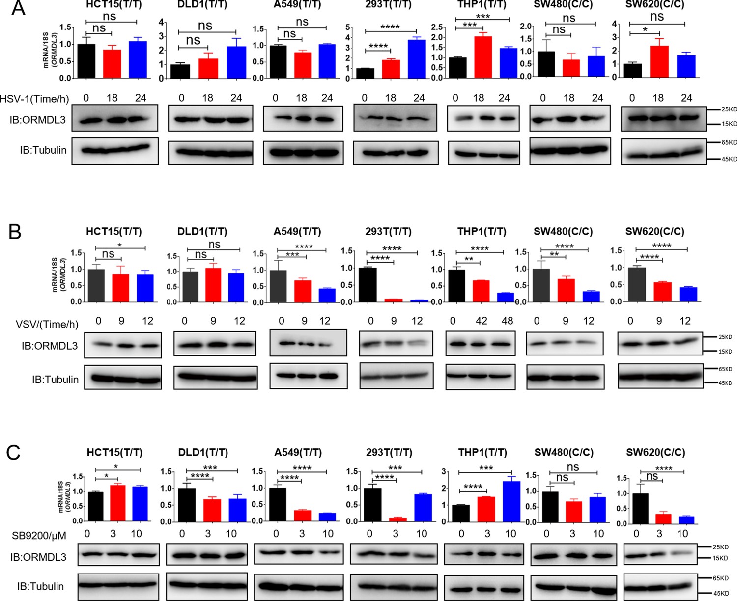
ORMDL3 expression under different treatments.
(A) Results of the WB and quantitative real-time PCR (qRT-PCR) assays showing ORMDL3 protein and RNA levels in indicated cell lines infected with herpes simplex virus-1 (HSV) (MOI = 0.1) for indicated times. (B) Results of the WB and qRT-PCR assays showing ORMDL3 protein and RNA levels in indicated cell lines infected with VSV-1 (MOI = 0.01) for indicated times. (C) Results of the WB and qRT-PCR assays showing ORMDL3 protein and RNA levels in indicated cell lines treated with RIG-I agonist SB9200 for indicated times. Data from three independent experiments are presented as mean ± SD and were analyzed by two-tailed Student’s t test (A-C), *p<0.05, **p<0.01, ***p<0.001, ****p<0.0001, and ns = no significance.
-
Figure 1—figure supplement 1—source data 1
Original files for western blots shown in Figure 1—figure supplement 1, indicating relevant bands.
- https://cdn.elifesciences.org/articles/101973/elife-101973-fig1-figsupp1-data1-v1.zip
-
Figure 1—figure supplement 1—source data 2
Original files for western blots shown in Figure 1—figure supplement 1.
- https://cdn.elifesciences.org/articles/101973/elife-101973-fig1-figsupp1-data2-v1.zip
Figure 2 with 1 supplement
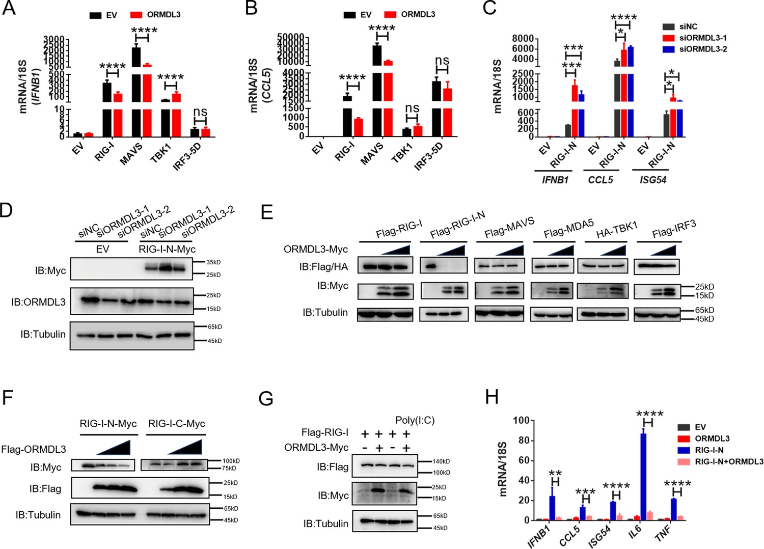
ORMDL3 regulates the protein abundance of RIG-I.
(A, B) Quantitative real-time PCR (qRT-PCR) analyses of the expression of the indicated mRNA levels in HEK293T cells transfected with empty vector (EV) or ORMDL3 plasmids combined with co-transfection of individual plasmids encoding EV, RIG-I, MAVS, TBK1, IRF3-5D (the constitutively activated form of IRF3). (C) Results of the qRT-PCR assays showing mRNA levels of IFNB1, CCL5, and ISG54 in HEK293T cells transfected with control or ORMDL3-specific siRNAs followed by secondary transfection with EV or RIG-I-N plasmids. (D) Results of the WB assays showing protein levels in HEK293T cells transfected with control or ORMDL3-specific siRNAs followed by secondary transfection with EV or RIG-I-N plasmids. (E) Immunoblot analysis of 293T cells transfected with individual plasmid encoding Flag-tagged RIG-I, RIG-I-N, MAVS, MDA5, IRF3, or HA-TBK1, in combination with increasing doses of ORMDL3-Myc plasmids. (F) Immunoblot analysis of 293T cells transfected with plasmid of RIG-I-N-Myc or RIG-I-C-Myc and increasing doses of Flag-ORMDL3 plasmid. (G) HEK293T cells were transfected with Flag-RIG-I and ORMDL3-Myc plasmids, as indicated, with or without poly(I:C) co-transfection. Cell lysates were immunoblotted with anti-Flag and anti-Myc antibodies. (H) Results of the qRT-PCR assays showing IFNB1, CCL5, ISG54, IL6, and TNF mRNA levels in HEK293T cells transfected with EV, ORMDL3, and RIG-I-N plasmids as indicated for 24 hr. Data from three independent experiments are presented as mean ± SD and were analyzed by two-tailed Student’s t test (A-C, H), *p<0.05, **p<0.01, ***p<0.001, ****p<0.0001, and ns = no significance.
-
Figure 2—source data 1
Original files for western blots shown in Figure 2, indicating relevant bands.
- https://cdn.elifesciences.org/articles/101973/elife-101973-fig2-data1-v1.zip
-
Figure 2—source data 2
Original files for western blots shown in Figure 2.
- https://cdn.elifesciences.org/articles/101973/elife-101973-fig2-data2-v1.zip
Figure 2—figure supplement 1
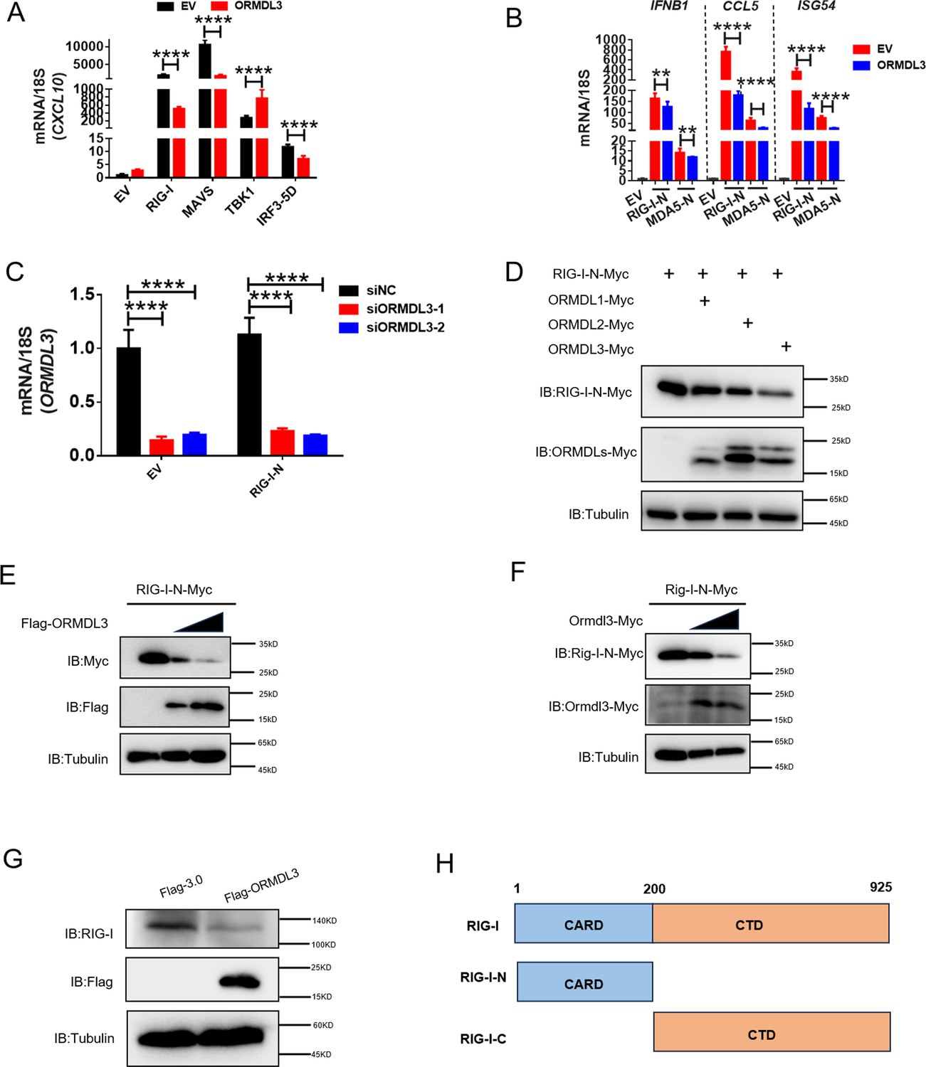
ORMDL3-mediated downregulation of RIG-I is conserved in both human and murine cells.
(A) Quantitative real-time PCR (qRT-PCR) analysis of CXCL10 when ORMDL3 was co-expressed with RIG-I, MAVS,TBK1, or IRF3-5D. (B) Results of the qRT-PCR assays showing IFNB1, CCL5, ISG54 mRNA level in HEK293T cells transfected with empty vector (EV) or ORMDL3 plasmids together with individual RIG-I-N or MDA5-N plasmids for 24 hr. (C) Results of the qRT-PCR assays showing ORMDL3 knockdown efficiency which links to Figure 2D. (D) Immunoblot assay of RIG-I-N expression in HEK293T co-transfected with ORMDLs family members. (E) Immunoblot assay of exogenous RIG-I-N expression in HEK293T cells transfected with human ORMDL3. (F) Immunoblot assay of exogenous Rig-I-N expression in HEK293T cells transfected with murine Ormdl3. (G) Immunoblot assay of endogenous RIG-I expression in HEK293T cells transfected with Flag-ORMDL3. (H) The diagram of RIG-I truncations. Data from three independent experiments are presented as mean ± SD and were analyzed by two-tailed Student’s t test (A-C), **p<0.01, ****p<0.0001.
-
Figure 2—figure supplement 1—source data 1
Original files for western blots shown in Figure 2—figure supplement 1, indicating relevant bands.
- https://cdn.elifesciences.org/articles/101973/elife-101973-fig2-figsupp1-data1-v1.zip
-
Figure 2—figure supplement 1—source data 2
Original files for western blots shown in Figure 2—figure supplement 1.
- https://cdn.elifesciences.org/articles/101973/elife-101973-fig2-figsupp1-data2-v1.zip
Figure 3 with 1 supplement
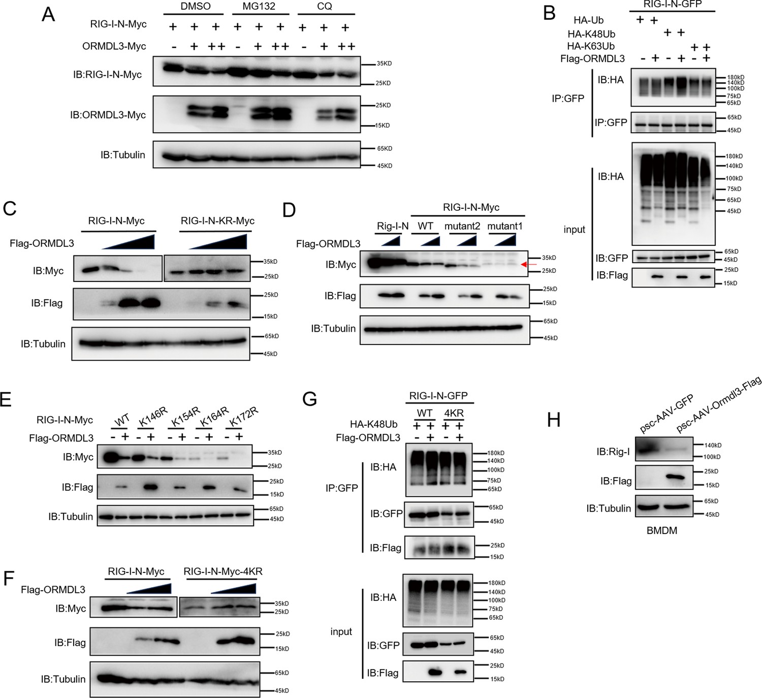
ORMDL3 promotes proteasomal degradation of RIG-I.
(A) HEK293T cells were transfected with plasmids encoding RIG-I-N-Myc together with increasing amounts of Flag-ORMDL3 plasmid treated with MG132 (10 μM) or chloroquine (CQ) (50 μM) for 6 hr and the cell lysates were analyzed by immunoblot. (B) HEK293T cells were transfected with the indicated plasmids, and cell lysates were immunoprecipitated with an anti-GFP antibody followed by immunoblots using anti-GFP and anti-HA antibodies. (C) HEK293T cells were transfected with RIG-I-N-Myc (WT or KR) and increasing doses of plasmid for Flag-ORMDL3. The expression levels of RIG-I-N-Myc were analyzed by immunoblot. (D) HEK293T cells were transfected with Rig-I-N, RIG-I-N-Myc (WT, KR, mutant1, or mutant2) and increasing doses of plasmid for Flag-ORMDL3. The expression levels of RIG-I-N-Myc were analyzed by immunoblot. (E) 293T cells were transfected with RIG-I-N-Myc (WT, K146R, K154R, K164R, or K172R) with or without Flag-ORMDL3. The expression levels of RIG-I-N-Myc and its mutant forms were analyzed by immunoblot. (F) HEK293T cells were transfected with RIG-I-N-Myc (WT or 4KR) and increasing doses of Flag-ORMDL3. The expression levels of RIG-I-N-Myc (WT or 4KR) were analyzed by immunoblot. (G) HEK293T cells were transfected with RIG-I-N-GFP (WT or 4KR) and HA-K48Ub in combination with EV or Flag-ORMDL3, and cell lysates were immunoprecipitated with an anti-GFP antibody followed by immunoblots using anti-GFP, anti-HA, and anti-Flag antibodies. (H) Bone marrow-derived primary macrophages (BMDM) were infected with psc-AAV-GFP or psc-AAV-Ormdl3-Flag virus, followed by immunoblot analysis of Rig-I, Flag, and Tubulin.
-
Figure 3—source data 1
Original files for western blots shown in Figure 3, indicating relevant bands.
- https://cdn.elifesciences.org/articles/101973/elife-101973-fig3-data1-v1.zip
-
Figure 3—source data 2
Original files for western blots shown in Figure 3.
- https://cdn.elifesciences.org/articles/101973/elife-101973-fig3-data2-v1.zip
Figure 3—figure supplement 1
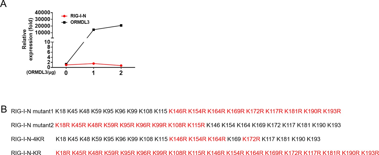
ORMDL3 promotes the degradation of RIG-I.
(A) Quantitative real-time PCR (qRT-PCR) assay of RIG-I-N mRNA levels in HEK293T cells transfected with RIG-I-N and increasing amounts of ORMDL3. (B) Different annotations of KR mutation of RIG-I-N.
Figure 4 with 1 supplement
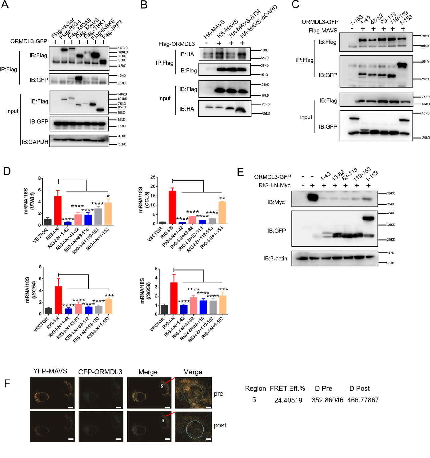
ORMDL3 interacts with signaling adaptor MAVS.
(A) HEK293T cells were transfected with empty vector (EV) or Flag-RIG-I/MAVS/MDA5/TBK1/IRF3/IKBKE with ORMDL3-GFP, and cell lysates were immunoprecipitated with an anti-Flag antibody followed by immunoblots using anti-GFP and anti-Flag antibodies. (B) HEK293T cells were transfected with different MAVS truncations in combination with EV or Flag-ORMDL3, and cell lysates were immunoprecipitated with anti-Flag antibody followed by immunoblots using anti-HA and anti-Flag antibodies. (C) HEK293T cells were transfected with EV or Flag-MAVS in combination with different ORMDL3 truncations, and cell lysates were immunoprecipitated with anti-Flag antibody followed by immunoblots using anti-GFP and anti-Flag antibodies. (D) HEK293T cells were transfected with EV or Flag-MAVS in combination with different ORMDL3 truncations, followed by quantitative real-time PCR (qRT-PCR) analysis of IFNB1, CCL5, ISG54, and ISG56. (E) HEK293T cells were transfected with RIG-I-N-Myc in combination with EV or different ORMDL3 truncations, followed by immunoblots using anti-GFP and anti-Myc antibodies. (F) FRET experiment of YFP-MAVS and CFP-ORMDL3 in HeLa cells. YFP-MAVS is the donor and CFP-ORMDL3 is the acceptor, and FRET efficiency is 24.40519%. Scale bars, 10 μm. Data from three independent experiments are presented as mean ± SD and were analyzed by two-tailed Student’s t test (D), *p<0.05, **p<0.01, ***p<0.001, ****p<0.0001.
-
Figure 4—source data 1
Original files for western blots shown in Figure 4, indicating relevant bands.
- https://cdn.elifesciences.org/articles/101973/elife-101973-fig4-data1-v1.zip
-
Figure 4—source data 2
Original files for western blots shown in Figure 4.
- https://cdn.elifesciences.org/articles/101973/elife-101973-fig4-data2-v1.zip
Figure 4—figure supplement 1
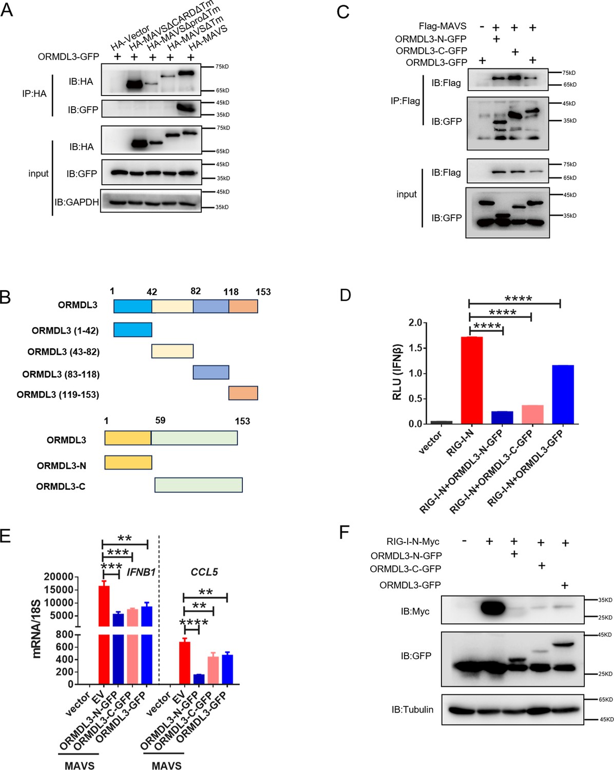
All truncations of ORMDL3 can inhibit type I interferon (type I IFN) production.
(A) HEK293T cells were transfected with the indicated plasmids, and cell lysates were immunoprecipitated with anti-HA antibody followed by immunoblots using anti-GFP and anti-HA antibodies. (B) Scheme of the truncations of ORMDL3. (C) HEK293T cells were transfected with the indicated plasmids, and cell lysates were immunoprecipitated with anti-Flag antibody, followed by immunoblots using anti-GFP and anti-Flag antibodies. (D) Results of the luciferase assays showing IFNβ-Luc activity in HEK293T cells co-transfected with ORMDL3 truncations and RIG-I-N. (E) Results of the quantitative real-time PCR (qRT-PCR) assays showing mRNA levels of IFNB1 and CCL5 in HEK293T cells co-transfected with ORMDL3 truncations and MAVS. (F) HEK293T cells were transfected with RIG-I-N-Myc and different truncation of ORMDL3 followed by immunoblot assay. Data from three independent experiments are presented as mean ± SD and were analyzed by two-tailed Student’s t test (D-E), **p<0.01, ***p<0.001, ****p<0.0001.
-
Figure 4—figure supplement 1—source data 1
Original files for western blots shown in Figure 4—figure supplement 1, indicating relevant bands.
- https://cdn.elifesciences.org/articles/101973/elife-101973-fig4-figsupp1-data1-v1.zip
-
Figure 4—figure supplement 1—source data 2
Original files for western blots shown in Figure 4—figure supplement 1.
- https://cdn.elifesciences.org/articles/101973/elife-101973-fig4-figsupp1-data2-v1.zip
Figure 5 with 1 supplement
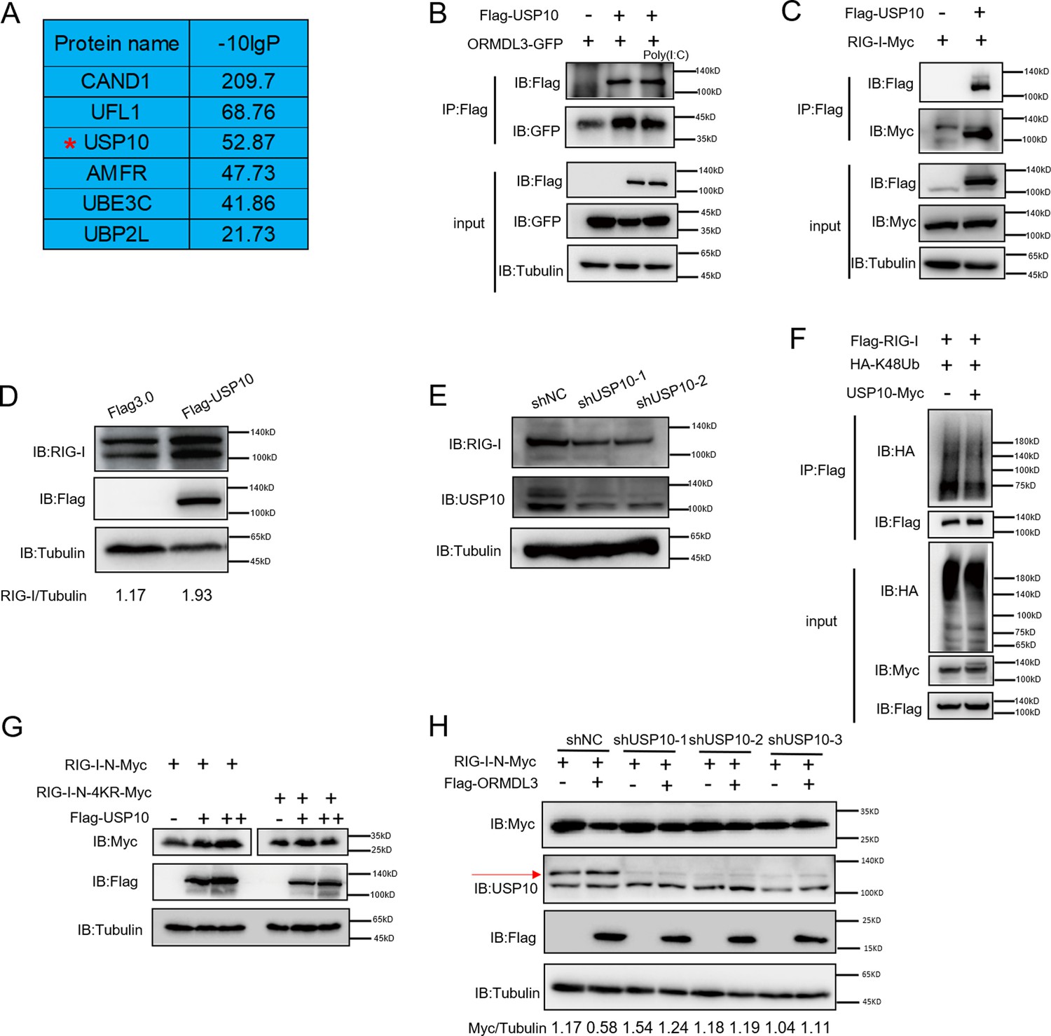
USP10 induces RIG-I stabilization.
(A) Candidate proteins interacted with ORMDL3 screened from mass spectrometry results. (B) HEK293T cells were transfected with ORMDL3-GFP and Flag-USP10 plasmids, as indicated, with or without poly(I:C) co-transfection. Cell lysates were immunoprecipitated with the anti-Flag antibody and immunoblotted with anti-Flag and anti-GFP antibodies. (C) HEK293T cells were transfected with RIG-I-Myc and EV or Flag-USP10 plasmids. Cell lysates were immunoprecipitated with the anti-Flag antibody and immunoblotted with anti-Flag and anti-Myc antibodies. (D) Immunoblot the protein level of RIG-I in USP10 stable overexpression HEK293T cell line. (E) Immunoblot the protein level of RIG-I in USP10 stable knockdown HEK293T cell lines. (F) Immunoprecipitation (IP) and immunoblot analysis of 293T cells transfected with vectors expressing Flag-RIG-I and HA-K48Ub with or without USP10-Myc. (G) HEK293T cells were transfected with RIG-I-N-Myc (WT or 4KR) and increasing doses of expression vector for Flag-USP10. The expression levels of RIG-I-N-Myc were analyzed by immunoblot. (H) USP10 stable knockdown HEK293T cell lines were transfected with RIG-I-N-Myc and Flag-ORMDL3 as indicated. The expression levels of RIG-I-N-Myc were analyzed by immunoblot.
-
Figure 5—source data 1
Original files for western blots shown in Figure 5, indicating relevant bands.
- https://cdn.elifesciences.org/articles/101973/elife-101973-fig5-data1-v1.zip
-
Figure 5—source data 2
Original files for western blots shown in Figure 5.
- https://cdn.elifesciences.org/articles/101973/elife-101973-fig5-data2-v1.zip
Figure 5—figure supplement 1
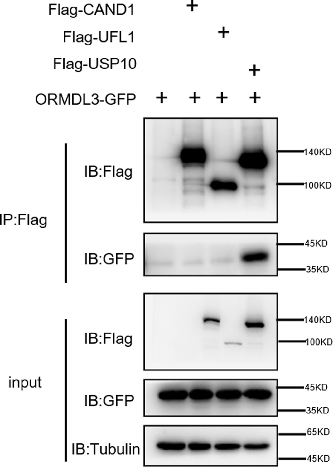
ORMDL3 interacts with USP10 but not CAND1 or UFL1.
HEK293T cells were transfected with the indicated plasmids, and cell lysates were immunoprecipitated with anti-Flag antibody followed by immunoblots using anti-GFP and anti-Flag antibodies.
-
Figure 5—figure supplement 1—source data 1
Original files for western blots shown in Figure 5—figure supplement 1, indicating relevant bands.
- https://cdn.elifesciences.org/articles/101973/elife-101973-fig5-figsupp1-data1-v1.zip
-
Figure 5—figure supplement 1—source data 2
Original files for western blots shown in Figure 5—figure supplement 1.
- https://cdn.elifesciences.org/articles/101973/elife-101973-fig5-figsupp1-data2-v1.zip
Figure 6
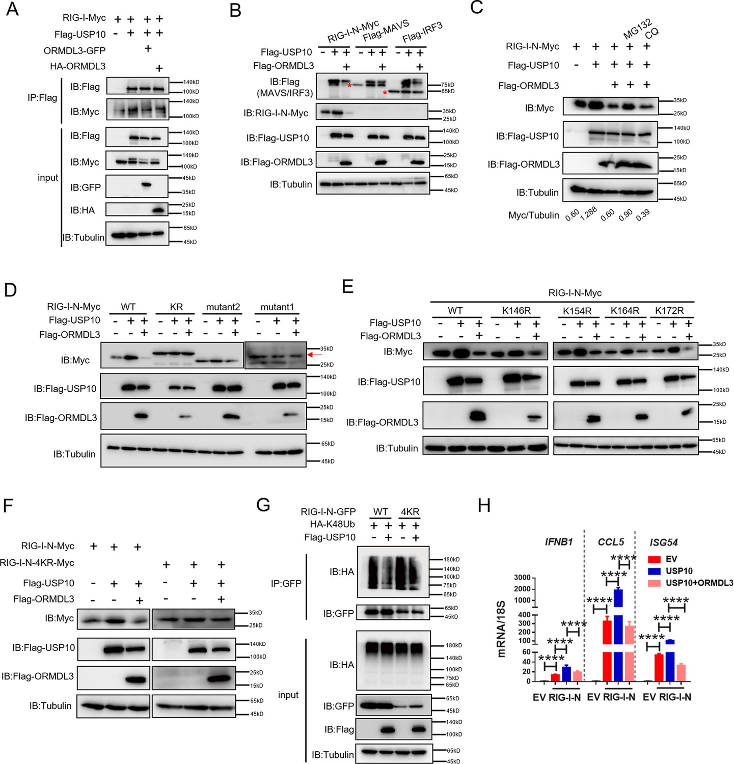
ORMDL3 disturbs USP10-induced RIG-I stabilization.
(A) HEK293T cells were transfected with Flag-USP10 and RIG-I-Myc plasmids, as indicated, with or without ORMDL3 co-transfection. Cell lysates were immunoprecipitated with the anti-Flag antibody and immunoblotted with anti-Flag and anti-Myc antibodies. (B) Immunoblot analysis of HEK293T cells transfected with Flag-USP10 and Flag-MAVS, Flag-IRF3, or RIG-I-N-Myc with or without ORMDL3 co-transfection using indicated antibodies. (C) HEK293T cells were transfected with plasmids encoding RIG-I-N-Myc together with Flag-USP10 with or without Flag-ORMDL3 plasmid followed by MG132 (10 μM) or chloroquine (CQ) (50 μM) treatment for 6 hr. The cell lysates were analyzed by immunoblot. (D) HEK293T cells were transfected with Flag-USP10 and RIG-I-N-Myc (WT, KR, mutant1, or mutant2) plasmids, as indicated, with or without Flag-ORMDL3 co-transfection. Cell lysates were immunoblotted with indicated antibodies. (E) HEK293T cells were transfected with RIG-I-N-Myc (WT, K146R, K154R, K164R, or K172R) and Flag-USP10 with or without Flag-ORMDL3. Cell lysates were immunoblotted with indicated antibodies. (F) HEK293T cells were transfected with Flag-USP10 and RIG-I-N-Myc (WT or 4KR) plasmids, as indicated, with or without Flag-ORMDL3 co-transfection. Cell lysates were immunoblotted with indicated antibodies. (G) Immunoprecipitation (IP) and immunoblot analysis of HEK293T cells transfected with vectors expressing RIG-I-N-GFP/RIG-I-N-4KR-GFP and HA-K48Ub with or without USP10 transfection. (H) HEK293T cells were transfected with Flag-USP10 and RIG-I-N-Myc, with or without ORMDL3 co-transfection followed by quantitative real-time PCR (qRT-PCR) analysis of IFNB1, CCL5, and ISG54. Data from three independent experiments are presented as mean ± SD and were analyzed by two-tailed Student’s t test (H), ****p<0.0001.
-
Figure 6—source data 1
Original files for western blots shown in Figure 6, indicating relevant bands.
- https://cdn.elifesciences.org/articles/101973/elife-101973-fig6-data1-v1.zip
-
Figure 6—source data 2
Original files for western blots shown in Figure 6.
- https://cdn.elifesciences.org/articles/101973/elife-101973-fig6-data2-v1.zip
Figure 7 with 3 supplements
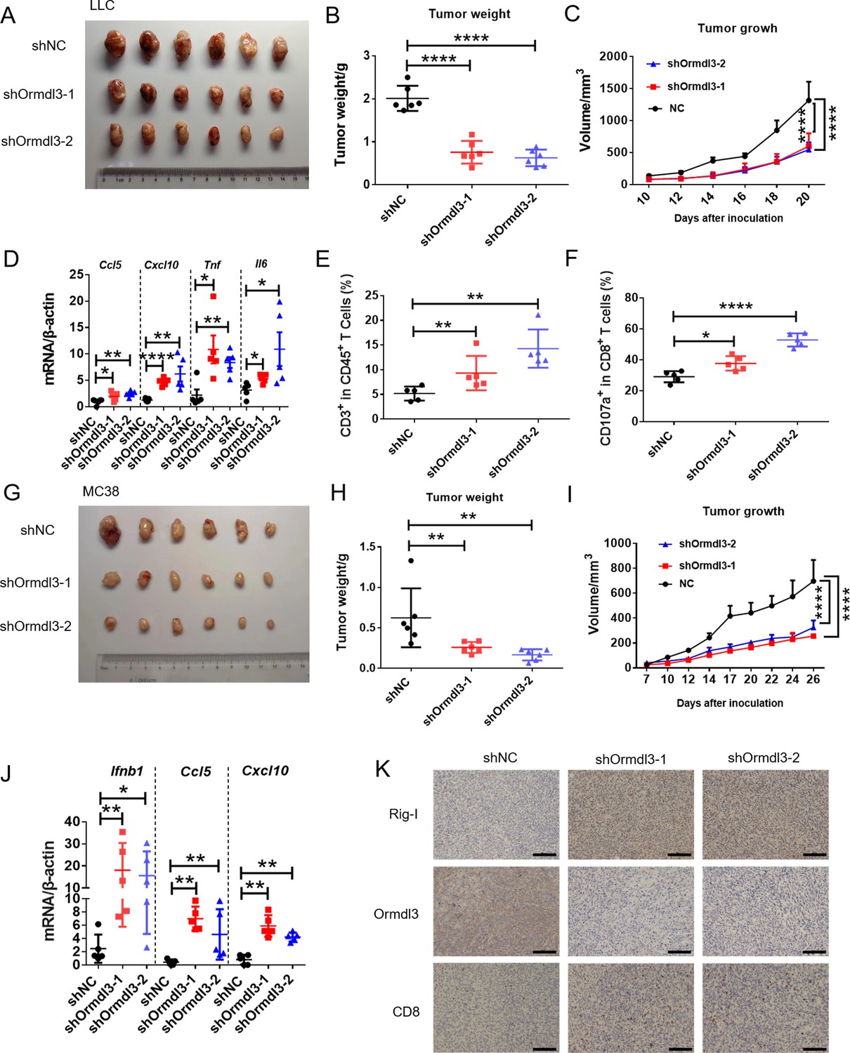
Knockdown of ORMDL3 in LLC/MC38 cells enhances anti-tumor immunity.
(A–C) Representative images (A), tumor weight (B), and tumor growth (C) of LLC tumors on day 21 after inoculation with 1.5×10⁶ LLC cells with or without Ormdl3 stable knockdown into C57BL/6 mice, (n=6). (D) Results of the quantitative real-time PCR (qRT-PCR) assays showing mRNA levels of Ccl5, Cxcl10, Tnf, and Il6 in LLC tumors, (n=5). (E, F) Flow cytometry assay of CD3+ T and CD107a+ CD8+ T cell percentages in indicated population, (n=5). (G–I) Representative images (G), tumor weight (H), and tumor growth (I) on day 27 after tumor inoculation with 5×10⁵ MC38 cells with or without Ormdl3 stable knockdown into C57BL/6 mice, (n=6). (J) Results of the qRT-PCR assays showing mRNA levels of Ifnb1, Ccl5, and Cxcl10 in MC38 tumors, (n=5). (K) Results of the immunohistochemistry (IHC) assay showing expression levels of Ormdl3, Rig-I, and CD8 in MC38 tumors. Scale bars, 50 μm. Data from three independent experiments are presented as mean ± SD and were analyzed by one-way ANOVA (B, D-F, H, and J) or two-way ANOVA (C ,and I), *p<0.05, **p<0.01, ****p<0.0001.
Figure 7—figure supplement 1
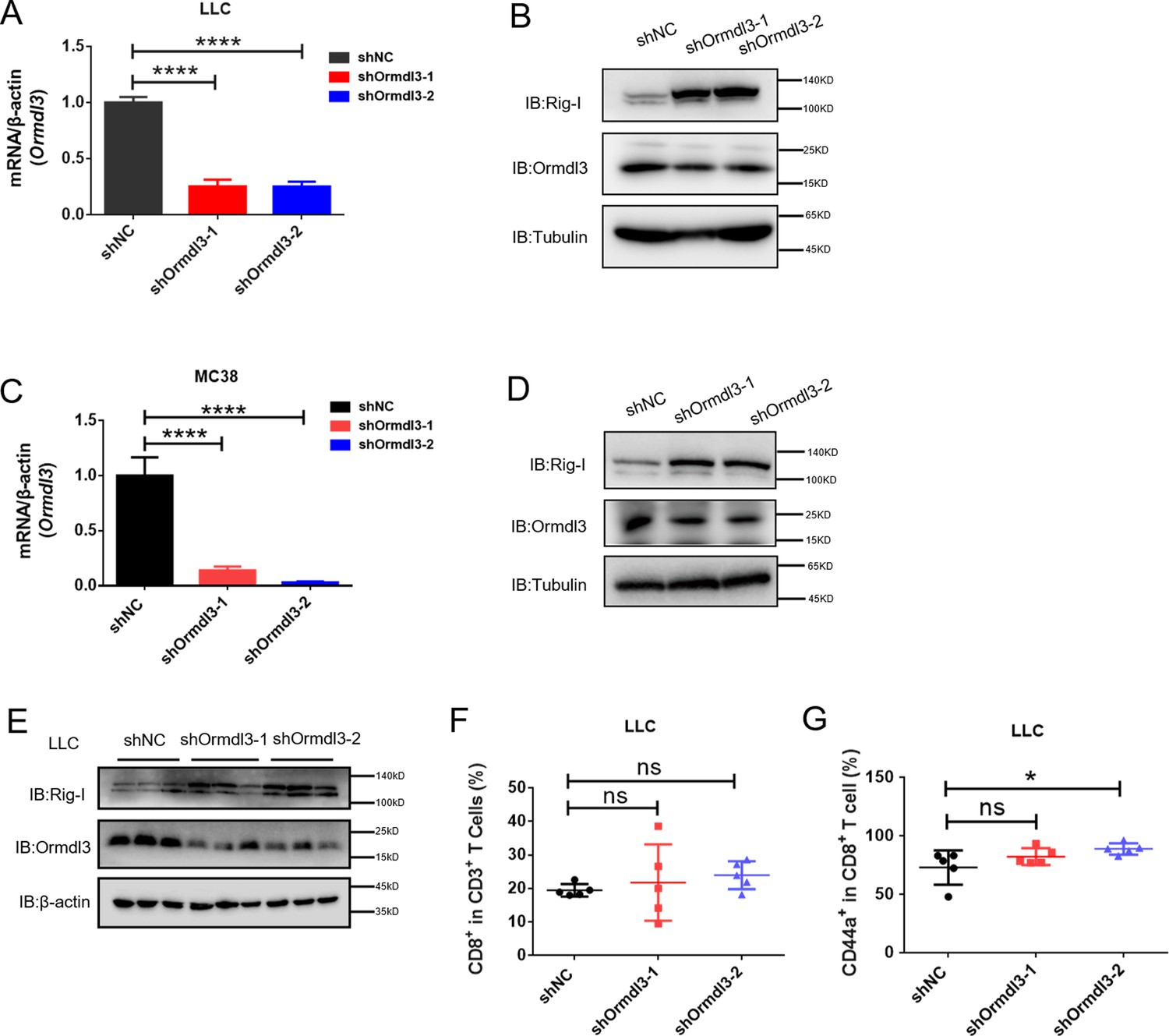
Inhibiting ORMDL3 increases the abundance of RIG-I.
(A) Quantitative real-time PCR (qRT-PCR) showing the shRNA knockdown efficiency of Ormdl3 in LLC lung cancer cells. (B) Immunoblot showing Ormdl3 knockdown and its effects on Rig-I abundance in LLC cells. (C) qRT-PCR showing the shRNA knockdown efficiency of Ormdl3 in MC38 colon cancer cells. (D) Immunoblot showing Ormdl3 knockdown and its effects on Rig-I abundance in MC38 cells. (E) Immunoblot assay of Ormdl3 and Rig-I protein levels in shNC, shOrmdl3-1, and shOrmdl3-2 in LLC tumors. (F, G) Flow cytometry assay of CD8+T and CD44+CD8+ T cell percentages in indicated populations (n=5). Data from three independent experiments are presented as mean ± SD and were analyzed by two-tailed Student’s t test (A, and C) or one way ANOVA (F-G),*p<0.05, ****p<0.0001, and ns = no significance.
-
Figure 7—figure supplement 1—source data 1
Original files for western blots shown in Figure 7—figure supplement 1, indicating relevant bands.
- https://cdn.elifesciences.org/articles/101973/elife-101973-fig7-figsupp1-data1-v1.zip
-
Figure 7—figure supplement 1—source data 2
Original files for western blots shown in Figure 7—figure supplement 1.
- https://cdn.elifesciences.org/articles/101973/elife-101973-fig7-figsupp1-data2-v1.zip
Figure 7—figure supplement 2
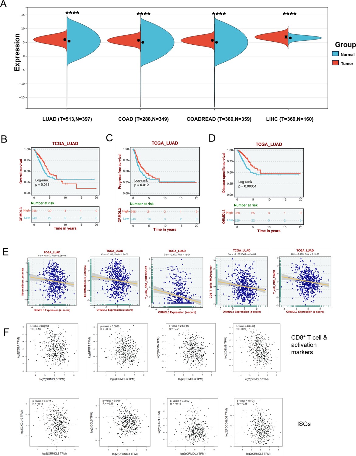
ORMDL3 level is associated with poor survival and reduced immune cell infiltration and interferon-stimulated gene (ISG) expression.
(A) ORMDL3 expression between tumor and adjacent normal tissues in The Cancer Genome Atlas (TCGA) pan-cancer cohorts. (B–D) Association of ORMDL3 level with overall survival (OS), progression-free survival (PFS), and disease-specific survival (DSS) in TCGA-lung adenocarcinoma (LUAD). (E) Correlation among ORMDL3 expression with stromal score and CD8+T cell infiltration in LUAD cohort from TCGA datasets. (F) Correlation among ORMDL3 expression with CD8+T and its activation markers (CD8A, PRF1, GZMA, GZMB) and ISGs (CCL5, CXCL10, CD274, and PDCD1LG2) in TCGA-LUAD from the GEPIA website (Tang et al., 2017). Data were analyzed by Unpaired Wilcoxon Rank Sum and Signed Rank Tests (A), ****p<0.0001.
Figure 7—figure supplement 3
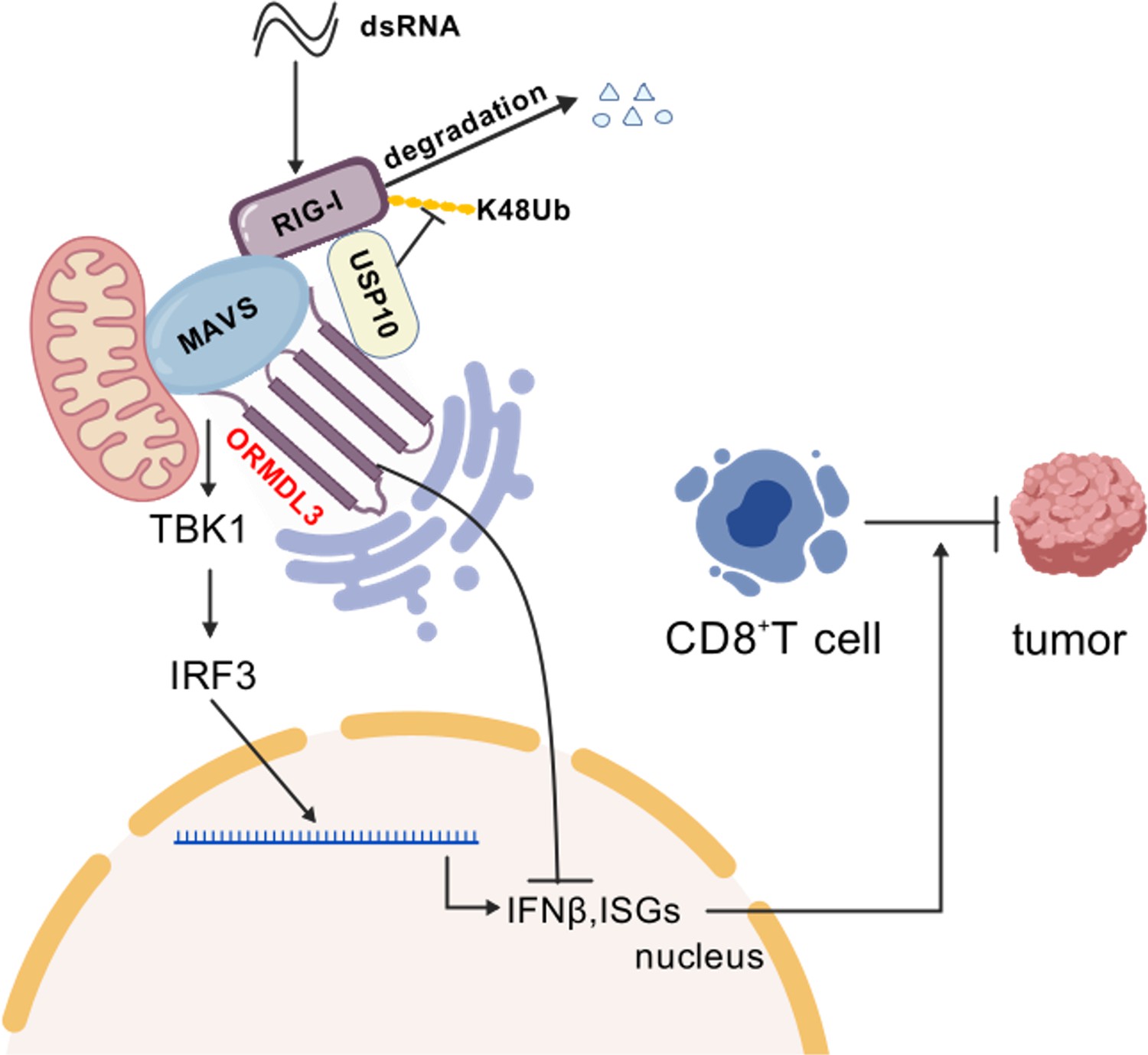
Schematic shows that ORMDL3 promotes the degradation of RIG-I and attenuates type I IFN in cancer.
The image was created with BioGDP.com (Jiang et al., 2025).
Author response image 1

PCR amplification of human TLR3 was conducted on cDNA derived from HEK293T and A549 cells (lanes 1 and 2, respectively), and PCR amplification of murine Tlr3 was performed on cDNA from BMDM (lane 3).
Human spleen cDNA (lane 4, TAKARA Human MTCTM Panel I, Cat# 636742) served as a positive control, and 18s rRNA was used as an internal control.
Tables
Key resources table
| Reagent type (species) or resource | Designation | Source or reference | Identifiers | Additional information |
|---|---|---|---|---|
| Cell line (Homo sapiens) | HEK293T | ATCC | Cat#CRL-11268 | RRID:CVCL_1926 |
| Cell line (Homo sapiens) | A549 | ATCC | Cat#CCL-185 | RRID:CVCL_0023 |
| Cell line (Homo sapiens) | HeLa | ATCC | Cat#CCL-2 | RRID:CVCL_0030 |
| Cell line (Homo sapiens) | DLD1 | ATCC | Cat#CCL-221 | RRID:CVCL_0248 |
| Cell line (Homo sapiens) | HCT15 | ATCC | Cat#CCL-225 | RRID:CVCL_0292 |
| Cell line (Homo sapiens) | SW480 | ATCC | Cat#CCL-228 | RRID:CVCL_0546 |
| Cell line (Homo sapiens) | SW620 | ATCC | Cat#CCL-227 | RRID:CVCL_0547 |
| Cell line (Homo sapiens) | THP1 | This paper | TIB-202 | Cell line maintained in Shuai Chen lab (Sun Yat-sen University Cancer Center) |
| Cell line (Mus musculus) | MC38 | This paper | Gift from Prof. Penghui Zhou (Sun Yat-sen University Cancer Center) | Cell line maintained in Shuai Chen lab (Sun Yat-sen University Cancer Center) |
| Cell line (Mus musculus) | LLC | This paper | Gift from Prof. Penghui Zhou (Sun Yat-sen University Cancer Center) | Cell line maintained in Shuai Chen lab (Sun Yat-sen University Cancer Center) |
| Sequence-based reagent | ORMDL3-shRNA-1 | This paper | PCR primers | CCGGCCCACAGAATGTGATAGTAATCTCGAGATTACTATCACATTCTGTGGGTTTTTG |
| Sequence-based reagent | ORMDL3-shRNA-2 | This paper | PCR primers | CCGGCATGGGCATGTATATCTTCCTCTCGAGAGGAAGATATACATGCCCATGTTTTTG |
| Sequence-based reagent | USP10 shRNA-1 | This paper | PCR primers | CCGGCCTATGTGGAAACTAAGTATTCTCGAGAATACTTAGTTTCCACATAGGTTTTTG |
| Sequence-based reagent | USP10 shRNA-2 | This paper | PCR primers | CCGGCCCATGATAGACAGCTTTGTTCTCGAGAACAAAGCTGTCTATCATGGGTTTTTG |
| Sequence-based reagent | USP10 shRNA-3 | This paper | PCR primers | CCGGCGACAAGCTCTTGGAGATAAACTCGAGTTTATCTCCAAGAGCTTGTCGTTTTTG |
| Sequence-based reagent | Ormdl3-shRNA-1 | This paper | PCR primers | CCGGCCAAGTATGACCAAGTCCATTCTCGAGAATGGACTTGGTCATACTTGGTTTTTG |
| Sequence-based reagent | Ormdl3-shRNA-2 | This paper | PCR primers | CCGGGCCGACTTGGAGTAGCTTGTACTCGAGTACAAGCTACTCCAAGTCGGCTTTTTG |
| Antibody | ORMDL3 (Rabbit, polyclonal) | Abcam | ab211522 | RRID:AB_3102000 WB (1:1000) |
| Antibody | ORMDL3 (Rabbit, polyclonal) | Abcam | ab107639 | RRID:AB_10863267 WB (1:1000) |
| Antibody | RIG-I (Mouse, monoclonal) | Santa Cruz | sc376845 | RRID:AB_2732794 WB (1:1000) |
| Antibody | Flag (Mouse, monoclonal) | Sigma | F1804# | RRID:AB_262044 WB (1:1000) |
| Antibody | GFP (Mouse, monoclonal) | Proteintech | 66002-1-Ig | RRID:AB_11182611 WB (1:1000) |
| Antibody | Myc (Mouse, monoclonal) | Proteintech | 60003-2-Ig | RRID:AB_2734122 WB (1:1000) |
| Antibody | Myc (Rabbit, polyclonal) | Proteintech | 10828-1-AP | RRID:AB_2148585 WB (1:1000) |
| Antibody | HA (Mouse, monoclonal) | Ray antibody | RM1004 | WB (1:1000) |
| Antibody | USP10 (Rabbit, polyclonal) | Abclonal | A13387 | RRID:AB_2760247 WB (1:1000) |
| Antibody | Tubulin (Mouse, polyclonal) | Fdbio | FD0064 | RRID:AB_3076327 WB (1:2000) |
| Antibody | GAPDH (Mouse, monoclonal) | Proteintech | 60004-1-Ig | RRID:AB_2107436 WB (1:2000) |
| Antibody | GAPDH (Rabbit, monoclonal) | ServiceBio | GB15004 | RRID:AB_2943040 WB (1:5000) |
| Antibody | β-Actin (Mouse, monoclonal) | Proteintech | 60008-1-Ig | RRID:AB_2289225 WB (1:2000) |
| Antibody | β-Actin (Rabbit, monoclonal) | ServiceBio | GB15003 | RRID:AB_3083699 WB (1:2000) |
| Antibody | IRF3 (Rabbit, monoclonal) | Cell Signaling Technology | # 4302 | RRID:AB_1904036 WB (1:1000) |
| Antibody | Anti-phospho-IRF-3 (Rabbit, monoclonal) | Cell Signaling Technology | #29047 | RRID:AB_2773013 WB (1:1000) |
| Antibody | CD3ε-APC (anti-Mouse) | BioLegend | 100235 | RRID:AB_2561455 Flow (1:300) |
| Antibody | CD4-Pacific blue (anti-Mouse) | BioLegend | 100428 | RRID:AB_493647 Flow (1:300) |
| Antibody | CD8-PE-cy7 (anti-Mouse) | BioLegend | 100721 | RRID:AB_312760 Flow (1:300) |
| Antibody | CD45-APC-cy7 (anti-Mouse) | BioLegend | 157024 | RRID:AB_2876533 Flow (1:300) |
| Antibody | CD44-FITC (anti-Mouse) | BioLegend | 103006 | RRID:AB_312957 Flow (1:300) |
| Antibody | CD107a-PE (anti-Mouse) | BioLegend | 121611 | RRID:AB_17320511 Flow (1:300) |
| Chemical compound, drug | Poly(I:C) (LMW) | Invivogen | tlrl-picw | |
| Chemical compound, drug | Poly(dG:dC) | Invivogen | tlrl-pgcn | |
| Chemical compound, drug | SB9200 | Bidepharm | CAS:942123-43-5 | |
| Software, algorithm | GraphPad Prism 7 | GraphPad | N/A | |
| Software, algorithm | ImageJ | https://imagej.net/Fiji/Downloads | N/A | |
| Software, algorithm | FlowJo10 | FlowJo | N/A | |
| Commercial assay or kit | Lipo293 | Beyotime | C0521 | |
| Commercial assay or kit | Lipofectamine 2000 | Thermo Fisher Scientific | Cat#11668019 | |
| Commercial assay or kit | Lipofectamine RNAiMAX | Thermo Fisher Scientific | Cat#13778075 | |
| Commercial assay or kit | Dual-Luciferase Reporter Assay System | Promega | Cat#E1960 |
Additional files
-
Supplementary file 1
Primers for qPCR.
Related to Materials and methods.
- https://cdn.elifesciences.org/articles/101973/elife-101973-supp1-v1.xlsx
-
MDAR checklist
- https://cdn.elifesciences.org/articles/101973/elife-101973-mdarchecklist1-v1.docx
Download links
A two-part list of links to download the article, or parts of the article, in various formats.
Downloads (link to download the article as PDF)
Open citations (links to open the citations from this article in various online reference manager services)
Cite this article (links to download the citations from this article in formats compatible with various reference manager tools)
ORMDL3 restrains type I interferon signaling and anti-tumor immunity by promoting RIG-I degradation
eLife 13:RP101973.
https://doi.org/10.7554/eLife.101973.3


