Gaskell, Langley, and the "para-sympathetic" idea
- Institut de Biologie de l’ENS (IBENS), Inserm, CNRS, École normale supérieure,PSL Research University, France
Figures
Figure 1
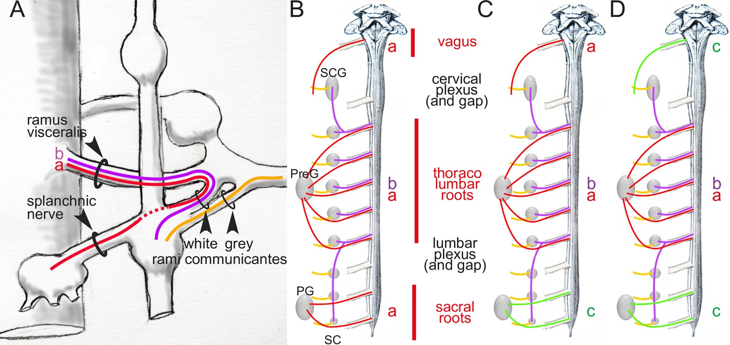
Arrangement of autonomic fibers according to Gaskell.
(A) The three types of involuntary fibers recognized by Gaskell: the myelinated fibers (purple and red) project from the CNS, either to the paravertebral chain, through the ramus visceralis and white ramus communicans (b, purple) or, bypassing the chain (stippled line), through the splanchnic nerves to prevertebral ganglia (a, red).Unmyelinated fibers (orange) emerge from the paravertebral chain and form the gray ramus communicans. (B) Pattern of projection (adapted from Gaskell, 1920) of involuntary fibers from the CNS in three outflows, cranial, thoracolumbar, and sacral, separated by the cervical and lumbar gaps (same color code as in A). The rostral and caudal parts of the paravertebral chain — superior cervical ganglion (SCG) and sacral chain (SC) — receive their rami viscerales (b, purple) not from the adjacent level of the spinal cord, but from the thoracic and lumbar levels, respectively. Consequently, at cranial and sacral levels, all rami viscerales are destined to distal ganglia, and thus are of the splanchnic type (a, red). (C, D) Reinterpretation of the pattern by Gaskell in 1916, in the light of physiological notions: the sacral splanchnic nerves (c, green) have been dissociated from the thoracolumbar ones (C), and finally likened to the cranial ones (D) (see text for details). PG: pelvic ganglion; PreG: prevertebral ganglion.
Figure 2

Categorization of autonomic nerves according to Gaskell and Langley (A), and according to genetically-defined cell types (B).
In (A) the vagus nerve and the sacral splanchnic are put in the same category (parasympathetic, green) despite their projections in different types of roots: dorso-ateral (and dedicated) for the vagus, ventral (and shared with somatic motor axons, black), for the sacral, as is the case at thoracolumbar level. In (B) the sacral splanchnic outflow is categorized as sympathetic (red) according to its transcriptionally defined cell types, both pre- and postganglionic (Alkaslasi et al., 2021; Blum et al., 2021; Espinosa-Medina et al., 2016; Sivori et al., 2023). CG: cardiac ganglion; DRG: dorsal root ganglion; HN: hypogastric nerve; IMG: inferior mesenteric ganglion; NG: nodose ganglion; PG: pelvic ganglion; PN: pelvic nerve. SCG: superior cervical ganglion; VN: vagus nerve. Green: parasympathetic; red: preganglionic sympathetic, to both para- and prevertebral ganglia; orange: postganglionic sympathetic neurons; black: somatic motoneurons.
Figure 3
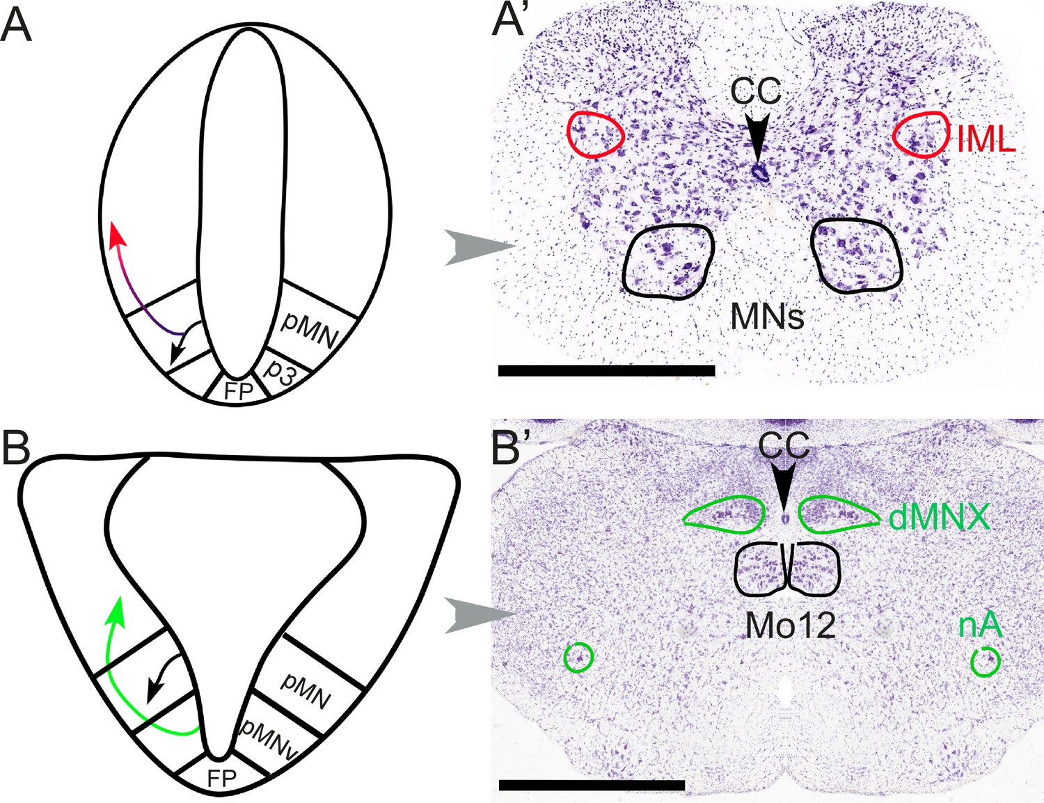
Different embryonic origins but similar migration pathways of preganglionic neurons at spinal and hindbrain levels.
At spinal levels (A, A’) — including sacral ones— preganglionic neurons (red) arise embryonically (A) from the same progenitor domain (pMN) as somatic motoneurons (black), with which they share an extensive transcriptional signature, but segregate from them to settle dorsally. In the adult (A’), they form the intermediolateral column (improperly called ‘parasympathetic nucleus’ at sacral levels). At hindbrain levels (B, B’), pre-ganglionic neurons (green) arise (B) in a progenitor domain (pMNv) different from that of somatic motoneurons (pMN), and have consequently different transcriptional signatures. However, they also migrate dorsally (B) to form (B’) preganglionic nuclei, such as the dorsal motor nucleus of the vagus nerve. The same pMNv domain produces branchiomotor neurons (for branchiomeric muscles), as in the nucleus ambiguus (nA). CC: central canal; dMNX: dorsal nucleus of the vagus nerve; FP: floor plate; IML: intermediolateral column; Mo12: hypoglossal nucleus; nA: nucleus ambiguus; p3: progenitor domain for spinal V3 interneurons (serially homologous with the pMNv domain of the hindbrain); pMN: progenitor domain for somatic motor neurons; pMNv: progenitor domain for visceral motor neurons (bulbar preganglionics and branchiomotor neurons). Scale bars: 1 mm.
Figure 4
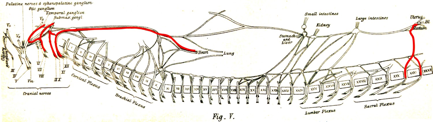
The visceral nerves of a dog as they appear in figure V, plate III, from Gaskell, 1886 (enhanced for clarity).
‘Vaso-inhibitory nerves’ are represented in red and are found at cranial and sacral levels.
Figure 5
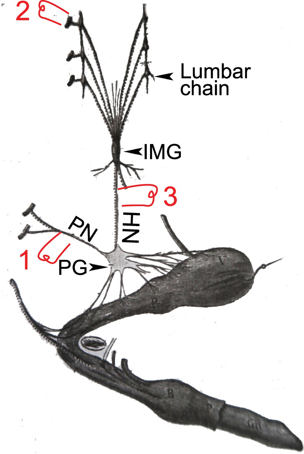
A dog’s penis and its innervation (reproduced and adapted from François-Franck, 1895).
HN: hypogastric nerve; IMG inferior mesenteric ganglion (or plexus); PG: pelvic ganglion (or hypogastric plexus); PN; pelvic nerve or nervus erigens of Eckhardt. 1, 2, & 3: electrostimulation sites used by Langley and others, see main text for details.
Figure 6
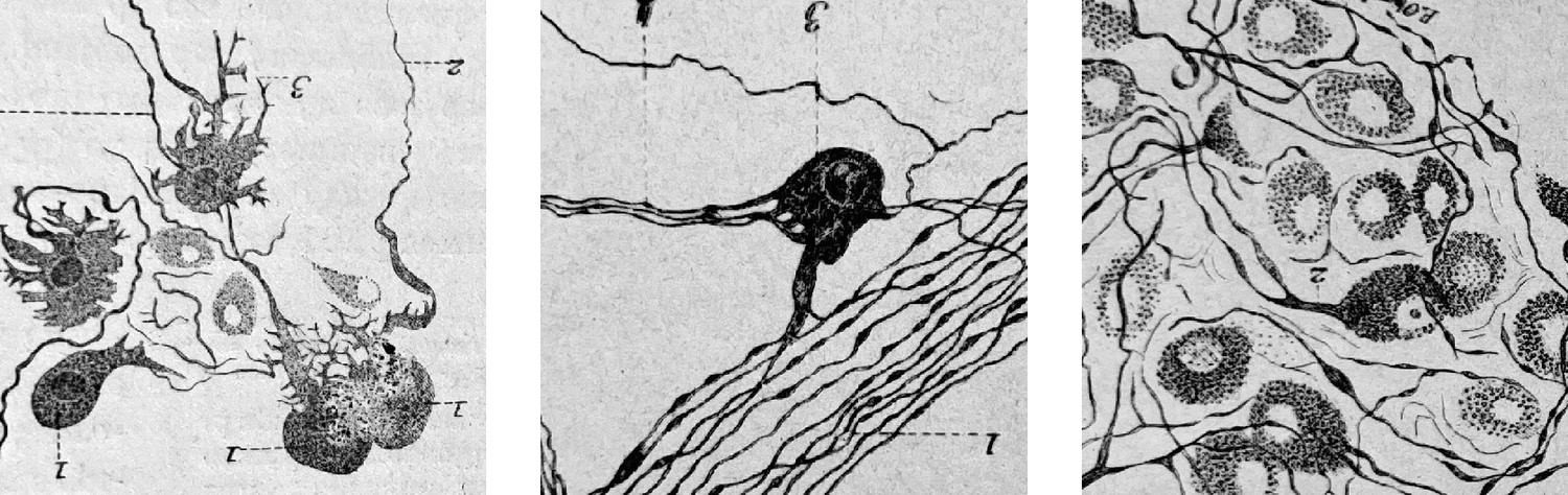
Cardiac ganglionic neurons of three morphological types recognized by Dogiel (reproduced in Testut, 1911, after Dogiel 1894).
Testut provides tens of references and comments: “Despite this body of work, the question of cardiac ganglia is far from settled, owing in part to the difficulty of the subject”. Not much has happened since, in the description of cardiac neuron types.
Figure 7
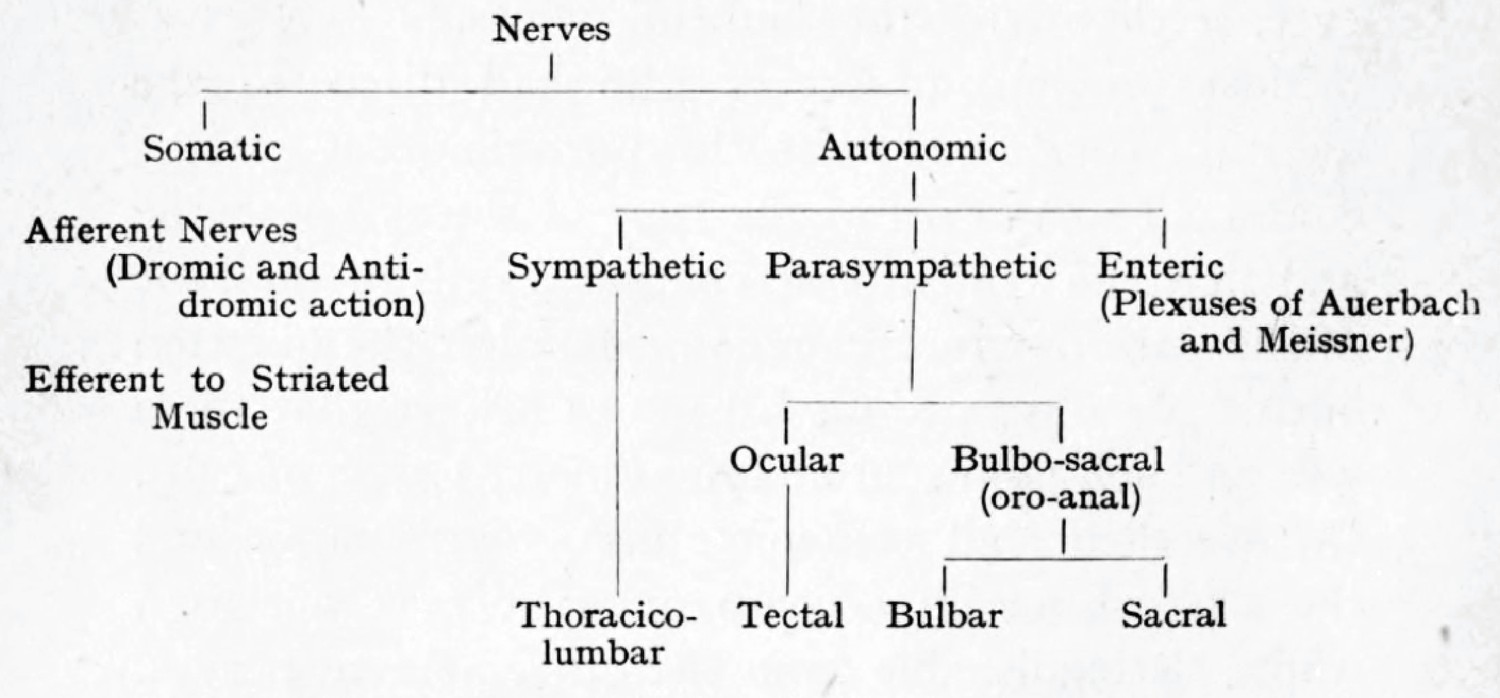
Langley’s cladogram of the peripheral nerves as it appears in The Autonomic nervous system Part I.
The “bulbo-sacral” outflow is unified across an anatomical gap that spans most of the spinal cord, but the ocular (or tectal) is in a separate subclass, despite being cranial, like the bulbar.
Figure 8
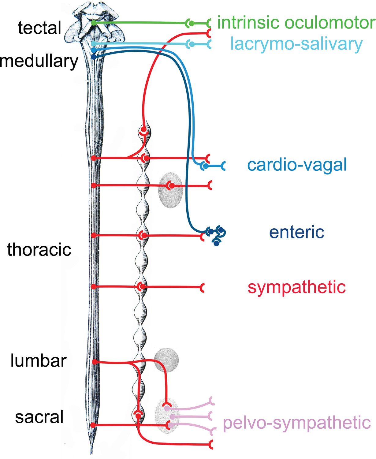
Provisional schematic of the polytomous, or mosaic autonomic outflow, based on published data (for the lumbosacral outflow, which was tentatively named ‘pelvosympathetic’; Sivori et al., 2023), unpublished data (for the oculomotor outflow), and educated guesses (for cardiac ganglia).
The lumbo-sacral outflow is distinguishable from the sympathetic (red) mostly at the level of post-ganglionic neurons, in the pelvic ganglion (pink). The genetic differences between the three medullary outflows (lacrymo-salivary, cardio-vagal, and enteric) are hypothetical at the preganglionic level (i.e. between the salivatory nuclei, the dorsal nucleus of the vagus nerve and the nucleus ambiguus). Note that the six outflows that are distinguished in this provisional scheme (including the pelvosympathetic one) form a series along the rostro-caudal axis of the central nervous system, restoring a link between development, cell types, and physiology.
Download links
A two-part list of links to download the article, or parts of the article, in various formats.
Downloads (link to download the article as PDF)
Open citations (links to open the citations from this article in various online reference manager services)
Cite this article (links to download the citations from this article in formats compatible with various reference manager tools)
Gaskell, Langley, and the "para-sympathetic" idea
eLife 14:e104826.
https://doi.org/10.7554/eLife.104826


