Layilin regulates Treg motility and suppressive capacity in skin
- Department of Dermatology, University of California, San Francisco, United States
- TRex Bio Inc, United States
- Department of Cellular and Molecular Pharmacology, University of California, San Francisco, United States
- Department of Surgery, University of California, San Francisco, United States
Figures
Figure 1 with 1 supplement
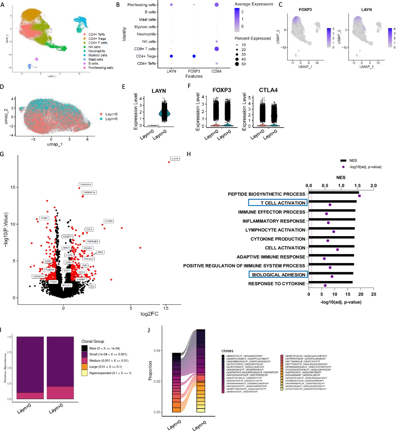
Layilin is preferentially expressed on regulatory T cells (Tregs) in skin and correlates with activation and motility signatures.
(A–C) Single-cell RNA-seq of FACS-purified CD45+ cells from healthy and psoriatic skin. n=5 healthy skins and 5 psoriatic skins. (A) Representative Uniform Manifold Approximation and Projection (UMAP) showing all clusters found. (B) Dot plot of LAYN, FOXP3, and CD8A expression by clusters. (C) UMAP of FOXP3 and LAYN expression in skin. (D) UMAP of Tregs subset showing in red LAYN non-expressing Tregs (LAYN<0) and blue LAYN-expressing Tregs (LAYN>0) based on gene count >0. (E, F) Violin plot of LAYN (E), FOXP3, and CTLA4 (F) in subset Tregs from skin followed by resclustering based on LAYN gene count >0. (G) Volcano plot of LAYN-expressing Tregs (right) compared to Layn-non-expressing Tregs (left) obtained from pseudo-bulk analysis performed on Tregs subset. (H) Gene set enrichment analysis of top enriched pathways in LAYN-expressing Tregs compared to LAYN-non-expressing Tregs associated with panel (F). (I, J) T cell receptor (TCR) analysis run on Treg cluster subset by LAYN expression. In (I), we show the relative abundance of TCR clonotype frequencies separated into five groups: rare being expressed between 0 and 104, small between 104 and 0.001, medium between 0.001 and 0.01, large between 0.01 and 0.1, and hyperexpanded between 0.1 and 1. (J) Representation of the top expressed clonotypes in a subset of Tregs from skin resclustered according to LAYN gene count >0.
Figure 1—figure supplement 1
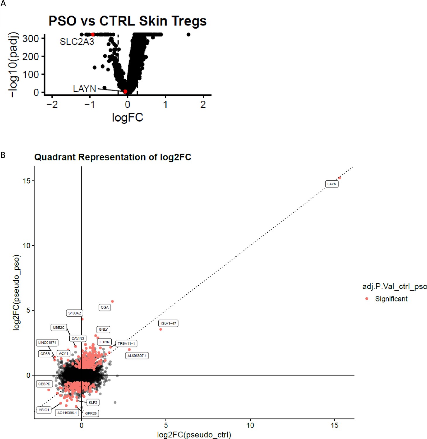
Layilin Expression on Tregs.
(A) Volcano plot comparing regulatory T cell (Treg) cluster from psoriasis to Treg cluster from healthy skin. (B) Quadrant representation of results from pseudo-bulk RNAseq of Treg cluster subclustered in LAYN-expressing and non-expressing Tregs. Red dots represent differentially expressed genes, and black dots are not differentially expressed ones. The left-upper quadrant corresponds to genes enriched only in psoriatic LAYN-expressing Tregs, the right-bottom quadrant are the ones only enriched in LAYN-expressing Tregs from healthy skin, and the upper-right quadrant corresponds to genes enriched in LAYN-expressing Tregs and common to both conditions.
Figure 2
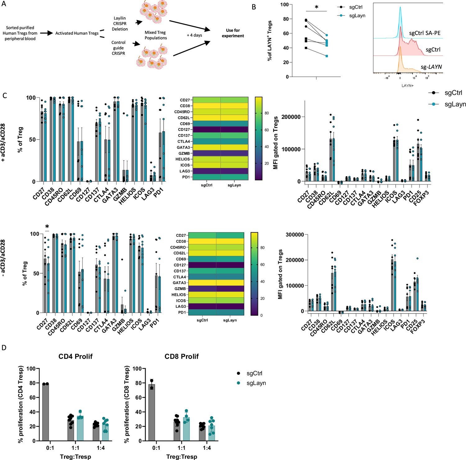
Layilin does not affect regulatory T cell (Treg) activation or suppression in vitro.
(A) Schematic experimental design strategy starting with the isolation of human Tregs from peripheral blood, activated and expanded using anti-CD3/anti-CD28 stimulation and IL-2 for 12 days. We deleted LAYN using CRISPR/Cas9, and after 4 days, cells were used for the experiment. (B) Frequency of LAYN+ Tregs gated on live CD8-CD4+CD25+FOXP3+ cells and representative expression of layilin. n=6 donors from two independent experiments. Significance was defined by a two‐tailed Student’s t‐test. *p<0.05. (C) Frequencies (on the left) and mean fluorescence intensity (MFI, on the right) of sg-LAYN and sg-CTRL Tregs gated on live CD8-CD4+CD25+FOXP3+ cells expressing the different activation Treg markers listed on the x-axis. The top-row graphs show results from cells stimulated with anti-CD3/anti-CD28; the bottom graphs show results obtained from cells without anti-CD3/anti-CD28 stimulation. n=6 donors from two independent experiments. Significance was defined by using a multiple-paired t‐test. (D) Proliferation percentage measured using cell trace violet for CD4+ T responder (left panel) or CD8+ T responder (right panel) after 3 days of suppression assay. Results are presented in the x-axis, and the different ratios of sg-LAYN and sg-CTRL Tregs and T responders used for the assay represent two independent experiments. Significance was defined by two‐way ANOVA, followed by Tukey’s multiple-comparisons test.
Figure 3 with 1 supplement
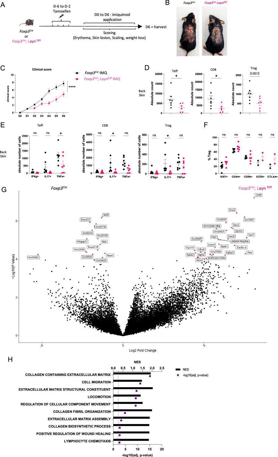
Layilin limits regulatory T cell (Treg)-mediated suppression of inflammation in vivo.
(A) Experimental design strategy of the imiquimod (IMQ) model. After 4 days of tamoxifen injection, mice were rested for 2 days before the start of the IMQ treatment. They received a daily dose of IMQ on shaved back skin and were scored for erythema, scaling, skin lesions, and weight loss. After 6 days of treatment, the mice were euthanized. (B) Representative pictures of mouse back skin after 5 days of treatment from each experimental group. (C) Overall clinical score over time. n=7 mice from three independent experiments. Significance was defined by two‐way ANOVA. ****p<0.0001. (D) Absolute count of Teff, CD8, and Tregs obtained from each experimental group, respectively, gated on live CD3+TCRβ+ and CD4+FOXP3- or CD8+, CD4+FOXP3+. n=7 and 6 mice per group from three independent experiments. Significance was defined by a two‐tailed Student’s t‐test. *p<0.05. (E) Absolute count of cells Teff, CD8, and Treg either IFNɣ+, IL17+, or TNFα+. n=7 and 6 mice per group from three independent experiments. Significance was defined by a multiple-paired t‐test. *p<0.05. (F) Frequencies of Tregs activation markers gated on live CD3+TCRβ+ CD4+FOXP3+ Tregs. n=7 and 6 mice per group from three independent experiments, except ICOS and CTLA from two mice and one experiment. Significance was defined by a multiple-paired t‐test. (G) Volcano plot of differential expression of genes from sorted Tregs gated on GFP+ from Foxp3Cre; Laynfl/fl (on the right) or Foxp3Cre (on the left) after 4 days of treatment. Red dots represent differentially expressed genes, and black dots are not differentially expressed ones. n=5 mice per group from two independent experiments. (H) Top deregulated pathways obtained from a gene set enrichment analysis comparing Foxp3Cre; Laynfl/fl to Foxp3Cre Tregs. The top axis and bar plot represent the normalized enrichment score (NES). The bottom axis and dot represent the adjusted p-value associated with pathways enrichment. n=5 mice per group from two independent experiments.
Figure 3—figure supplement 1
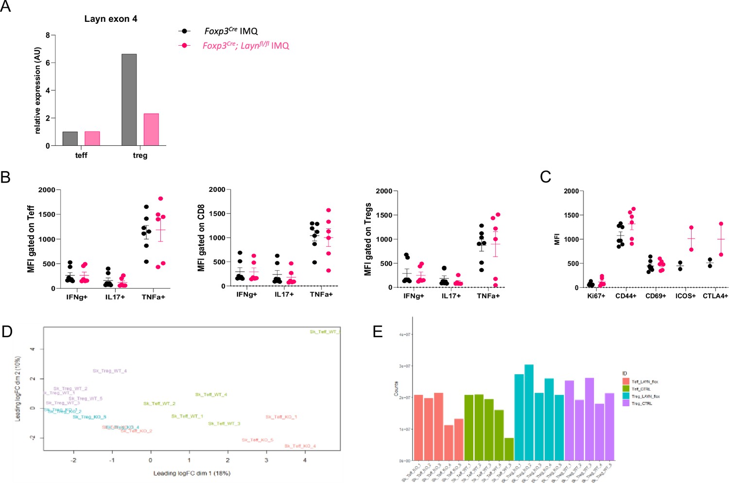
Immunophenotyping of imiquimod experiments in mice with Layilin-defieicnt Tregs.
(A) qRT-PCR of Layn from sorted regulatory T cells (Tregs) and Teff cells isolated respectively by gating on live CD45+CD3+CD4+CD8-CD25hiCD62LhiGFP+ and CD45+CD3+CD4+CD8-CD25loCD62LhiGFP-. Tregs and Teff have been isolated from back skin of Foxp3Cre; Laynfl/fl or Foxp3Cre mice after 4 days of treatment. Each bar corresponds to five mice mixed to create a representative sample for each group. (B) Mean fluorescence intensity (MFI) of cells Teff, CD8, and Treg either IFNɣ+, IL17+, or TNFα+. n=7 and 6 mice per group from three independent experiments. Significance was defined by a multiple-paired t‐test. (C) MFI of Tregs activation markers gated on live CD3+TCRβ+ CD4+FOXP3+ Tregs. n=7 and 6 mice per group from three independent experiments, except ICOS and CTLA from two mice and one experiment. Significance was defined by a multiple-paired t‐test. (D) Quality control of samples repartition used for bulk RNAseq in Figure 5. (E) Quality check of bulk RNAseq representing total of counts for each sample. In (F) and (G), n=5 mice per group from two independent experiments.
Figure 4 with 1 supplement
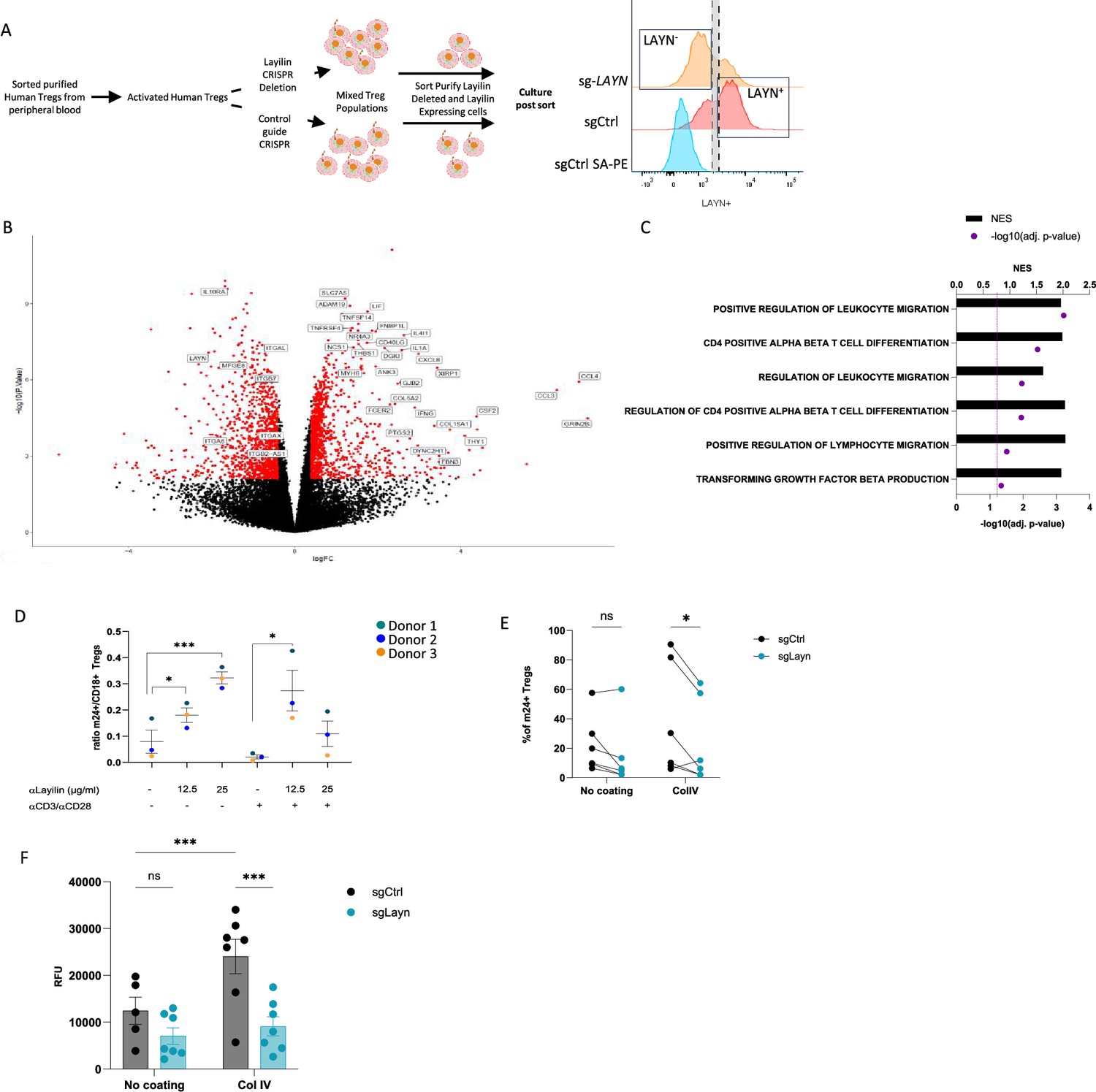
Layilin enhances regulatory T cell (Treg) adhesion and lymphocyte function-associated antigen-1 (LFA-1) activation.
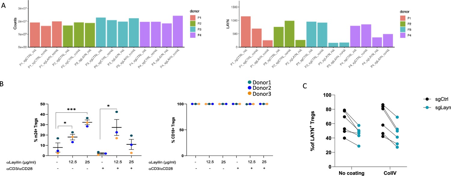
(A) The experimental design strategy starts with the isolation of human Tregs from peripheral blood, which were activated and expanded using anti-CD3/anti-CD28 stimulation and IL-2 for 12 days. We deleted LAYN using a CRISPR/Cas9; after 4 days, cells were sorted by flow cytometry based on LAYN expression, as represented by the representative histogram of LAYN expression on sgCtrl (stained with or without primary antibody detecting layilin followed by streptavidin-PE staining) or sg-LAYN Tregs, to obtain a pure population of LAYN+ or LAYN- Tregs. After sorting, cells were placed in culture for follow-up experiments presented in panels (B), (C), and (F). (B) Volcano plot of differential genes expressed in LAYN+ (on the left) or LAYN- (on the right) Tregs put in culture for 24 hours in the presence of anti-CD3/anti-CD28. Red dots represent differentially expressed genes, and black dots are not differentially expressed ones. n=4 paired donors for each group. (C) Top deregulated pathways obtained from a gene set enrichment analysis comparing LAYN+ to LAYN- Tregs. The top axis and bar plot represent the normalized enrichment score (NES). The bottom axis and dot represent the adjusted p-value associated with pathways enrichment. n=4 paired donors for each group. (D) Ratio of m24+ to total CD18+ Tregs gated on live CD8-CD4+CD25+FOXP3+ cells after 3 days culture with or without anti-CD3/anti-CD28 stimulation and in the presence of a layilin-crosslinking antibody as described on the x-axis for 20 minutes during LFA-1 activation staining. n=3 donors. Significance was defined by one‐way ANOVA, followed by Dunnett’s multiple-comparisons test. *p<0.05, ***p<0.001. (E) Frequency of m24+ sg-LAYN and sg-CTRL Tregs gated on live CD8-CD4+CD25+FOXP3+ cells after 24 hours of culture in a non-coated plate or collagen IV-coated plate. n=6 donors from two independent experiments. Significance was defined by two‐way ANOVA, followed by Sidak’s multiple-comparisons test. *p<0.05. (F) Numbers of cells attached to the bottom of the well are measured by the cell titer blue (expressed by relative fluorescence unit). n=7 donors from two independent experiments. Significance was defined by two‐way ANOVA. ns, nonsignificant, ***p<0.001.
Figure 4—figure supplement 1
Characterization of human ex vivo experiments.
(A) Quality check of bulk RNAseq representing total counts for each sample (left panel) and LAYN count for each sample demonstrating CRISPR efficiency (right panel). n=4 paired donors for each group. (B) On the left panel, we represent the frequency of total m24+ regulatory T cells (Tregs) gated on live CD8-CD4+CD25+FOXP3 + cells after 3 days of culture with or without anti-CD3/anti-CD28 stimulation and in the presence of a layilin-crosslinking antibody as described on the x-axis for 20 minutes during lymphocyte function-associated antigen-1 (LFA-1) activation staining. The right panel represents the frequency of CD18+ Tregs. n=3 donors. Significance was defined by one‐way ANOVA, followed by Dunnett’s multiple-comparisons test. *p<0.05, ***p<0.001. (C) Frequency of LAYN+ Tregs gated on live CD8-CD4+CD25+FOXP3+ cells to measure CRISPR efficiency after 24 hours of culture in a non-coated plate or collagen IV-coated plate. n=6 donors from two independent experiments.
Figure 5
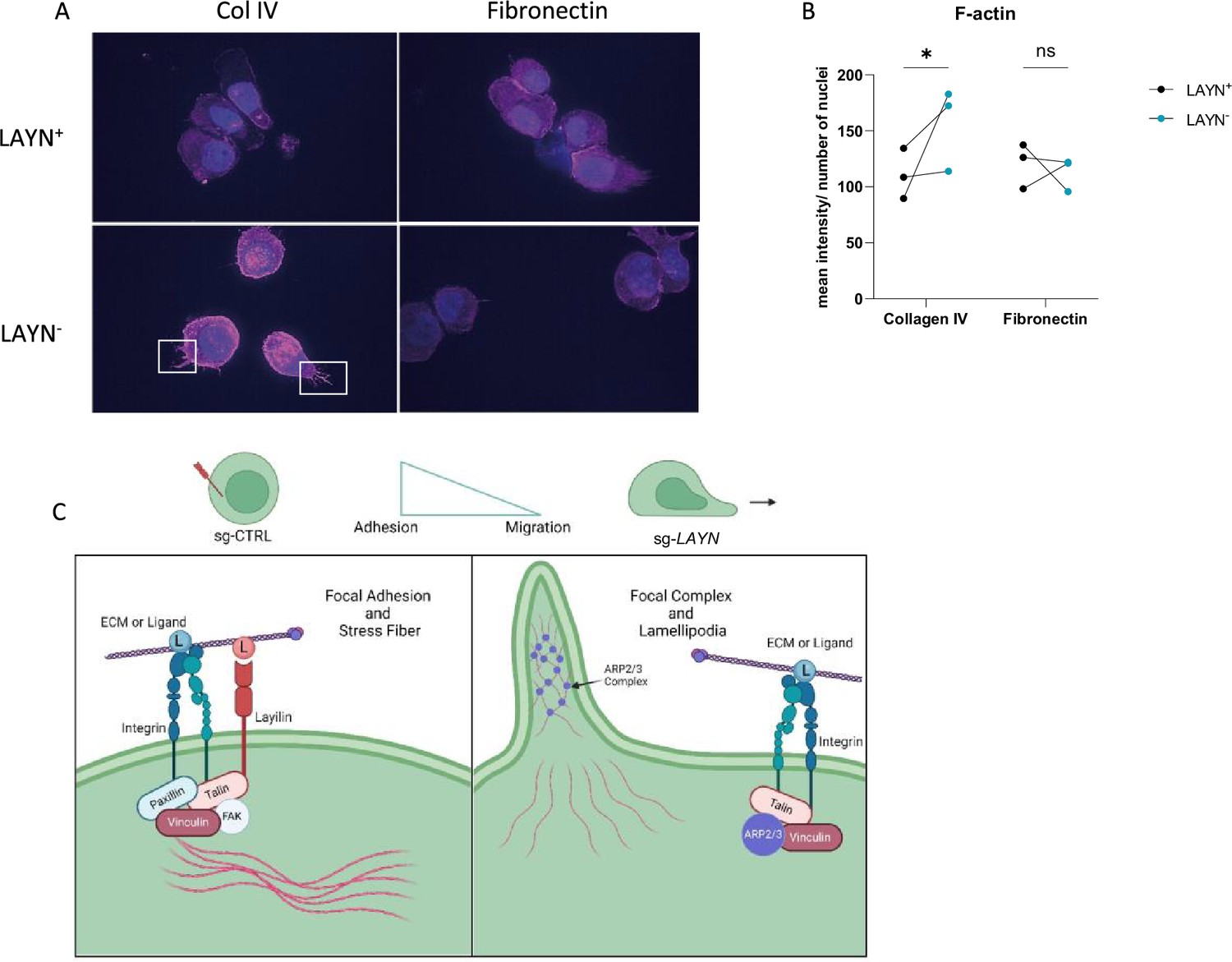
Layilin induces cytoskeleton changes in regulatory T cells (Tregs) indicative of reduced motility.
(A) Representative pictures of LAYN+ (on the top) or LAYN- (on the bottom), Tregs phalloidin staining (in pink) and nuclei (in blue) after a 48-hour culture on collagen IV (left) or fibronectin (right)-coated plates. White boxes highlight protuberances at the membranes. (B) Quantification of phalloidin staining associated with pictures in (A). n=3 donors; 10 pictures were quantified for each donor, and experiments were repeated independently twice. Significance was defined by two‐way ANOVA, followed by Sidak’s multiple-comparisons test. *p<0.05. (C) Graphical hypotheses of layilin pathway mechanism of action. In the presence of layilin, it interacts with lymphocyte function-associated antigen-1 (LFA-1) and maybe other integrins and stabilizes the formation of a focal adhesion complex, leading to the anchoring mechanism of the cells. Without layilin, integrins are less prone to adhesion mechanisms, and the ARP3 complex may act in lamellipodia formation, leading to more migratory Tregs.
Additional files
-
MDAR checklist
- https://cdn.elifesciences.org/articles/105277/elife-105277-mdarchecklist1-v1.docx
-
Source code 1
R analysis scripts used in this study.
- https://cdn.elifesciences.org/articles/105277/elife-105277-code1-v1.zip
Download links
A two-part list of links to download the article, or parts of the article, in various formats.
Downloads (link to download the article as PDF)
Open citations (links to open the citations from this article in various online reference manager services)
Cite this article (links to download the citations from this article in formats compatible with various reference manager tools)
Layilin regulates Treg motility and suppressive capacity in skin
eLife 14:RP105277.
https://doi.org/10.7554/eLife.105277.2


