Δ133p53α and Δ160p53α isoforms of the tumor suppressor protein p53 exert dominant-negative effect primarily by co-aggregation
- Department of Cell and Molecular Biology, Uppsala University, Biomedical Center, Sweden
- Department of Medical Biochemistry and Microbiology, Uppsala University, Biomedical Center, Sweden
Figures
Figure 1 with 2 supplements
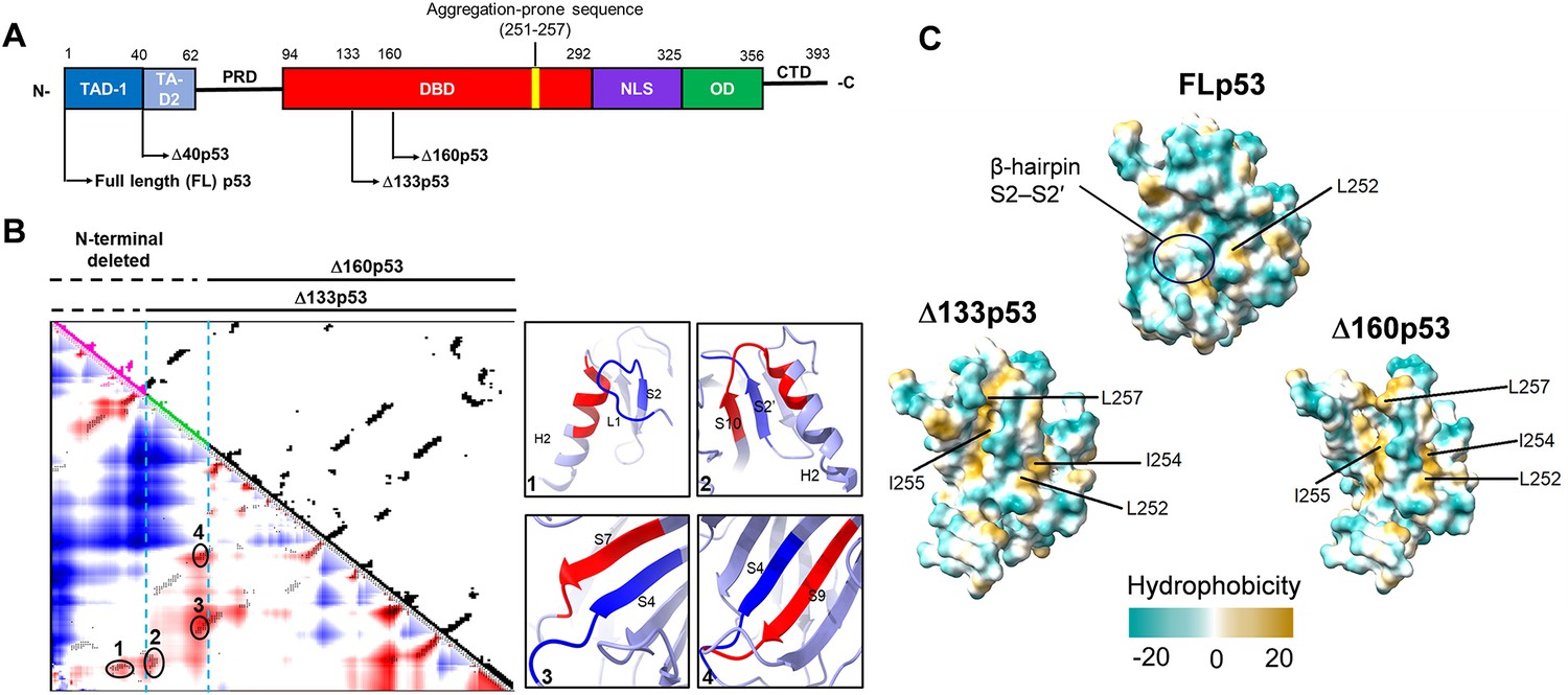
The Δ133p53 and Δ160p53 proteins exhibit structural destabilization.
(A) Schematic domain structures of full-length (FL) p53 and its isoforms. TAD, transactivation domain; PRD, proline-rich domain; DBD, DNA-binding domain; NLS, nuclear localization sequence; OD, oligomerization domain; CTD, C-terminal domain. Δ133p53 lacks the entire TAD and approximately 20% of the DBD. Δ160p53 lacks the entire TAD and approximately 33% of the DBD. An aggregation region ILTIITL (residues 251–257) is indicated in the DBD domain. (B) Contact density map (C-α 8 Å) of the core domain of FLp53 (PDB ID: 3KMD) using CWView (Vehlow et al., 2011). The residues 94–132 and 133–159 were colored magenta and green, respectively. Higher densities are shown in red, and low densities are shown in blue. Among these contacts, four higher-density contacts were selected and shown in oval circles on the map. The corresponding secondary structures of the contacts between the deleted region (blue) and the region (red) in the loop-sheet-helix (LSH) motif or the β-sandwich are shown on the right. (C) Comparison analysis of surface hydrophobicity of the core domain in FLp53 (PDB ID: 3KMD) and its isoforms Δ133p53 and Δ160p53 by ChimeraX (Goddard et al., 2018; Meng et al., 2023). The exposed hydrophobic residues in the aggregation region ILTIITL (residues 251–257) are labeled. The color scale of hydrophobicity is indicated with the maximum and minimum values in dark cyan and dark goldenrod, respectively. The β-hairpin S2–S2′ was shown in an oval circle.
Figure 1—figure supplement 1
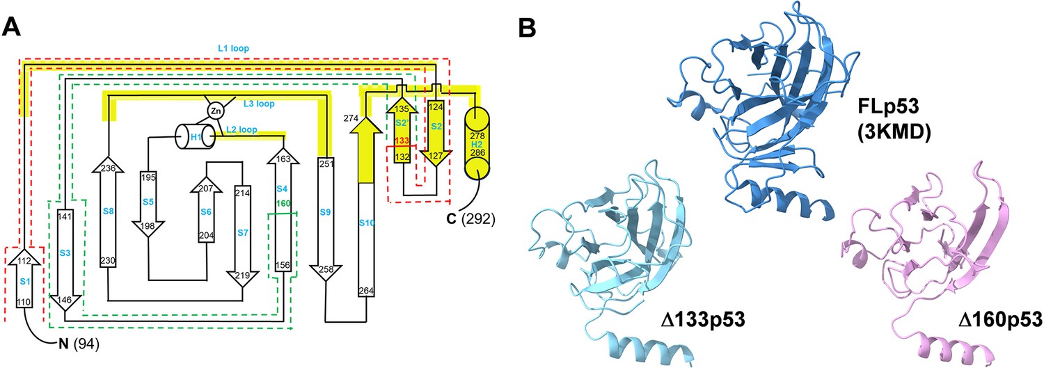
Core domain structures of full-length (FL) p53 and its isoforms Δ133p53 and Δ160p53.
(A) Topology diagram of the p53 DNA-binding domain based on prior research (Cho et al., 1994). Residues S94 to K132 are outlined with red dash rectangles. Furthermore, the residues M133 to A159 are outlined with green dash rectangles. The β-strands (S), α helices (H), loops (L), and the zinc atom (Zn) are labeled. The DNA-binding surface is emphasized in yellow and includes two large loops (L2 and L3) and the loop-sheet-helix (LSH) motif (L1, β-hairpin S2–S2′, H2, and the C-terminal residues of S10). (B) Comparison of core domain structures. The core domain structures of FLp53 (A chain, residues 92–291, PDB ID: 3KMD) and its isoforms Δ133p53 (residues 133–291, predicted by AlphaFold2) and Δ160p53 (residues 160–291, predicted by AlphaFold2) (Mirdita et al., 2022). Each structure is presented in the same orientation to facilitate direct comparison. The structures are analyzed by ChimeraX (Goddard et al., 2018; Meng et al., 2023).
Figure 1—figure supplement 2
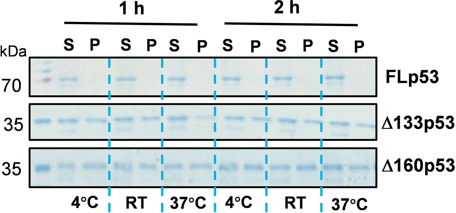
Aggregation propensity of FLp53 and its Δ133p53 and Δ160p53 isoforms by sedimentation analysis in SDS-PAGE.
Purified protein FLp53 and its isoforms Δ133p53 and Δ160p53 at a concentration of 1 μM incubated in 20 µl of reaction buffer (20 mM Tris-HCl pH 7.9, 150 mM NaCl, 5 mM DTT, 5% glycerol) at 4°C, room temperature (RT) (approximately 23°C), or 37°C for 1 hr or 2 hr. After incubation, the samples were centrifuged at 20,000×g for 30 min. The pellets were resuspended in 20 µl buffer. Both the supernatant (S) and pellet (P) fractions were analyzed by SDS-PAGE. While the FLp53 protein was detected only in the soluble supernatant fraction irrespective of temperature, a significant amount of Δ133p53 and Δ160p53 proteins was detected in the insoluble pellet fraction. This result suggests that the Δ133p53 and Δ160p53 isoforms are inherently destabilized and have much higher aggregation propensity than FLp53. FLp53 protein was sourced from Sigma (catalog number P6247), while Δ133p53 and Δ160p53 proteins were purified in our lab using multiple column chromatography.
-
Figure 1—figure supplement 2—source data 1
Original SDS-PAGE gel images corresponding to Figure 1—figure supplement 2, labeled.
- https://cdn.elifesciences.org/articles/106469/elife-106469-fig1-figsupp2-data1-v1.zip
-
Figure 1—figure supplement 2—source data 2
Original SDS-PAGE gel images corresponding to Figure 1—figure supplement 2.
- https://cdn.elifesciences.org/articles/106469/elife-106469-fig1-figsupp2-data2-v1.zip
Figure 2 with 1 supplement
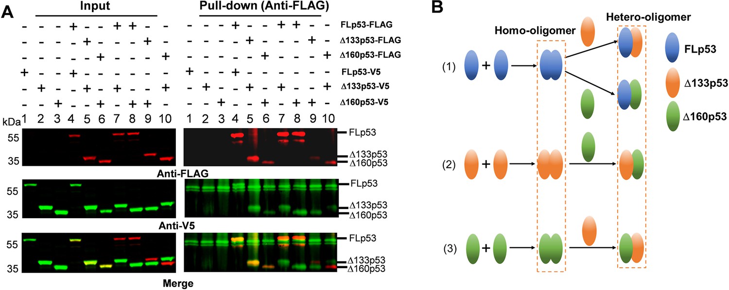
Complex formation among FLp53 and its isoforms Δ133p53 and Δ160p53.
(A) Co-immunoprecipitation of FLp53-FLAG, Δ133p53-FLAG, and Δ160p53-FLAG along with the FLp53-V5, Δ133p53-V5, and Δ160p53-V5 proteins in H1299 cells. The complexes were isolated with anti-FLAG antibody-coupled magnetic beads. Western blot analysis was done with the anti-FLAG and anti-V5 antibodies. The merged image corresponds to the overlay between the FLAG and V5 antibody signals. Input corresponds to 5% of the whole-cell lysate. (B) A summary of FLp53, Δ133p53, and Δ160p53 oligomerization based on data shown in panel A.
-
Figure 2—source data 1
Original membranes corresponding to Figure 2A, labeled.
- https://cdn.elifesciences.org/articles/106469/elife-106469-fig2-data1-v1.zip
-
Figure 2—source data 2
Original membranes corresponding to Figure 2A.
- https://cdn.elifesciences.org/articles/106469/elife-106469-fig2-data2-v1.zip
Figure 2—figure supplement 1
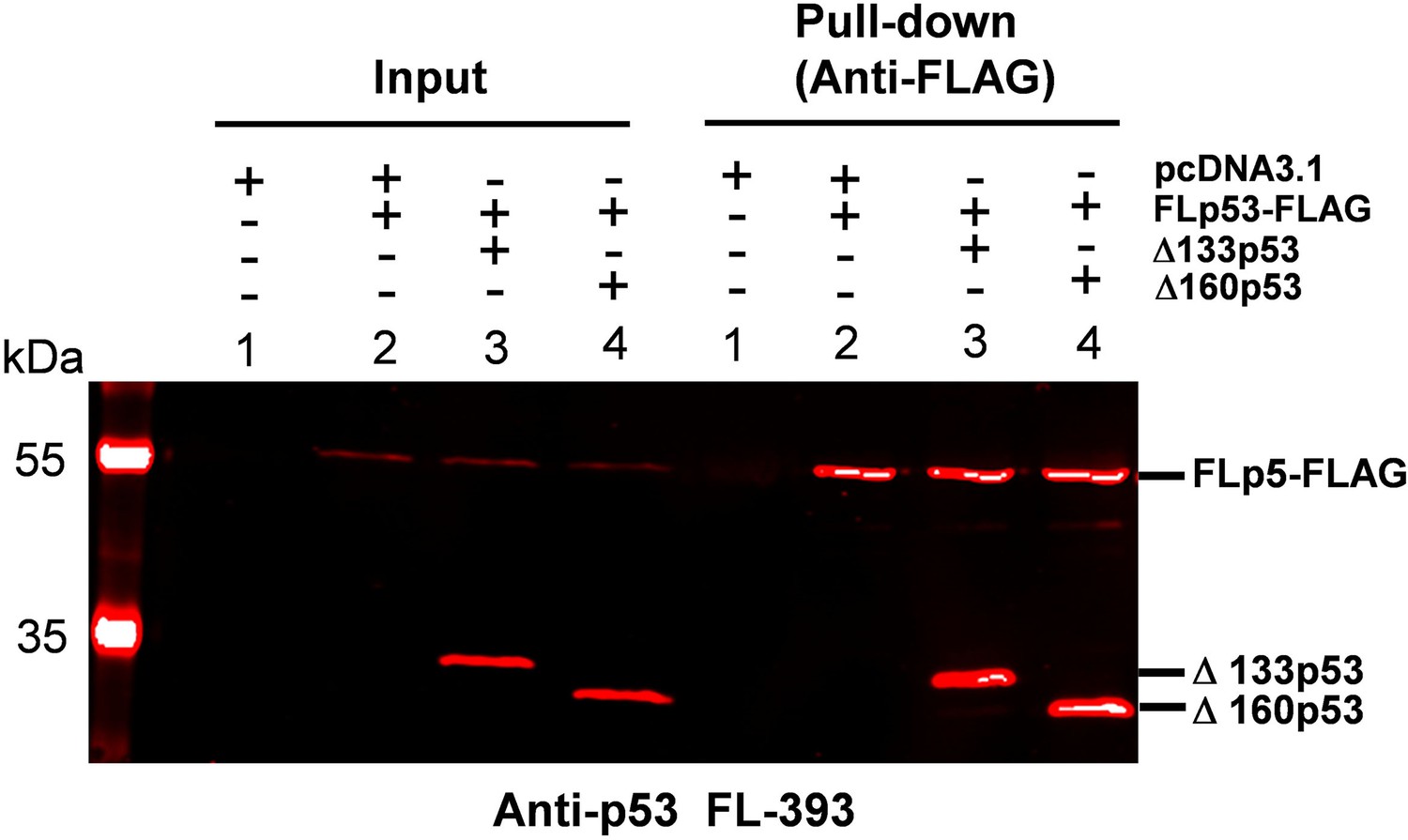
Hetero-oligomers formation between full-length p53 (FLp53) and its isoforms Δ133p53 or Δ160p53.
Co-immunoprecipitation of FLAG-tagged FLp53 with untagged Δ133p53 and Δ160p53 in H1299 cells. The complexes were isolated with anti-FLAG antibody-coupled magnetic beads. Western blot analysis was performed using the anti-p53 FL-393 antibody, which targets amino acids 1–393 corresponding to the FLp53 protein. Input corresponds to 5% of the whole-cell lysate.
-
Figure 2—figure supplement 1—source data 1
Original membranes corresponding to Figure 2—figure supplement 1, labeled.
- https://cdn.elifesciences.org/articles/106469/elife-106469-fig2-figsupp1-data1-v1.zip
-
Figure 2—figure supplement 1—source data 2
Original membranes corresponding to Figure 2—figure supplement 1.
- https://cdn.elifesciences.org/articles/106469/elife-106469-fig2-figsupp1-data2-v1.zip
Figure 3
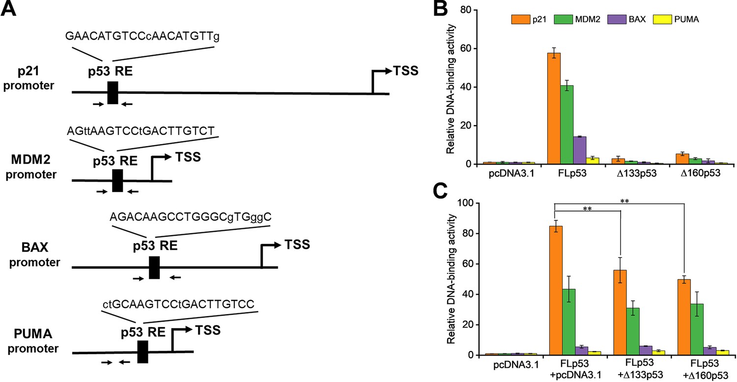
Δ133p53 and Δ160p53 negatively influence FLp53’s DNA-binding activity.
(A) Schematic representation of the promoters of p53 target genes (p21, MDM2, BAX, and PUMA). An arrow denotes the transcription start site (TSS). The DNA sequences of p53 response elements (REs) are shown, with uppercase and lowercase letters indicating matched and mismatched bases, respectively, in relation to the canonical p53 RE sequence (RRRCWWGYYY). Paired arrows highlight the regions subjected to quantitative PCR (qPCR) amplification. Specifically, the distal (5') p53 RE within the p21 gene promoter was analyzed. (B) Relative DNA binding of the FLp53-FLAG, Δ133p53-FLAG, and Δ160p53-FLAG proteins to p53 target genes (p21, MDM2, BAX, and PUMA) in H1299 cells. (C) Relative DNA binding of the FLp53-FLAG protein to the p53 target gene promoters in the presence of the V5-tagged protein Δ133p53 or Δ160p53 at a 1:1 ratio. Chromatin immunoprecipitation (ChIP)-qPCR assay data are shown as relative enrichment of promoter sequences of the target genes after normalization to the control, pcDNA3.1 plasmid transfected cells. Data are represented as the mean of technical triplicates ± standard deviation (SD), **p<0.01 (Student’s t-test).
-
Figure 3—source data 1
Chromatin immunoprecipitation (ChIP)-quantitative PCR (qPCR) assay data corresponding to Figure 3B and C.
- https://cdn.elifesciences.org/articles/106469/elife-106469-fig3-data1-v1.xlsx
Figure 4 with 1 supplement
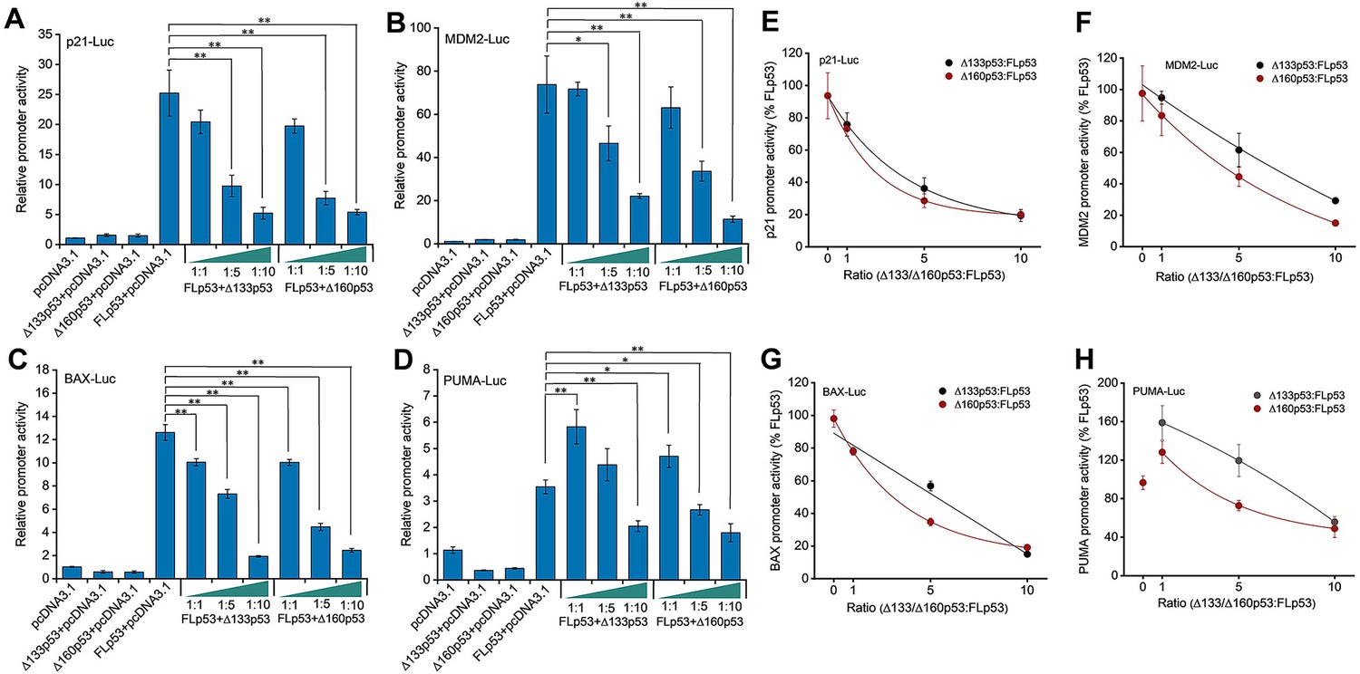
Impact of Δ133p53 and Δ160p53 on FLp53-activated transactivation.
(A–D) H1299 cells were transfected with luciferase reporter plasmids driven by p21 (A), MDM2 (B), BAX (C), and PUMA (D) promoters, along with vectors expressing FLp53, Δ133p53, or Δ160p53. To evaluate the influence of the isoforms on FLp53’s transactivation capability, co-expression was performed at ratios of 1:1, 1:5, and 1:10 relative to FLp53. Basal promoter activity was determined by transfecting cells with the empty vector, pcDNA3.1. Relative promoter activity is shown after normalization to the pcDNA3.1-treated sample activity. Data represent mean values ± standard deviation (SD) (n=3). *p<0.05; **p<0.01 (Student’s t-test). (E–H) The transcriptional activity of FLp53 on the p21 (E), MDM2 (F), BAX (G), and PUMA (H) promoters was inhibited by Δ133p53 and Δ160p53. Inhibition curve fitting was performed using the exponential function ExpDec1 in Origin 2018 software.
-
Figure 4—source data 1
Luciferase reporter assay data corresponding to Figure 4A–D.
- https://cdn.elifesciences.org/articles/106469/elife-106469-fig4-data1-v1.xlsx
-
Figure 4—source data 2
Data corresponding to Figure 4E–H, showing the inhibition of FLp53’s transcriptional activity by Δ133p53 and Δ160p53.
- https://cdn.elifesciences.org/articles/106469/elife-106469-fig4-data2-v1.xlsx
Figure 4—figure supplement 1
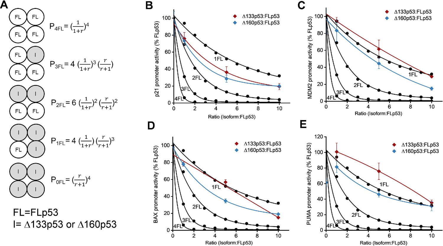
Predictive inhibitory capacity of Δ133p53 and Δ160p53 isoforms in hetero- or homo-tetrameric complexes.
(A) Mathematical modeling of tetramer formation probabilities (Chan et al., 2004). The probabilities (p) of tetramer formation involving different combinations of FLp53(FL) and its isoforms (I) Δ133p53 or Δ160p53 are expressed. The ratio r is defined as the concentration of Δ133p53 or Δ160p53 relative to the concentration of FLp53. (B–E) Prediction of isoform/FLp53 hetero-tetramer formation based on the data from Figure 4. The promoter activity of p53 target genes p21 (B), MDM2 (C), BAX (D), and PUMA (E) was shown. For PUMA, promoter activities are normalized to the activity when Δ133p53 and FLp53 are co-transfected at a 1:1 ratio. The predicted inhibition curves (black dots) are derived from the tetramer formation probabilities as shown in panel A. We hypothesize that the transcription factor is fully active only when it forms a tetramer consisting of 4FL; then, the final activity A equals P4FL. Thus, A4FL = P4FL; A3FL = P4FL + P3FL; A2FL = P4FL + P3FL + P2FL; A1FL = P4FL + P3FL + P2FL + P1FL. The experimental inhibition curves with Δ133p53 (red) or Δ160p53 (blue) were all above the theoretical inhibition curves for two isoform molecules per tetramer. Our analyses indicate that the Δ133p53 or Δ160p53 isoforms can exert an inhibitory effect on FLp53 function only when present in the tetramer in higher proportion than FLp53.
Figure 5 with 1 supplement
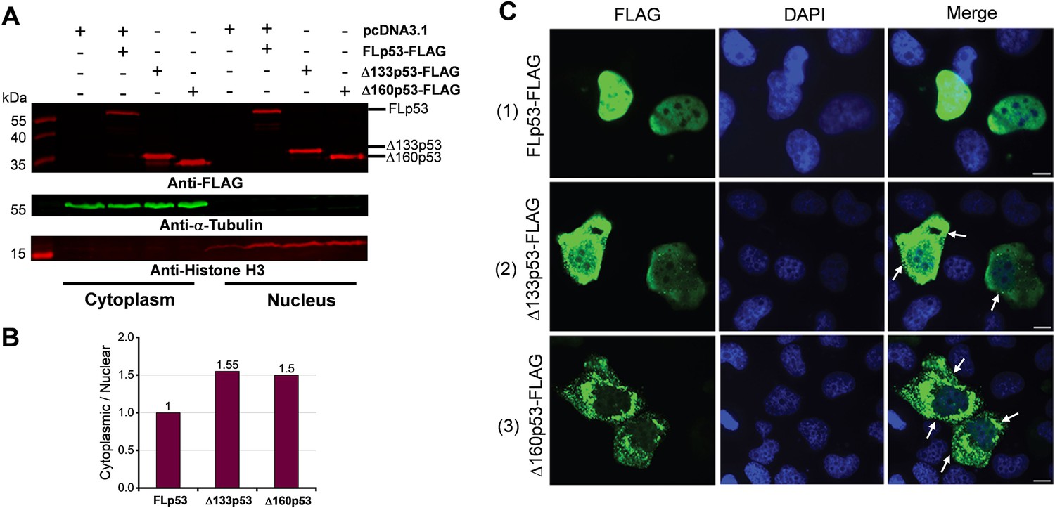
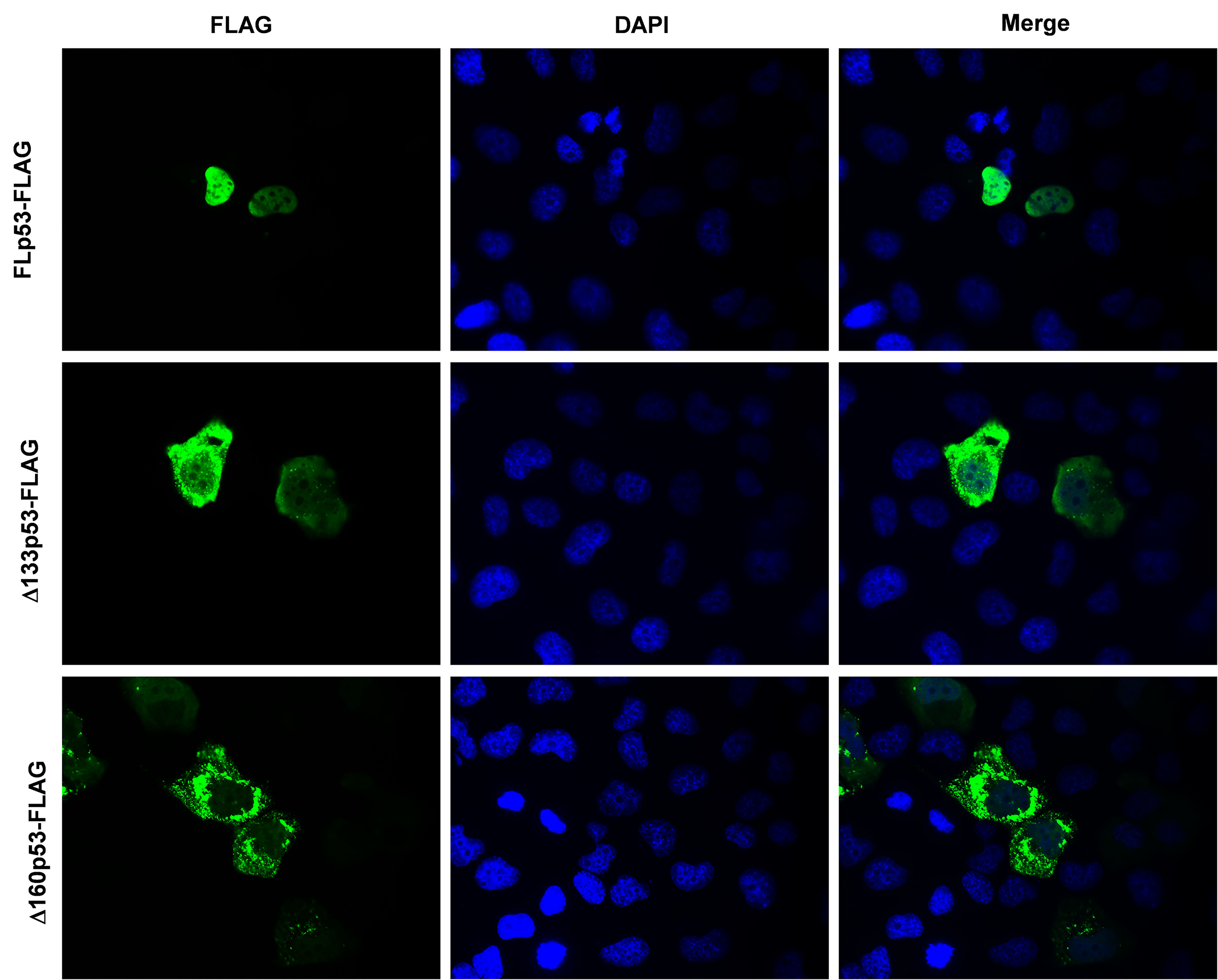
Subcellular distribution of the FLp53, Δ133p53, and Δ160p53 proteins.
(A) Western blot analysis of subcellular fractionation of H1299 cells transfected with the FLAG-tagged FLp53, Δ133p53, or Δ160p53 proteins. Representative immunoblots indicate the presence of these proteins in different cellular fractions. α-Tubulin (cytoplasmic marker) and Histone H3 (nuclear marker) were used to ensure fraction integrity. (B) Quantitative bar graph summarizing the cytoplasm-to-nucleus ratio of protein levels. FLAG-tagged protein levels were normalized to their respective fractionation markers: α-Tubulin for the cytoplasmic fraction and Histone H3 for the nuclear fraction. The relative ratios of Δ133p53 and Δ160p53 protein levels are displayed, with the FLp53 cytoplasm-to-nucleus ratio set as 1. (C) Immunofluorescence analysis of H1299 cells expressing FLAG-tagged FLp53, Δ133p53, and Δ160p53. Cell nuclei are visualized with DAPI (blue). The punctate spots (white arrows) indicating protein aggregates are marked. Scale bar, 50 μm.
-
Figure 5—source data 1
Original membranes corresponding to Figure 5A, labeled.
- https://cdn.elifesciences.org/articles/106469/elife-106469-fig5-data1-v1.zip
-
Figure 5—source data 2
Data corresponding to Figure 5B, showing the cytoplasm-to-nucleus ratio of protein levels.
- https://cdn.elifesciences.org/articles/106469/elife-106469-fig5-data2-v1.xlsx
-
Figure 5—source data 3
Original immunofluorescence images corresponding to Figure 5C.
- https://cdn.elifesciences.org/articles/106469/elife-106469-fig5-data3-v1.jpg
-
Figure 5—source data 4
Original membranes corresponding to Figure 5A.
- https://cdn.elifesciences.org/articles/106469/elife-106469-fig5-data4-v1.zip
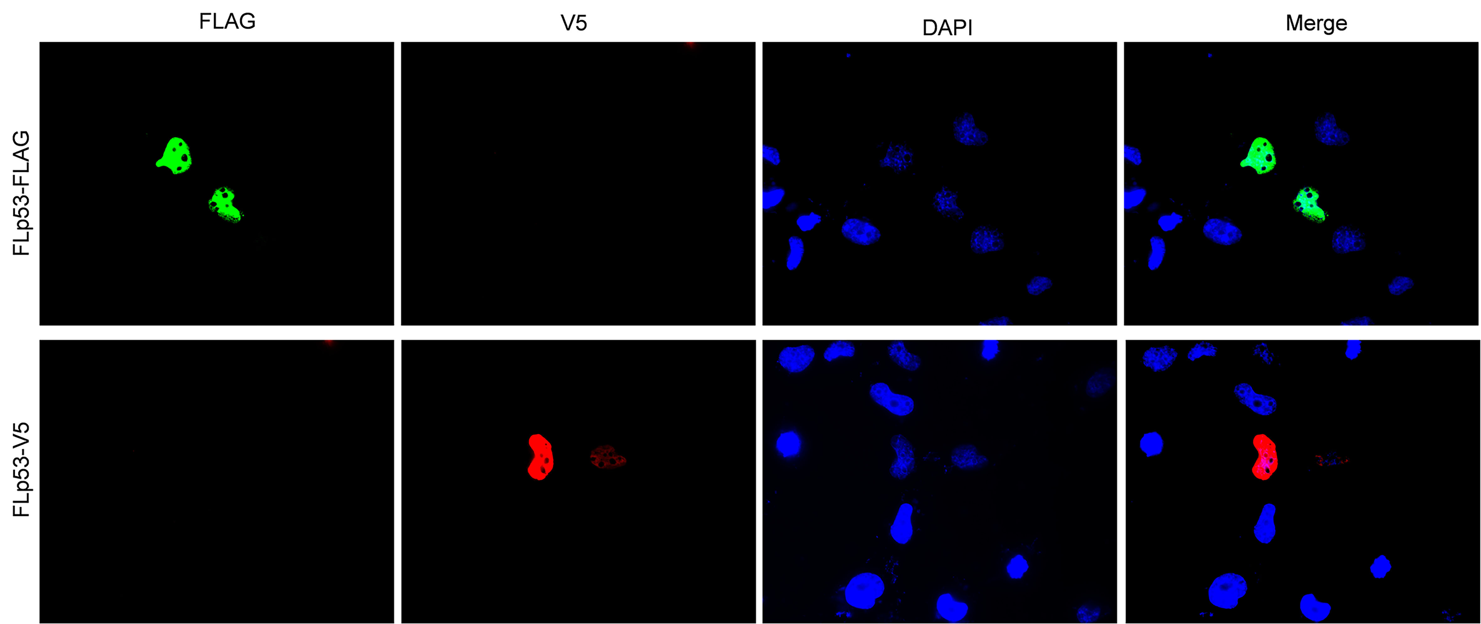
Figure 5—figure supplement 1
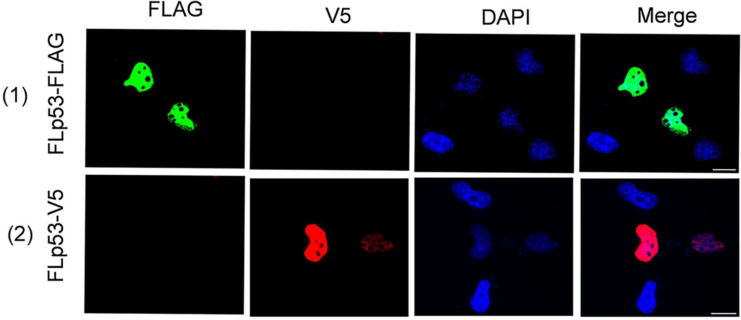
Immunofluorescence analysis of full-length p53 (FLp53) localization in H1299 cells.
FLp53 protein was expressed in H1299 cells. To rule out the possibility that epitope tags might affect protein localization, H1299 cells expressing either FLp53-tagged or V5-tagged FLp53 were analyzed. Cell nuclei were visualized with DAPI (blue). The co-aggregation of isoforms with FLp53 in cytoplasm (white arrows) and nucleus (bright green arrows) is indicated. Scale bar, 50 μm.
-
Figure 5—figure supplement 1—source data 1
Original immunofluorescence images corresponding to Figure 5—figure supplement 1.
- https://cdn.elifesciences.org/articles/106469/elife-106469-fig5-figsupp1-data1-v1.jpg
Figure 6 with 3 supplements
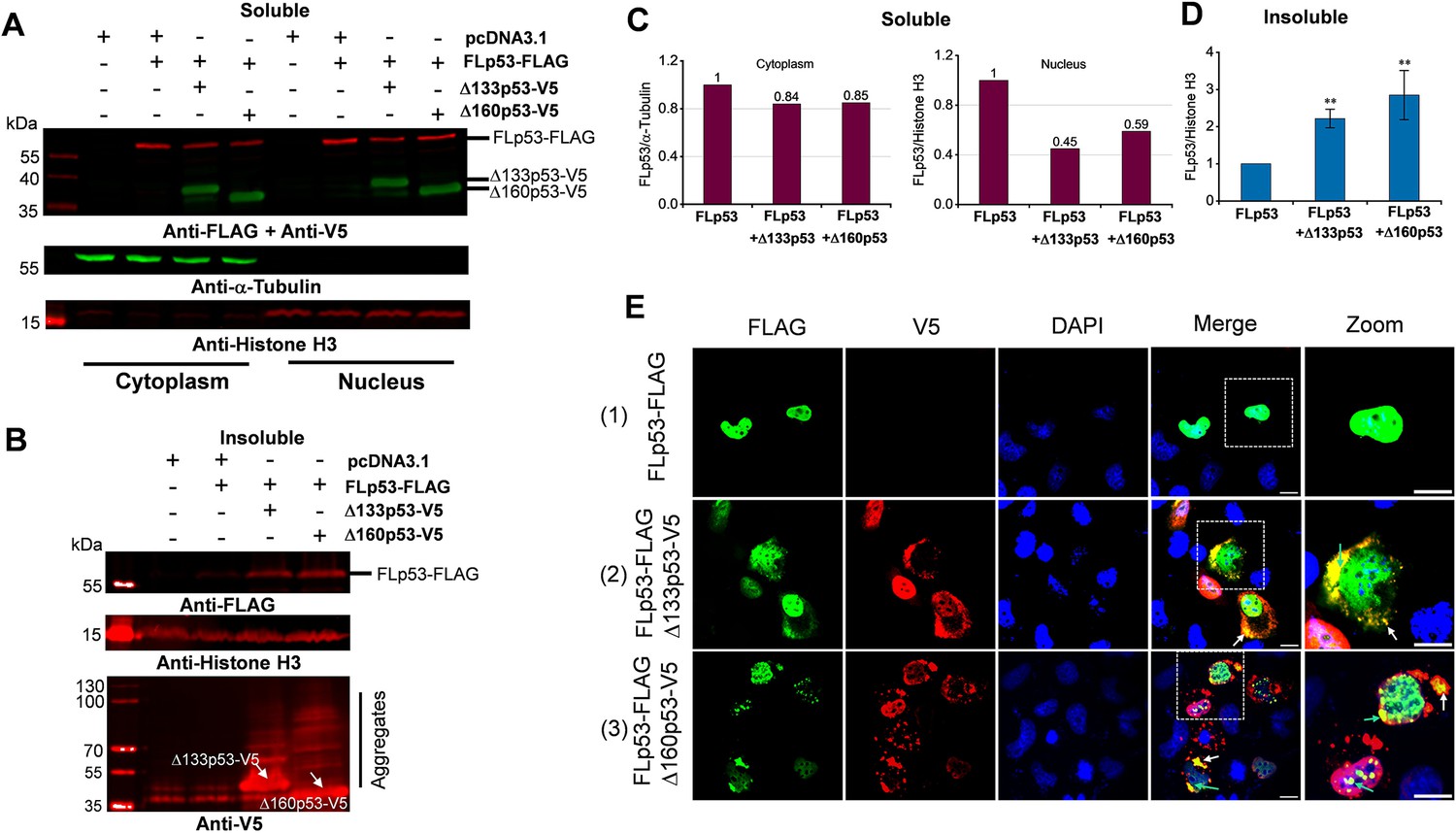
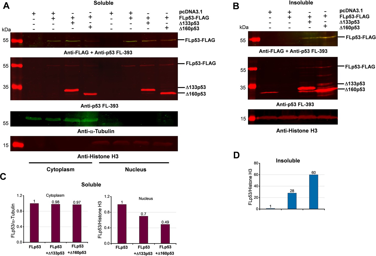
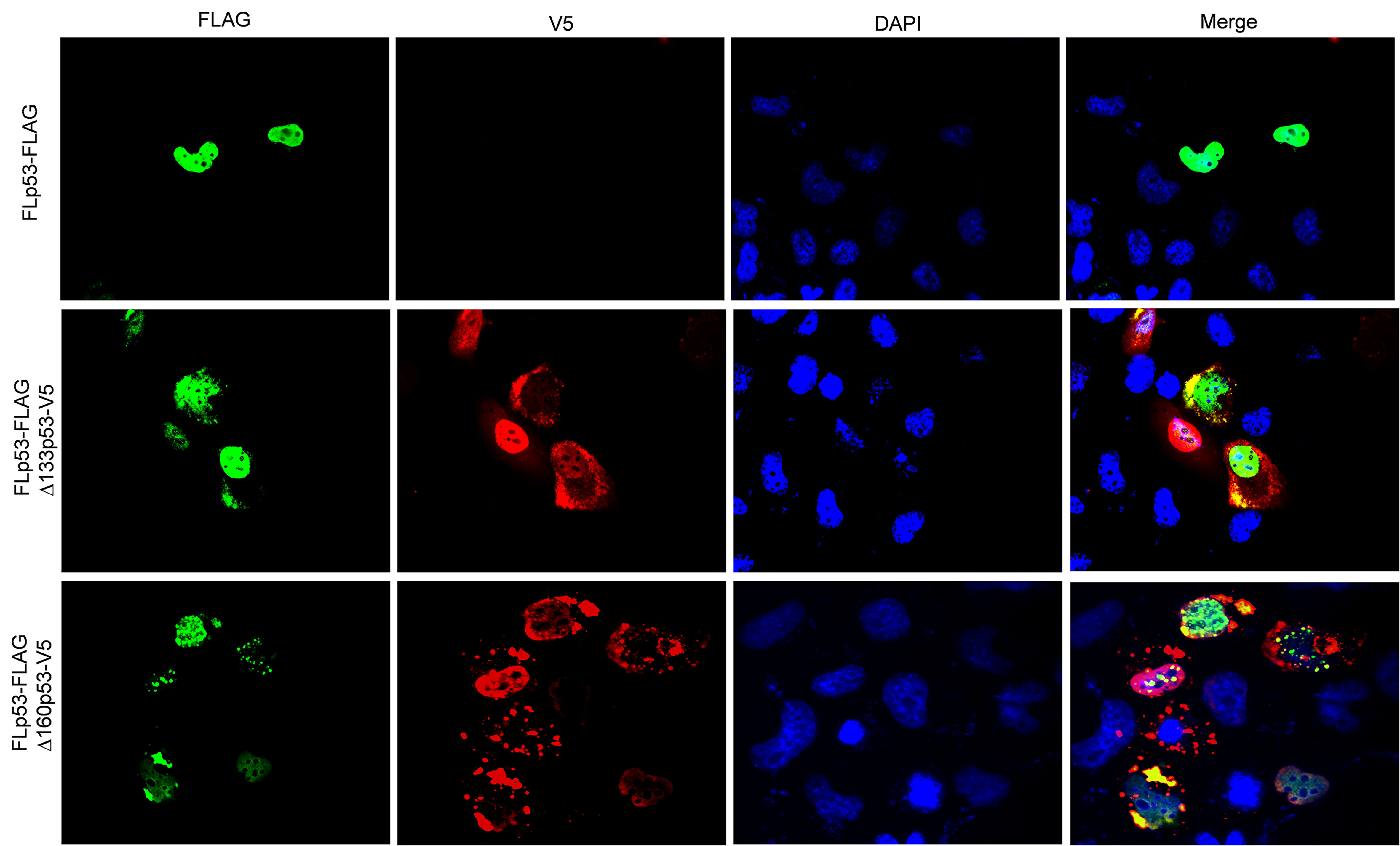
Induction of full-length p53 (FLp53) aggregation by Δ133p53 and Δ160p53 isoforms.
(A, B) Western blot analysis of soluble cytoplasmic and nuclear subcellular fractions (A) and insoluble nuclear fraction (B). Biochemical fractionation of H1299 cells transfected with the FLAG-tagged FLp53 and V5-tagged Δ133p53 or Δ160p53 at a 1:5 ratio. The aggregated monomers of V5-tagged Δ133p53 and Δ160p53 (indicated by white arrows), as well as the higher molecular weight aggregates, are shown. This figure shows one representative experiment; n=3. (C) Quantitative bar graph summarizing the levels of the soluble FLp53-FLAG protein in the cytoplasmic and nuclear fractions. The FLp53-FLAG protein levels were normalized to the corresponding fractionation marker (α-Tubulin or Histone H3). Relative accumulation of the FLp53-FLAG in Δ133p53-V5 or Δ160p53-V5 expressing cells is shown after considering the FLp53-FLAG protein expressing sample as 1. (D) Quantitative bar graph summarizing the levels of the FLp53-FLAG protein in the insoluble nuclear fraction relative to the Histone H3. Data normalization as in panel C, data represent mean values ± standard deviation (SD). **p<0.01 (Student’s t-test). (E) Immunofluorescence analysis of H1299 cells transfected with FLp53-FLAG alone or in combination with V5-tagged isoforms Δ133p53 and Δ160p53 expressing plasmids at a concentration ratio of 1:5. Cell nuclei were visualized with DAPI (blue). The co-aggregation of isoforms with FLp53 in the cytoplasm (white arrows) and nucleus (bright green arrows) is indicated. Scale bar, 50 μm.
-
Figure 6—source data 1
Original membranes corresponding to Figure 6A, labeled.
- https://cdn.elifesciences.org/articles/106469/elife-106469-fig6-data1-v1.zip
-
Figure 6—source data 2
Original membranes corresponding to Figure 6B, labeled.
- https://cdn.elifesciences.org/articles/106469/elife-106469-fig6-data2-v1.zip
-
Figure 6—source data 3
Data corresponding to Figure 6C, showing the levels of the soluble FLp53-FLAG protein in the cytoplasmic and nuclear fractions.
- https://cdn.elifesciences.org/articles/106469/elife-106469-fig6-data3-v1.xlsx
-
Figure 6—source data 4
Data corresponding to Figure 6D, showing the levels of the FLp53-FLAG protein in the insoluble nuclear fraction relative to the histone H3.
- https://cdn.elifesciences.org/articles/106469/elife-106469-fig6-data4-v1.xlsx
-
Figure 6—source data 5
Original immunofluorescence images corresponding to Figure 6E.
- https://cdn.elifesciences.org/articles/106469/elife-106469-fig6-data5-v1.jpg
-
Figure 6—source data 6
Original membranes corresponding to Figure 6A and B.
- https://cdn.elifesciences.org/articles/106469/elife-106469-fig6-data6-v1.zip
Figure 6—figure supplement 1
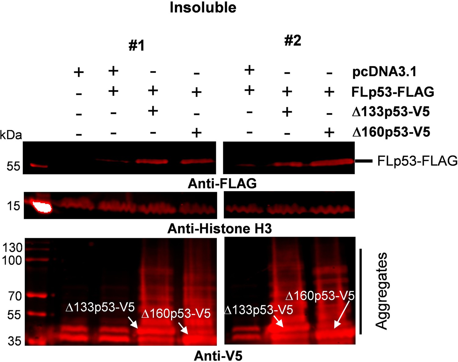
Western blot analysis of insoluble nuclear fraction of H1299 cells transfected with the FLAG-tagged full-length p53 (FLp53) and V5-tagged Δ133p53 or Δ160p53 at a ratio of 1:5.
The insoluble nuclear fraction was run in SDS-PAGE and blotted with anti-FLAG and anti-V5 antibodies. Histone H3 was employed as a nuclear marker. While the anti-FLAG antibody showed a single band of FLp53-FLAG, multiple bands of diverse molecular weight were seen with anti-V5 antibody signifying the aggregated variants of V5-tagged Δ133p53 and Δ160p53 (indicated by white arrows, as well as marked in the margin).
-
Figure 6—figure supplement 1—source data 1
Original membranes corresponding to Figure 6—figure supplement 1, labeled.
- https://cdn.elifesciences.org/articles/106469/elife-106469-fig6-figsupp1-data1-v1.zip
-
Figure 6—figure supplement 1—source data 2
Original membranes corresponding to Figure 6—figure supplement 1.
- https://cdn.elifesciences.org/articles/106469/elife-106469-fig6-figsupp1-data2-v1.zip
Figure 6—figure supplement 2
Induction of full-length p53 (FLp53) aggregation by Δ133p53 and Δ160p53 isoforms.
(A, B) Western blot analysis of soluble cytoplasmic and nuclear subcellular fractions (A) and insoluble nuclear fraction (B). Biochemical fractionation of H1299 cells transfected with the FLAG-tagged FLp53 and untagged Δ133p53 or Δ160p53 at a 1:5 ratio. (C) Quantitative bar graph summarizing the levels of the soluble FLp53-FLAG protein in the cytoplasmic and nuclear fractions. The FLp53-FLAG protein levels were normalized to the corresponding fractionation marker (α-Tubulin or Histone H3). Relative accumulation of the FLp53-FLAG in Δ133p53 or Δ160p53 expressing cells is shown after considering the FLp53-FLAG protein expressing sample as 1. (D) Quantitative bar graph summarizing the levels of the FLp53-FLAG protein in the insoluble nuclear fraction relative to the Histone H3. Data normalization as in panel C.
-
Figure 6—figure supplement 2—source data 1
Original membranes corresponding to Figure 6—figure supplement 2A, labeled.
- https://cdn.elifesciences.org/articles/106469/elife-106469-fig6-figsupp2-data1-v1.zip
-
Figure 6—figure supplement 2—source data 2
Original membranes corresponding to Figure 6—figure supplement 2B, labeled.
- https://cdn.elifesciences.org/articles/106469/elife-106469-fig6-figsupp2-data2-v1.zip
-
Figure 6—figure supplement 2—source data 3
Data corresponding to Figure 6—figure supplement 2C, showing the levels of the soluble FLp53-FLAG protein in the cytoplasmic and nuclear fractions.
- https://cdn.elifesciences.org/articles/106469/elife-106469-fig6-figsupp2-data3-v1.xlsx
-
Figure 6—figure supplement 2—source data 4
Data corresponding to Figure 6—figure supplement 2D, showing the levels of the FLp53-FLAG protein in the insoluble nuclear fraction relative to Histone H3.
- https://cdn.elifesciences.org/articles/106469/elife-106469-fig6-figsupp2-data4-v1.xlsx
-
Figure 6—figure supplement 2—source data 5
Original membranes corresponding to Figure 6—figure supplement 2A and B.
- https://cdn.elifesciences.org/articles/106469/elife-106469-fig6-figsupp2-data5-v1.zip
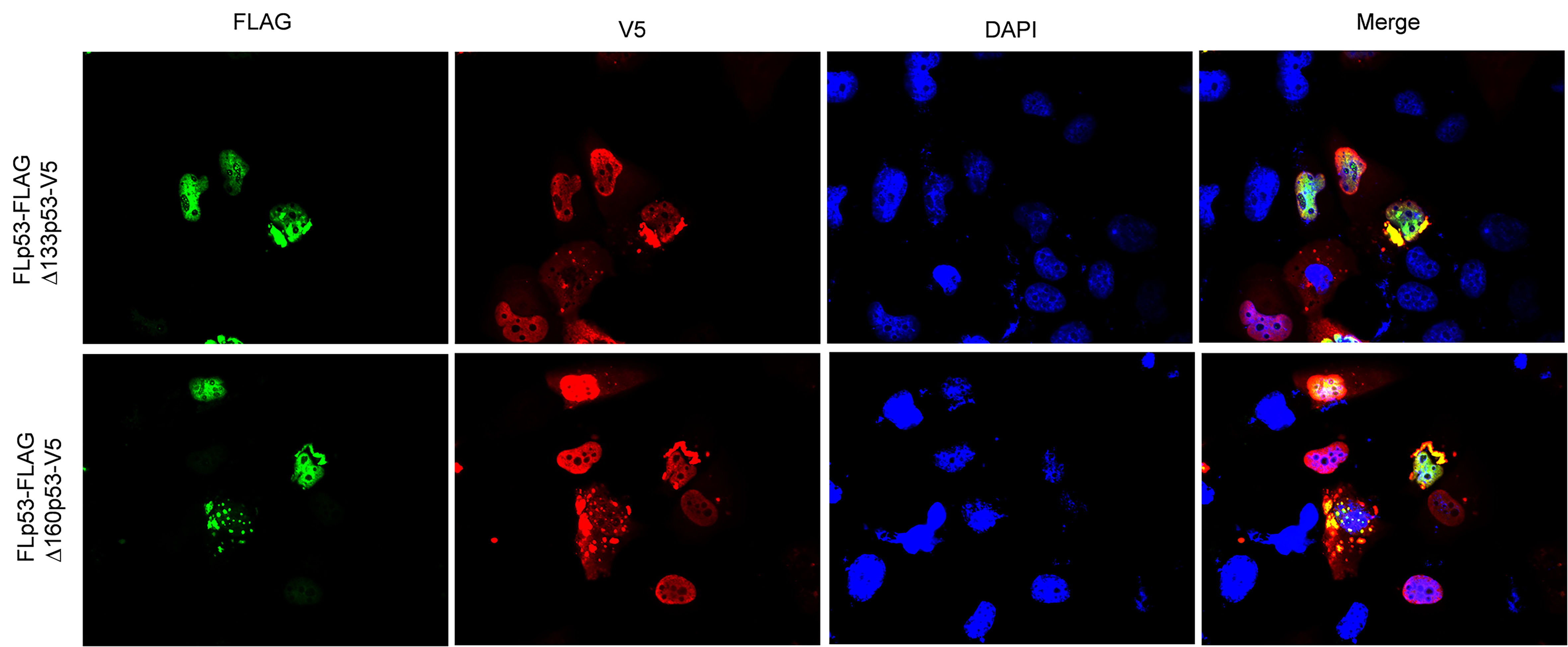
Figure 6—figure supplement 3
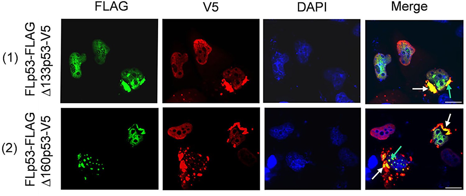
Immunofluorescence analysis of full-length p53 (FLp53) and its isoforms Δ133p53 and Δ160p53 localization in H1299 cells.
H1299 cells expressing FLAG-tagged FLp53 in combination with V5-tagged isoforms Δ133p53 and Δ160p53 at a ratio of 1:5. Cell nuclei were visualized with DAPI (blue). The co-aggregation of isoforms with FLp53 in cytoplasm (white arrows) and nucleus (bright green arrows) is indicated. Scale bar, 50 μm.
-
Figure 6—figure supplement 3—source data 1
Original immunofluorescence images corresponding to Figure 6—figure supplement 3.
- https://cdn.elifesciences.org/articles/106469/elife-106469-fig6-figsupp3-data1-v1.jpg
Figure 7

Overexpression of Δ133p53 and Δ160p53 blocks pro-apoptotic activity of full-length p53 (FLp53).
Caspase-3 and -7 activities were detected in H1299 cells co-transfected with plasmids expressing FLp53 and either Δ133p53 or Δ160p53 at a 1:5 ratio. Experiments were performed with the plasmids expressing FLAG-tagged and untagged FLp53, Δ133p53, and Δ160p53. The relative activities of caspase-3 and -7 were normalized to those of the control vector (transfected only with the pcDNA3.1 plasmid). Data represent mean values ± standard deviation (SD). **p<0.01 (Student’s t-test).
-
Figure 7—source data 1
Data corresponding to Figure 7, showing caspase-3 and -7 activities of H1299 cells co-transfected with plasmids expressing FLp53 and either Δ133p53 or Δ160p53 at a 1:5 ratio.
- https://cdn.elifesciences.org/articles/106469/elife-106469-fig7-data1-v1.xlsx
Figure 8
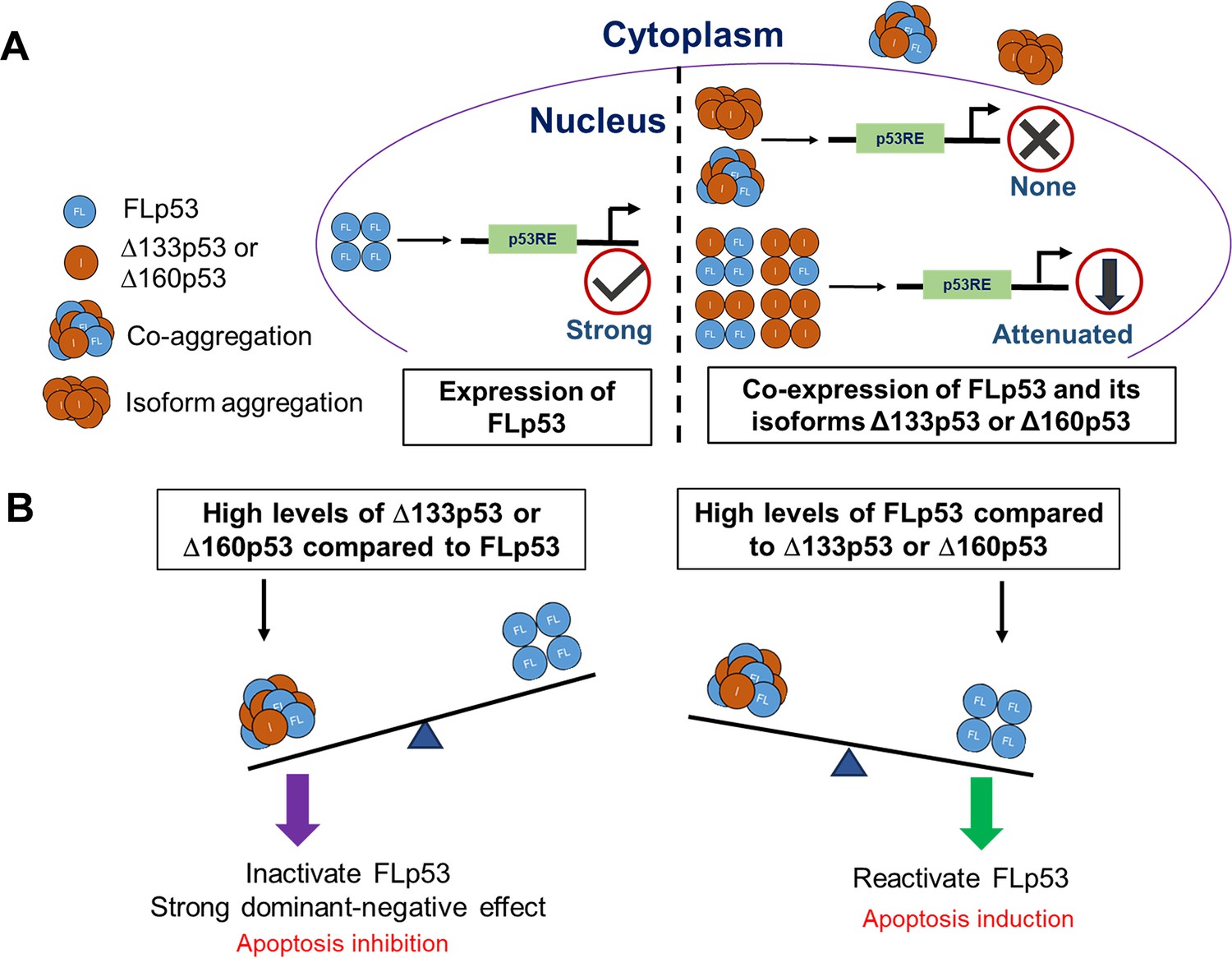
Schematic representation of the underlying mechanisms by which Δ133p53 and Δ160p53 exert a dominant-negative effect on full-length p53 (FLp53).
(A) When expressed alone, FLp53 demonstrates strong transcriptional activity, but when expressed together with the Δ133p53 or Δ160p53 isoforms, FLp53 activity gets impaired due to the dominant-negative effect exerted by the isoforms. The Δ133p53 or Δ160p53 isoforms either attenuate FLp53’s transcription activity by hetero-tetramerization at a ratio higher than 1:1 or abolish it completely by aggregation. Since Δ133p53 and Δ160p53 exhibit much higher propensity for aggregation than FLp53, they sequester FLp53 in the cytoplasmic aggregates, leading to a reduction in the amount of functional FLp53 in the nucleus. Aggregation also limits the availability of these isoforms in the nucleus. Thus, co-aggregation is the primary reason for the dominant-negative effect of the Δ133p53 or Δ160p53 isoforms on FLp53. It is important to note that the numbers of tetramers and aggregates depicted do not represent the actual quantities present in the cytoplasm and nucleus. (B) The balance between the expression level of the FLp53 and its isoforms Δ133p53 and Δ160p53 regulates FLp53 activity. Increased levels of Δ133p53 and Δ160p53 relative to FLp53 lead to a stronger dominant-negative effect due to co-aggregation and hetero-tetramerization, resulting in the inhibition of apoptosis. Increased levels of FLp53 compared to the isoforms Δ133p53 and Δ160p53 in cancer cells restore the function of FLp53, leading to the induction of apoptosis.
Author response image 1

Comparative analysis of protein expression in A549 and H1299 cells.
(A) A549 cells (p53 wild-type) were treated with etoposide to induce endogenous wild-type p53 expression. To assess the effects of FLp53 and its isoforms Δ133p53 and Δ160p53 on endogenous wild-type p53 aggregation, A549 cells were transfected with 2 μg of V5-tagged p53 expression plasmids, with or without etoposide (20μM for 8h) treatment. Western blot analysis was done with the anti-V5 (rabbit) to detect V5-tagged proteins and anti-DO-1 (mouse), the latter detects both endogenous wild-type p53 and V5-tagged FLp53. The merged image corresponds to the overlay between the V5 and DO1 antibody signals. (B) H1299 cells (p53-null) were transfected with 2 μg V5tagged p53 expression plasmids or the empty vector control pcDNA3.1. Western blot analysis was done with the anti-V5 (mouse) antibody.
Author response image 2
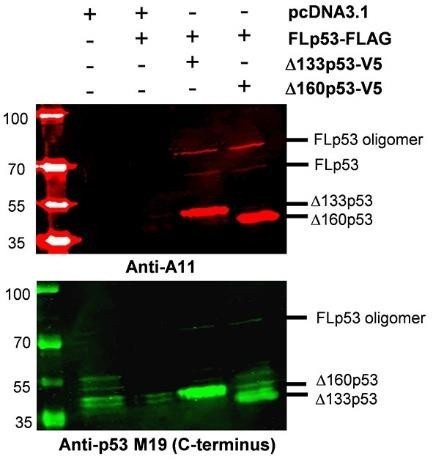
Induction of FLp53 Aggregation by p53 Isoforms Δ133p53 and Δ160p53.
H1299 cells transfected with the FLAG-tagged FLp53 and V5-tagged Δ133p53 or Δ160p53 at a 1:5 ratio. The cells were subjected to subcellular fractionation, and the resulting insoluble nuclear pellet was resuspended in RIPA buffer. The samples were heated at 95°C until the pellet was completely dissolved, and then analyzed by Western blotting. Immunoprecipitation was performed using the A11 antibody, which specifically recognizes amyloid protein aggregates, and the anti-p53 M19 antibody, which detects FLp53 as well as its isoforms Δ133p53 and Δ160p53.
Additional files
-
Supplementary file 1
Sequences of oligonucleotides used in the work.
- https://cdn.elifesciences.org/articles/106469/elife-106469-supp1-v1.docx
-
MDAR checklist
- https://cdn.elifesciences.org/articles/106469/elife-106469-mdarchecklist1-v1.docx
Download links
A two-part list of links to download the article, or parts of the article, in various formats.
Downloads (link to download the article as PDF)
Open citations (links to open the citations from this article in various online reference manager services)
Cite this article (links to download the citations from this article in formats compatible with various reference manager tools)
Δ133p53α and Δ160p53α isoforms of the tumor suppressor protein p53 exert dominant-negative effect primarily by co-aggregation
eLife 14:RP106469.
https://doi.org/10.7554/eLife.106469.2


