
A GCN1-independent activator of the kinase GCN2

- Cambridge Institute for Medical Research (CIMR), University of Cambridge, United Kingdom
- Division of Cancer Research, School of Medicine, Ninewells Hospital, United Kingdom
- The ALBORADA Drug Discovery Institute, University of Cambridge, United Kingdom
- Department of Medicine, University of Cambridge, United Kingdom
- Signalling Programme, The Babraham Institute, Babraham Research Campus, United Kingdom
- Domainex Ltd, Churchill Building, Chesterford Research Park, United Kingdom
- Cancer Research UK Cambridge Institute, University of Cambridge, United Kingdom
- Department of Pharmacology, University of Cambridge, United Kingdom
- Royal Papworth Hospital NHS Foundation Trust, United Kingdom
Figures
Figure 1

Chemical screen of novel GCN2-specific ISR-activating molecules.
(A) Schematic of ATF4::NanoLuc reporter, exploiting the upstream open reading frames (uORFs). Short uORFs 1, 2, and 3 precede the ATF4 translation start site, with uORF3 overlapping the ATF4 coding sequence, but out-of-frame. During the ISR, skipping of uORF3 leads to increased translation initiation at the ATF4 initiator AUG. (B) Relative Light Unit (RLU) of ATF4 signal in CHO stable cell-line overexpressing ATF4::NanoLuc treated with 1 mM histidinol, 2.5 µg/ml tunicamycin, or 1 µM latrunculin A (n = 3 biological repeats; ± SEM). (C) Chemical screen of 6461 small molecules tested with CHO stable cell-line overexpressing ATF4::NanoLuc. The molecules were treated alone or with submaximal ISR activation with 0.3 mM histidinol. All molecules were displayed along the x-axis based on their Z-score of relative reporter signal when treated with compound alone (pale symbols). Z-score for treatment with compound and histidinol is also plotted at the same x-axis position (dark symbols). Molecules with Z-score >3 (i.e. over 3 standard deviations from the mean reporter signal) were identified as hits: ISR activator in orange and ISR augmenter (exaggerating ISR activation by histidinol) in green. (D) Pipeline of molecule short-listing, primary screening using CHO ATF4::NanoLuc reporter (153 hits), NanoLuc alone control (134 hits), selection based on cytotoxicity (130 hits), orthogonal CHO ATF4::FLuc reporter (89 hits), and subsequent GCN2 specificity in GCN2 null reporter cells (30 hits) and compound quality control via liquid chromatography–mass spectrometry (LC–MS; 16 hits), finally validation by GCN2-specific FACS to a short list of three compounds.
-
Figure 1—source data 1
Results of chemical screen.
The molecules were treated alone or with submaximal ISR activation with 0.3 mM histidinol. Z-score for treatment with compound and for compound plus histidinol are shown: xlsx format.
- https://cdn.elifesciences.org/articles/108590/elife-108590-fig1-data1-v1.xlsx
Figure 2

Shortlist and characterisation of top hits.
Representative dot plot of flow cytometry data in CHO CHOP::GFP and XBP1::Turquoise dual-reporter wild-type, Eif2ak4Δ, and Ppp1r15aΔ cells under 18-hr treatment of 0.75 mM histidinol (blue), 2 μg/ml tunicamycin (orange), and untreated control (grey) (n = 2 biological repeats). Bi-exponential display was used for the plots. The x-axis represents fluorescence signals at 530 nm for GFP (CHOP reporter) and y-axis represents fluorescence signals at 450 nm for turquoise (XBP1/IRE1 reporter). (B) Representative bi-exponential displayed dot plot of flow cytometry data in CHO CHOP::GFP and XBP1::Turquoise dual-reporter wild-type, Eif2ak4Δ, and Ppp1r15aΔ cells under 18-hr treatment of 30 µM compound 18, 20 µM compound 20, and 7.5 µM compound 21 alone (magenta) or in combination with 0.75 mM histidinol (yellow) (n = 6 biological repeats). Histidinol alone is shown in blue and untreated in grey. The x-axis represents fluorescence signals at 530 nm for GFP (CHOP reporter) and y-axis represents fluorescence signals at 450 nm for turquoise (XBP1/IRE1 reporter). Structures of compounds 18, 20, and 21 shown on the right. (C) Quantification of B (n = 6 biological repeats); Two-way ANOVA with Tukey’s multiple comparisons test was performed and the CHOP-GFP signals (530 nm) in different treatment groups were compared. *p ≤ 0.05, **p ≤ 0.01, ***p ≤ 0.001, ****p ≤ 0.0001. (D) Box and whisker plot displaying mRNA fold-change of ISR effector CHOP and PPP1R15A after 4- and 8-hr treatment of 30 µM compound 18, 10 µM compound 20, and 10 µM compound 21 as detected by quantitative real-time PCR (qPCR; n = 3 biological repeats; ± SEM), two-way ANOVA was performed, *p ≤ 0.05, **p ≤ 0.01. (E) Wild-type CHO cells stably expressing the ATF4::nanoLuc-PEST reporter were treated with Nano-Glo and either (left) 13 μM compound 20 or (right) 13 μM compound 21. Median bioluminescence (fold-change normalised to DMSO control) ± 95% confidence. Representative experiment (n = 4 technical repeats). (F) Representative immunoblots of lysates from wild-type or Eif2ak4−/− CHO cells treated with 10 μM compound 20 or 7.5 μM compound 21 for the indicated times. Immediately before harvesting, cells were treated with 10 μg/ml puromycin to label newly synthesised polypeptides. ‘-’ indicates cells not incubated with puromycin. ‘U’ cells were treated with puromycin but without test compound. ‘CHX’ represents the cycloheximide control (100 μg/ml). Molecular size in kDa. (G) Quantification of puromycinylated proteins normalised to GAPDH. Mean ± SEM. CHO WT (black) and Eif2ak4−/− cells turquoise. N = 4 independent experiments. Two-way ANOVA with Šídák’s multiple comparisons test; ***p ≤ 0.001.
-
Figure 2—source data 1
Uncropped immunoblots for Figure 2F.
Representative immunoblots of lysates from wild-type or Eif2ak4−/− CHO cells treated with 10 μM compound 20 or 7.5 μM compound 21 for the indicated times. Immediately before harvesting, cells were treated with 10 μg/ml puromycin to label newly synthesised polypeptides. ‘-’ indicates cells not incubated with puromycin. ‘U’ cells were treated with puromycin but without test compound. ‘CHX’ represents the cycloheximide control (100 μg/ml). Molecular size in kDa.
- https://cdn.elifesciences.org/articles/108590/elife-108590-fig2-data1-v1.pdf
-
Figure 2—source data 2
Original western blots of Figure 2F.
- https://cdn.elifesciences.org/articles/108590/elife-108590-fig2-data2-v1.zip
-
Figure 2—source data 3
Numerical data used to generate Figure 2.
- https://cdn.elifesciences.org/articles/108590/elife-108590-fig2-data3-v1.xlsx
Figure 3

Compounds 18, 20, and 21 display cell type-specific differences in ISR activation.
(A) Normalised dose-dependent fold-change in ATF4 expression by neratinib in HeLa WT, HeLa EIF2AK4Δ, and CHO WT ATF4::NanoLuc reporter cells after 16-hr treatment (n = 3 biological repeats; mean ± SEM). DMSO was used as vehicle. (B) Normalised dose-dependent fold-change in ATF4 expression by compounds 18, 20, and 21 in CHO WT ATF4::NanoLuc reporter cells after 16-hr treatment (n = 3 biological repeats; mean ± SEM). EC50 was estimated using 4-parameter non-linear regression. DMSO treatment was used as vehicle. (C) Normalised dose-dependent fold-change in ATF4 expression by compounds 18, 20, and 21 in HeLa (black) and 293T (blue) WT cells transiently transfected with ATF4::NanoLuc reporter after 16-hr treatment (n = 3 biological repeats; ± SEM). DMSO treatment was used as vehicle. (D) Fold-change of ATF4::NanoLuc reporter signal in HEK293T, HeLa and CHO cells transiently transfected with reporter and treated for 20 hr with 3 mM histidinol. Fold-change calculated relative to vehicle control (n = 3 biological repeats; mean ± SEM). (E) Relative Light Unit (RLU) of ATF4 signal in CHO WT ATF4::NanoLuc reporter cells treated with 1, 3, 10, and 30 µM Compounds 18, 20, and 21 alone (black) or in combination with 1 µM GCN2iB (pink) for 18 hr (n = 3 biological repeats; ± SEM). 0.01, 0.03, 0.1, and 0.3% DMSO served as vehicle and 3 mM histidinol as positive control. Two-way ANOVA was performed; ***p ≤ 0.001, ****p ≤ 0.0001.
-
Figure 3—source data 1
Data of cell type differences.
(Tab A) Normalised dose-dependent fold-change in ATF4 expression by neratinib in HeLa WT, HeLa EIF2AK4Δ, and CHO WT ATF4::NanoLuc reporter cells after 16-hr treatment. DMSO was used as vehicle. (Tab B) Normalised dose-dependent fold-change in ATF4 expression by compounds 18, 20, and 21 in CHO WT ATF4::NanoLuc reporter cells after 16 hr. DMSO treatment was used as vehicle. (Tab C) Normalised dose-dependent fold-change in ATF4 expression by compounds 18, 20, and 21 in HeLa and 293T WT cells transiently transfected with ATF4::NanoLuc reporter after 16-hr treatment. DMSO treatment was used as vehicle. (Tab D) Fold-change of ATF4::NanoLuc reporter signal in HEK293T, HeLa, and CHO cells transiently transfected with reporter and treated for 20 hr with 3 mM histidinol. Fold-change calculated relative to vehicle control. (Tab E) Relative Light Unit (RLU) of ATF4 signal in CHO WT ATF4::NanoLuc reporter cells treated with 1, 3, 10, and 30 µM compounds 18, 20, and 21 alone (black) or in combination with 1 µM GCN2iB (pink) for 18 hr. 0.01, 0.03, 0.1, and 0.3% DMSO served as vehicle and 3 mM histidinol as positive control.
- https://cdn.elifesciences.org/articles/108590/elife-108590-fig3-data1-v1.xlsx
Figure 4

Compound 20 displays GCN1 independence.
(A) Relative Light Unit (RLU) of ATF4 signal in 293T WT (black) and GCN1Δ (magenta) cells transiently transfected with ATF4::NanoLuc reporter for 16-hr treatment with compound 20 (10 and 20 µM), GCN2iB (5, 10, 35, and 75 nM), and neratinib (75, 200, 500, and 1000 nM) (n = 3 biological repeats; ± SEM). Two-way ANOVA was performed; *p ≤ 0.05, **p ≤ 0.01, ***p ≤ 0.001, ****p ≤ 0.0001. (B) Normalised fold-change in ATF4 signal in CHO WT (black) and Gcn1−/− (blue) ATF4::NanoLuc reporter cells for 18-hr treatment with compound 20 (1, 3, 10, and 30 µM) and neratinib (75, 200, and 500 nM) (n = 3 biological repeats; ± SEM). Two-way ANOVA; *p ≤ 0.05, **p ≤ 0.01. (C) Normalised fold-change in ATF4 signal in CHO WT (black) and Gcn1−/− (blue) ATF4::NanoLuc reporter cells treated for 19 hr with a panel of ATP-competitive kinase inhibitors reported to activate GCN2 (0.1, 0.3, 1, 3, and 10 µM) (n = 3–9 biological repeats; mean ± SEM). Two-way ANOVA was performed; *p ≤ 0.05, **p ≤ 0.01, ***p ≤ 0.001, ****p ≤ 0.0001. (D) Normalised fold-change in ATF4 signal in CHO WT (purple) and Gcn2−/− (blue) ATF4::NanoLuc reporter cells treated for 19 hr with a panel of ATP-competitive kinase inhibitors reported to activate GCN2 (at 1 and 3 µM, sunitinib used at 3 and 10 µM, AZD1175 used at 0.3 and 1 µM, gefitinib and erlotinib used at 3 and 10 µM). DMSO was used as vehicle control (n = 3 biological repeats; mean ± SEM).
-
Figure 4—source data 1
Compound 20 displays GCN1 independence.
(Tab A) Relative Light Unit (RLU) of ATF4 signal in 293T WT (black) and GCN1Δ (magenta) cells transiently transfected with ATF4::NanoLuc reporter for 16-hr treatment with compound 20 (10 and 20 µM), GCN2iB (5, 10, 35, and 75 nM), and neratinib (75, 200, 500, and 1000 nM). (Tab B) Normalised fold-change in ATF4 signal in CHO WT (black) and Gcn1−/− (blue) ATF4::NanoLuc reporter cells for 18-hr treatment with compound 20 (1, 3, 10, and 30 µM) and neratinib (75, 200, and 500 nM). (Tab C) Normalised fold-change in ATF4 signal in CHO WT (black) and Gcn1−/− (blue) ATF4::NanoLuc reporter cells treated for 19 hr with a panel of ATP-competitive kinase inhibitors reported to activate GCN2 (0.1, 0.3, 1, 3, and 10 µM). (Tab D) Normalised fold-change in ATF4 signal in CHO WT (purple) and Gcn2−/− (blue) ATF4::NanoLuc reporter cells treated for 19 hr with a panel of ATP-competitive kinase inhibitors reported to activate GCN2 (at 1 and 3 µM, sunitinib used at 3 and 10 µM, AZD1175 used at 0.3 and 1 µM, gefitinib and erlotinib used at 3 and 10 µM). DMSO was used as vehicle control.
- https://cdn.elifesciences.org/articles/108590/elife-108590-fig4-data1-v1.xlsx
Figure 5

Compounds 18 and 20 directly bind to GCN2 in cellulo and in vitro.
(A) Illustration of the NanoBRET assay. Left panel displays BRET signal at baseline with tracer binding to GCN2; right panel displays reduced BRET signal due to the addition of a competitive compound that outcompetes the tracer from binding to GCN2. (B) BRET ratio in CHO and 293T WT cells with 2-hr treatment of 3% DMSO, 100 µM compounds 18, 20, and 21 or 0.5 µM GCN2iB (n = 3 biological repeats; ± SEM). Two-way ANOVA was performed; *p ≤ 0.05, **p ≤ 0.01, ***p ≤ 0.001, ****p ≤ 0.0001. (C) Schematic of GST–GCN2 fusion protein (residues 585–1005) and representative immunoblot of in vitro kinase assay. Purified GST–GCN2 was incubated with eIF2α-NTD (residues 2–187) using a ramp of ATP concentrations (0, 0.01, 0.03, 0.1, 0.3, 1, 3, and 10 mM) (n = 3 biological repeats). Total and serine51 phosphorylated eIF2α were detected. (D) Representative immunoblot of in vitro kinase assay showing inhibition of eIF2α-NTD (residues 2–187) phosphorylation. Purified GST–GCN2 and eIF2α-NTD were incubated with neratinib (0.075, 0.68, 6.67, 10, and 20 µM) and compound 18 (100–500 µM), compound 20 (100, 200, and 500 µM), and compound 21 (100–500 µM) (n = 3 biological repeats). 5% DMSO was used as vehicle control. Total and serine51 phosphorylated eIF2α were detected. Molecular size in kDa. (E) Representative immunoblot of in vitro kinase assay (n = 3 biological repeats) showing inhibition of eIF2α-NTD (residues 2–187) phosphorylation by 300 µM compound 18 using a ramp of ATP concentrations (0.01, 0.03, 0.1, 0.3, 1, 3, and 10 mM); 3% DMSO was used as vehicle control. Bottom panel showed Michaelis–Menten kinetics analysis of DMSO control (black) vs compound 18 (green). Molecular size in kDa. (F) Representative immunoblot of in vitro kinase assay (n = 3 biological repeats) showing inhibition of eIF2α-NTD (residues 2–187) phosphorylation by 100 µM compound 20 using a ramp of ATP concentrations (0.01, 0.03, 0.1, 0.3, 1, 3, and 10 mM); 3% DMSO was used as vehicle control. Bottom panel showed Michaelis–Menten kinetics analysis of DMSO control (black) vs compound 20 (green). Molecular size in kDa. (G) Schematic of full-length GCN2 protein and representative immunoblot of in vitro kinase assay. Purified full-length GCN2 was incubated with full-length eIF2a using a ramp of compound 20 or tRNA concentrations (0, 0.001, 0.005, 0.01, 0.05, 0.13, 0.4, 1.2, 3.7, 11, 33, and 100 µM) (n = 3 biological repeats). Total and serine51 phosphorylated eIF2a, as well as total and threonine899 phosphorylated GCN2 were detected. On the right showed quantification of serine51 phosphorylated eIF2a with a ramp of tRNA (black) vs compound 20 (green). Molecular size in kDa.
-
Figure 5—source data 1
Uncropped immunoblots for Figure 5C–G.
(C) Purified GST–GCN2 was incubated with eIF2a-NTD (residues 2–187) using a ramp of ATP concentrations (0, 0.01, 0.03, 0.1, 0.3, 1, 3, and 10 mM) (n = 3 biological repeats). Total and serine51-phosphorylated eIF2a were detected. (D) Representative immunoblot of in vitro kinase assay showing inhibition of eIF2a-NTD (residues 2–187) phosphorylation. Purified GST–GCN2 and eIF2a-NTD were incubated with neratinib (0.075, 0.68, 6.67, 10, and 20 µM) and compound 18 (100–500 µM), compound 20 (100, 200, and 500 µM), and compound 21 (100–500 µM) (n = 3 biological repeats). 5% DMSO was used as vehicle control. Total and serine51-phosphorylated eIF2a were detected. Molecular size in kDa. (E) Representative immunoblot of in vitro kinase assay (n = 3 biological repeats) showing inhibition of eIF2a-NTD (residues 2–187) phosphorylation by 300 µM compound 18 using a ramp of ATP concentrations (0.01, 0.03, 0.1, 0.3, 1, 3, and 10 mM); 3% DMSO was used as vehicle control. (F) Representative immunoblot of in vitro kinase assay (n = 3 biological repeats) showing inhibition of eIF2a-NTD (residues 2–187) phosphorylation by 100 µM compound 20 using a ramp of ATP concentrations (0.01, 0.03, 0.1, 0.3, 1, 3, 10 mM); 3% DMSO was used as vehicle control. (G) Purified full-length GCN2 was incubated with full-length eIF2a using a ramp of compound 20 or tRNA concentrations (0, 0.001, 0.005, 0.01, 0.05, 0.13, 0.4, 1.2, 3.7, 11, 33, and 100 µM) (n = 3 biological replicates). Total and serine51 phosphorylated eIF2a, as well as total and threonine899 phosphorylated GCN2, were detected.
- https://cdn.elifesciences.org/articles/108590/elife-108590-fig5-data1-v1.pdf
-
Figure 5—source data 2
Orginal western blots of Figure 5C–G.
- https://cdn.elifesciences.org/articles/108590/elife-108590-fig5-data2-v1.zip
-
Figure 5—source data 3
Compounds 18 and 20 directly bind to GCN2 in cellulo and in vitro.
(Tab B) BRET ratio in CHO and 293T WT cells with 2-hr treatment of 3% DMSO, 100 µM compounds 18, 20, and 21 or 0.5 µM GCN2iB. (Tab E) Michaelis–Menten kinetics analysis of DMSO control vs compound 18 showing inhibition of eIF2α-NTD (residues 2–187) phosphorylation by 300 µM compound 18 using a ramp of ATP concentrations (0.01, 0.03, 0.1, 0.3, 1, 3, and 10 mM); 3% DMSO was used as vehicle control. (Tab F) Michaelis–Menten kinetics analysis of DMSO control (black) vs compound 20 (green) showing inhibition of eIF2α-NTD (residues 2–187) phosphorylation by 100 µM compound 20 using a ramp of ATP concentrations (0.01, 0.03, 0.1, 0.3, 1, 3, and 10 mM); 3% DMSO was used as vehicle control. (Tab G) Quantification of serine51 phosphorylated eIF2a with a ramp of tRNA (black) vs compound 20. Purified full-length GCN2 was incubated with full-length eIF2a using a ramp of compound 20 or tRNA concentrations (0, 0.001, 0.005, 0.01, 0.05, 0.13, 0.4, 1.2, 3.7, 11, 33, and 100 µM). Total and serine51-phosphorylated eIF2a, as well as total and threonine899-phosphorylated GCN2 were detected.
- https://cdn.elifesciences.org/articles/108590/elife-108590-fig5-data3-v1.xlsx
Figure 6

Compound activity across eIF2α kinases using LanthaScreen in vitro kinase activity assay.
Left panels: dose-dependent inhibition of GCN2, PERK, HRI, or PKR with positive control drug GCN2iB or GSK2606414. Right panels: effect of concentration ranges of compounds 18, 20, and 21 in this system revealed weak inhibition of GCN2, HRI, and PKR (n = 2 biological repeats). Low ATP concentrations were used to accentuate inhibition, and this masks the stimulatory effects on GCN2.
-
Figure 6—source data 1
Compound activity across eIF2α kinases using LanthaScreen in vitro kinase activity assay.
Four tabs per panel ‘1’ showing dose-dependent inhibition of GCN2, PERK, HRI, or PKR with positive control drug GCN2iB or GSK2606414. Panels 2–4 for each panel show the effect of concentration ranges of compounds 18, 20, and 21 in this system revealed weak inhibition of GCN2, HRI, and PKR (in duplicate, representative of n = 2 biological replicates).
- https://cdn.elifesciences.org/articles/108590/elife-108590-fig6-data1-v1.xlsx
Figure 7

Structural modelling.
(A) Illustration of GCN2 kinase domain from up (N-terminus) to down (C-terminus) with illustrative ligand (yellow) in ATP-binding pocket. (B) 2D ligand interaction diagram of neratinib, (D) compound 18, and (F) compound 20. Violet, green, and red coloured lining indicate hydrogen bond, π–π, and cation–π interactions, respectively. Polar and hydrophobic residues are highlighted in blue and green, respectively. Mesh representation of the ATP-binding pocket in GCN2 kinase domain was shown with ligand (yellow): (C) neratinib, (E) compound 18, and (G) compound 20. Key residues surrounding the ATP-binding pocket, including the DFG motif (D866–G868) and EYC residues (E803–C805) following the gatekeeper M802 were labelled red. Figures adapted from crystal structure 6N3N.
Author response image 1

ISR activation by compound 20 and GC-7366 in CHO cells.
Normalised fold-change in ATF4 signal in CHO ATF4::NanoLuc reporter cells treated for 19 hours with Compound 20 or HC-7366. DMSO was used as vehicle control. (representative experiment, mean ± SEM, n=3 technical replicates).
Tables
Appendix 1—table 1
Structure-activity analysis of compound 20 and related analogues.
| Structure of compound 18 analogue and fragment | ID | ISR activation | GCN2 specificity |
|---|---|---|---|
NC(=O)c1ccc(cc1)S(=O)(=O)NCc1ccco1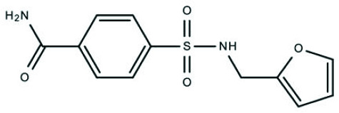 | Z33546257 | - | NA |
CC(C)c1nnc(s1)NC(=O)c1cc2c(nn(c2s1)-c1ccccc1)C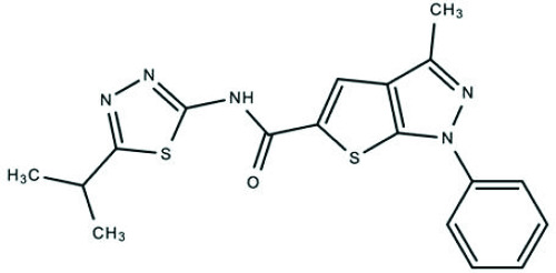 | Z56783036 | - | NA |
CC(C)Cn1nc(c2cc(sc12)C(=O)Nc1cc(ccc1C)S(=O)(=O)C)C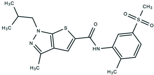 | Z88478561 | - | NA |
Cc1nn(c2sc(cc12)C(=O)Nc1cccc(c1)CO)-c1ccccc1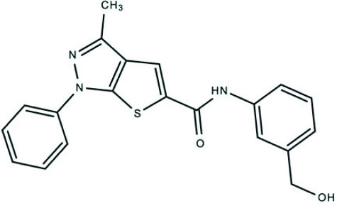 | Z28913272 | + | NA |
Cc1nn(c2sc(cc12)C(=O)Nc1ccncc1)-c1ccccc1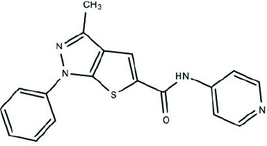 | Z85517350 | ++ | NA |
Cc1nn(c2sc(cc12)C(=O)Nc1ccc(cc1)S(=O)(=O)N)-c1ccc(cc1)Cl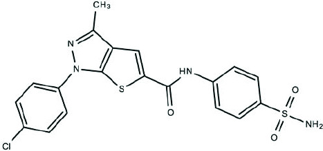 | Z27682945 | + | NA |
COc1ccc(cn1)CNC(=O)c1cc2c(nn(c2s1)-c1ccc(cc1)F)C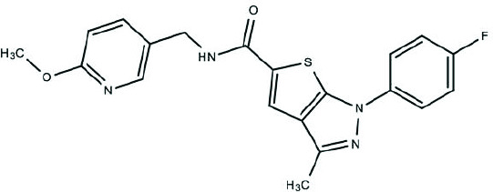 | Z226209130 | - | NA |
Cc1nn(c2sc(cc12)C(=O)Nc1cccnc1)Cc1ccccc1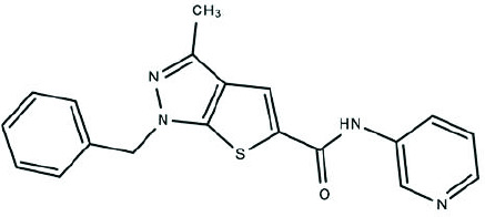 | Z117416778 | + | NA |
Cc1nn(c2sc(cc12)C(=O)Nc1cccc(c1)S(=O)(=O)NCc1ccco1)C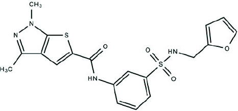 | Z228719934 | - | NA |
Cc1nn(-c2ccccc2)c2sc(C(=O)Nc3ccc4c(c3)OCCO4)cc12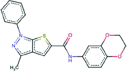 | BCC0077119 | + | + |
O=C(Nc1ccccc1)c1ccc(S(=O)(=O)NCc2ccco2)cc1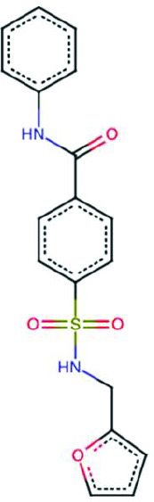 | BCC0112913 | - | NA |
Cc1nn(-c2ccccc2)c2sc(C(=O)Nc3cccc(O)c3)cc12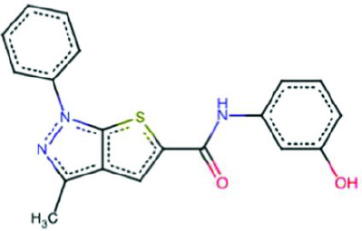 | BCC0106222 | ++ | - |
CC(=O)c1cccc(NC(=O)c2cc3c(C)nn(-c4ccccc4)c3s2)c1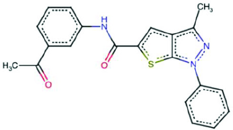 | BCC0104214 | + | NA |
Cc1c2cc(C(=O)NCc3ccco3)sc2nn1-c1ccccc1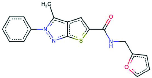 | BCC0040387 | - | NA |
Cc1nn(-c2ccccc2)c2sc(C(=O)Nc3ccccc3Br)cc12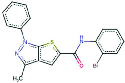 | BCC0113320 | + | + |
Cc1nn(-c2ccccc2)c2sc(C(=O)Nc3nccs3)cc12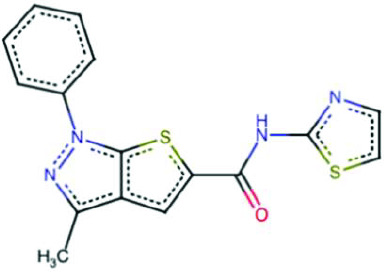 | BCC0073142 | - | NA |
Cc1nn(-c2ccccc2)c2sc(C(=O)Nc3cccc(C(F)(F)F)c3)cc12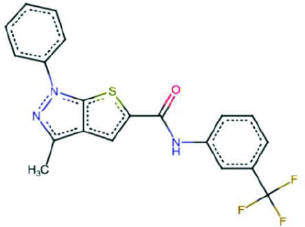 | BCC0043997 | - | NA |
Cc1nn(-c2ccccc2)c2sc(C(=O)Nc3ccccc3[N+](=O)[O-])cc12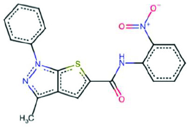 | BCC0073242 | - | NA |
Cc1nn(-c2ccccc2)c(Cl)c1C(=O)NCc1ccco1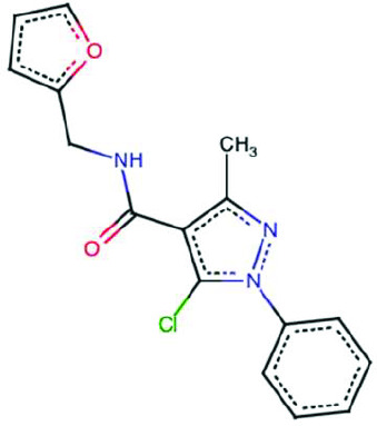 | BCC0104192 | - | NA |
Cc1nn(-c2ccccc2)c2sc(C(=O)NN3C(=O)c4ccccc4C3=O)cc12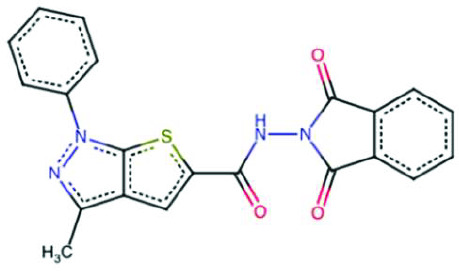 | BCC0074259 | - | NA |
| Structure of compound 21 analogue and fragment | ID | ISR activation | GCN2 specificity |
CC(C)(CNC(=O)c1ccc(cc1)S(=O)(=O)C)c1ccc(cc1)Br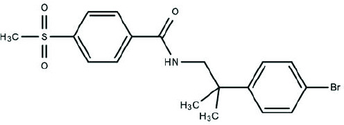 | Z423087112 | - | NA |
Cc1ccc(cc1)-c1nnc([nH]1)NC(=O)c1cccc(c1)S(=O)(=O)C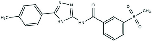 | Z119766318 | - | NA |
COc1cc(cc(c1)-c1cnc2c(c(nn2c1)C)S(=O)(=O)N)OC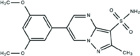 | Z3464816516 | - | NA |
COc1ccc(cc1OC)-c1cnc2c(c(nn2c1)C)S(=O)(=O)N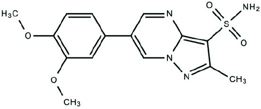 | Z3464816527 | - | NA |
| Structure of compound 20 analogue and fragment | ID | ISR activation | GCN2 specificity |
Oc1ccc(c(c1)O)-c1csc(n1)Nc1ccccc1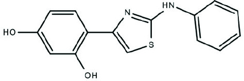 | Z48853885 | - | NA |
COc1ccc(cc1)Nc1nc(cs1)-c1ccc(s1)CNC(=O)C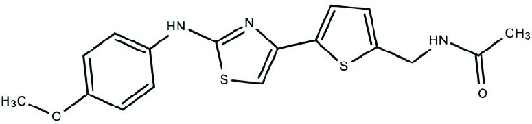 | Z73485298 | - | NA |
CC(=O)NCc1ccc(s1)-c1csc(n1)Nc1ccc(cc1)F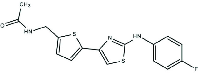 | Z118606822 | - | NA |
CCOc1ccc(cc1)Nc1nc(cs1)-c1cnc(c1)C(=O)N1CCCC1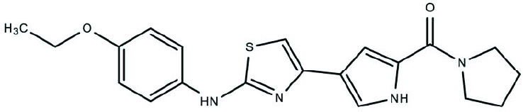 | Z134826202 | ++ | NA |
FC(F)(F)c1ccccc1Nc1nc(cs1)-c1cnc(c1)C(=O)N1CCCC1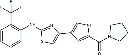 | Z134826094 | + | NA |
COc1cccc(c1)Nc1nc(cs1)-c1cnc(c1)C(=O)N1CCCC1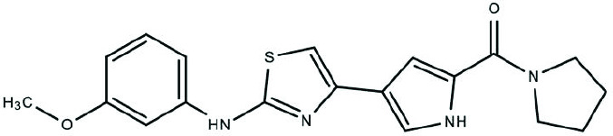 | Z134826982 | ++ | NA |
Oc1ccc(c(c1)O)-c1csc(n1)Nc1ccccn1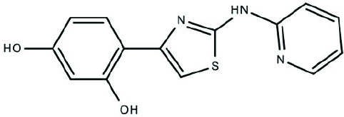 | Z238766124 | + | NA |
COc1ccccc1Nc1nc(cs1)-c1ccc(o1)CNC(=O)C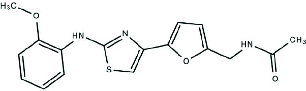 | Z48856426 | - | NA |
CC(=O)NCc1ccc(o1)-c1csc(n1)Nc1cc(cc(c1)C)C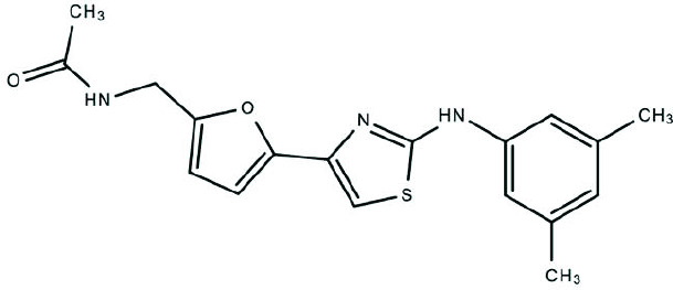 | Z48859481 | - | NA |
CC(=O)NCc1ccc(o1)-c1csc(n1)Nc1ccc(c(c1)Cl)F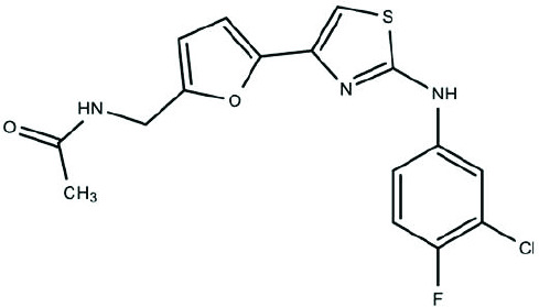 | Z48857131 | - | NA |
COc1ccc(cc1)Nc1nc(cs1)-c1cc(n(c1C)-c1nnc[nH]1)C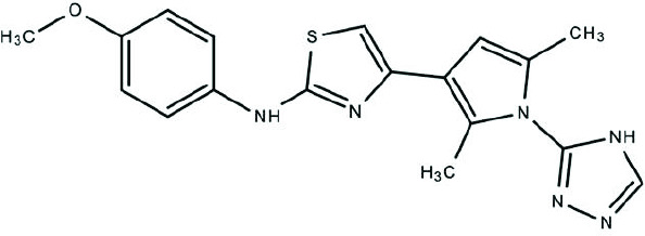 | Z204017924 | - | NA |
Cc1cc(c(n1-c1nnc[nH]1)C)-c1csc(n1)Nc1ccccc1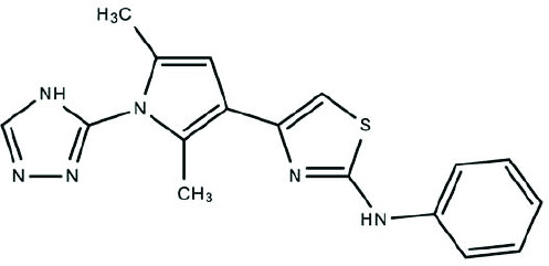 | Z204003168 | - | NA |
CN1C(=C(C(=O)N(C)C1=O)c1csc(n1)Nc1ccc(cc1)F)N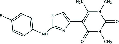 | Z118606410 | - | NA |
CN1C(=C(C(=O)N(C)C1=O)c1csc(n1)Nc1ccccc1)N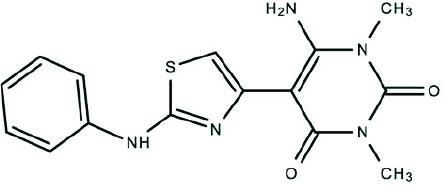 | Z94733799 | - | NA |
Br.CC(=O)Nc1nc(c(s1)-c1csc(n1)Nc1ccccc1)C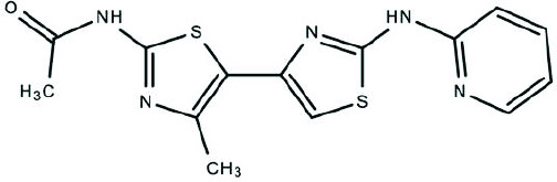 | Z2854692688 | - | NA |
Br.CC(=O)Nc1nc(c(s1)-c1csc(n1)Nc1ccccn1)C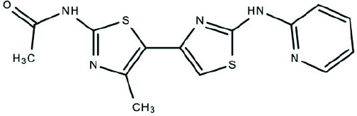 | Z2239043599 | - | NA |
Cc1cc(c(n1CC(F)(F)F)C)-c1csc(n1)Nc1ccccn1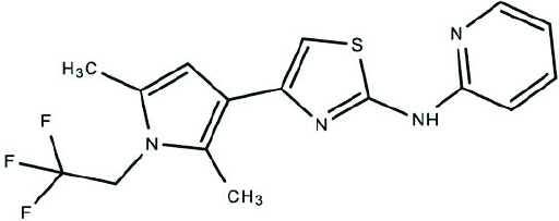 | Z238766162 | + | NA |
Cl.Cc1cc(c([nH]1)C)-c1csc(n1)Nc1ccccn1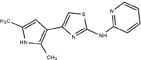 | Z381026350 | - | NA |
Cl.Cc1cc(c(n1C1CC1)C)-c1csc(n1)Nc1ccccn1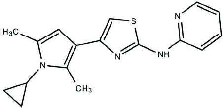 | Z357935644 | - | NA |
Cl.Cc1cc(c(n1C)C)-c1csc(n1)Nc1ccccn1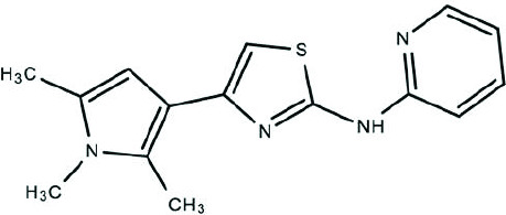 | Z381543540 | - | NA |
Cc1nccc(n1)NCCCc1nc(cs1)-c1ccccn1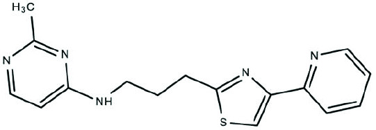 | Z2582963583 | - | NA |
Cc1nc(C)c(-c2csc(Nc3ncccn3)n2)s1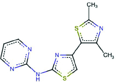 | BCC0083926 | - | NA |
Oc1ccc(-c2csc(Nc3ncccn3)n2)cc1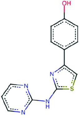 | BCC0089540 | + | + |
Cc1nc(N)sc1-c1csc(Nc2ccc([N+](=O)[O-])cc2)n1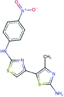 | BCC0084097 | ++ | - |
Cc1cc(-c2csc(Nc3ncccn3)n2)ccc1F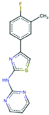 | BCC0080200 | - | NA |
CC(C)c1ccc(-c2csc(Nc3ncccn3)n2)cc1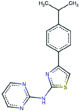 | BCC0080152 | - | NA |
Cc1cccnc1Nc1nc(-c2sc(N)nc2C)cs1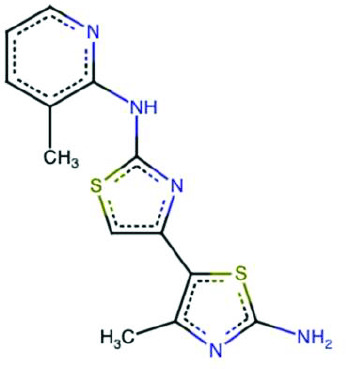 | BCC0084738 | + | + |
Cc1cccc(Nc2nc(-c3sc(N)nc3C)cs2)n1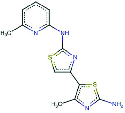 | BCC0084842 | - | NA |
Clc1ccc(-c2csc(Nc3ncccn3)n2)cc1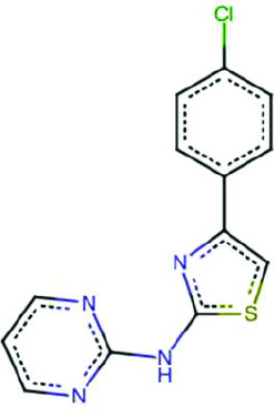 | BCC0075509 | - | NA |
COc1ccc(-c2csc(Nc3ncccn3)n2)cc1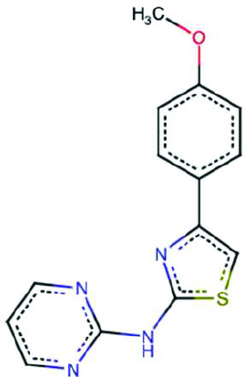 | BCC0057431 | - | NA |
Cc1ccnc(Nc2nc(-c3sc(N)nc3C)cs2)c1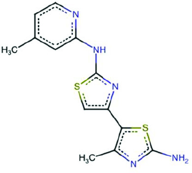 | BCC0083923 | + | + |
Cc1nc(N)sc1-c1csc(Nc2cccnc2)n1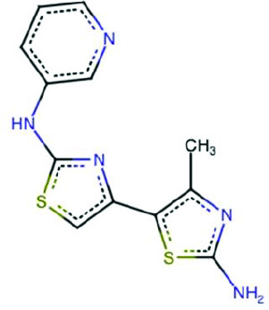 | BCC0084758 | - | NA |
Cc1nc(N)sc1-c1csc(Nc2ccccc2F)n1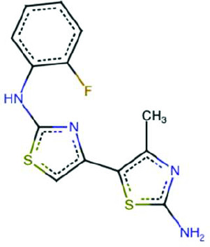 | BCC0084762 | + | - |
Cc1nc(N)sc1-c1csc(Nc2cccc(O)c2)n1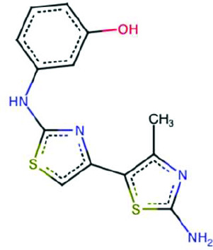 | BCC0105427 | - | NA |
Cc1ccc(C)c(Nc2nc(-c3sc(N)nc3C)cs2)c1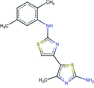 | BCC0084833 | - | NA |
Cc1nc(N)sc1-c1csc(Nc2cccc(C(=O)O)c2)n1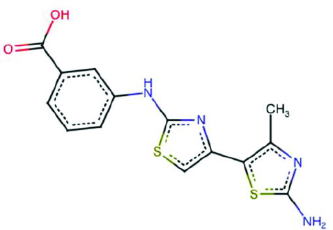 | BCC0057014 | - | NA |
Cc1cc(O)ccc1Nc1nc(-c2sc(N)nc2C)cs1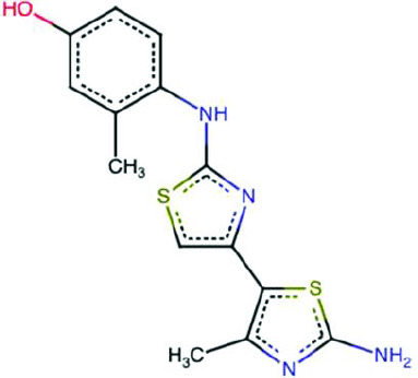 | BCC0084814 | - | NA |
CCOc1ccc(Nc2nc(-c3sc(N)nc3C)cs2)cc1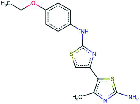 | BCC0087543 | + | - |
Cc1nc(N)sc1-c1csc(Nc2ccccc2[N+](=O)[O-])n1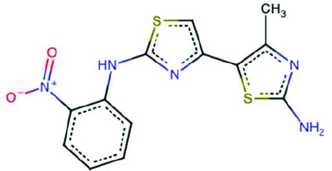 | BCC0084744 | - | NA |
Cc1nc(C)c(-c2csc(Nc3ccccn3)n2)s1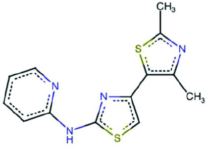 | BCC0079350 | ++ | - |
Cc1nc(N)sc1-c1csc(NCCc2ccccc2)n1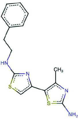 | BCC0089615 | - | NA |
c1cnc(Nc2nc(-c3cccs3)cs2)nc1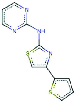 | BCC0079876 | - | NA |
Cc1ccc(C)c(-c2csc(Nc3ncccn3)n2)c1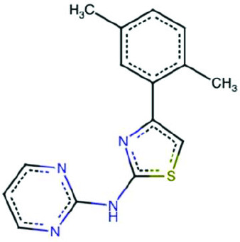 | BCC0080199 | - | NA |
Cc1nc(N)sc1-c1csc(N/N=C/c2cccnc2)n1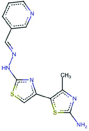 | BCC0041121 | - | NA |
Cc1nc(N)sc1-c1csc(Nc2ccc(O)c(C(=O)O)c2)n1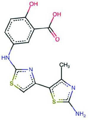 | BCC0089519 | - | NA |
Fc1ccc(-c2csc(Nc3ncccn3)n2)cc1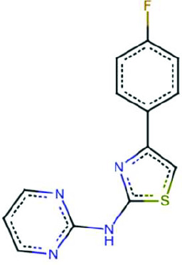 | BCC0084830 | - | NA |
Cc1cc(C)nc(NS(=O)(=O)c2ccc(Nc3nc(-c4sc(N)nc4C)cs3)cc2)n1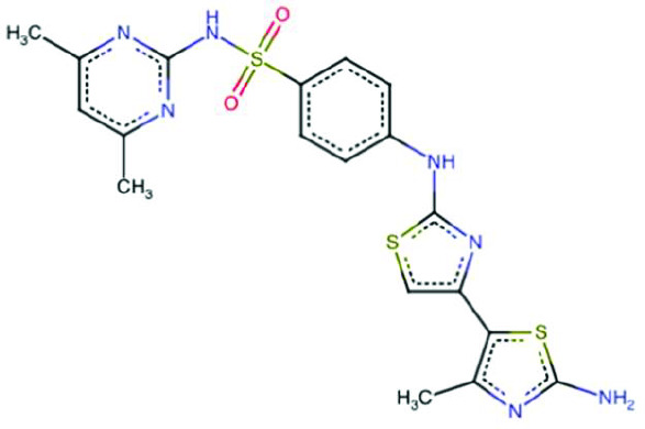 | BCC0087668 | - | NA |
-
NA = not applicable; assay not performed due to the inactivity of the analogue or fragment.
Additional files
Download links
A two-part list of links to download the article, or parts of the article, in various formats.
Downloads (link to download the article as PDF)
Open citations (links to open the citations from this article in various online reference manager services)
Cite this article (links to download the citations from this article in formats compatible with various reference manager tools)
A GCN1-independent activator of the kinase GCN2
eLife 14:RP108590.
https://doi.org/10.7554/eLife.108590.3






