The Ionotropic Receptors IR21a and IR25a mediate cool sensing in Drosophila
- Brandeis University, United States
- Harvard University, United States
- University of Miami, United States
- University of Lausanne, Switzerland
Figures
Figure 1 with 2 supplements

Dorsal Organ Cool Cells (DOCCs) express Ir21a-Gal4.
(a) First/second instar larval anterior. Each Dorsal Organ Ganglion (grey) contains three DOCCs (blue). Anterior-Posterior axis denoted by double-headed arrow. (b,c) Ir21a-Gal4;UAS-GFP (Ir21a>GFP) labels larval DOCCs. Arrows denote cell bodies and arrowheads dendritic bulbs. (d) Temperature responses of Ir21a-Gal4;UAS-GCaMP6m-labeled DOCCs. Left panels, raw images; right panels, colors reflect fluorescence intensity. Arrows denote cell bodies. (e) Fluorescence quantified as percent change in fluorescence intensity compared to minimum intensity. n=22 cells (from 6 animals). (f,g) Temperature-responses of Ir21a-Gal4;R11F02-Gal4;UAS-GCaMP6m-labeled DOCCs. n=26 (7). Scale bars, 10 microns. Traces, average +/- SEM. Figure 1—figure supplement 1 provides an example of the 3-D imaging stacks used for calcium imaging data acquisition.
Figure 1—figure supplement 1
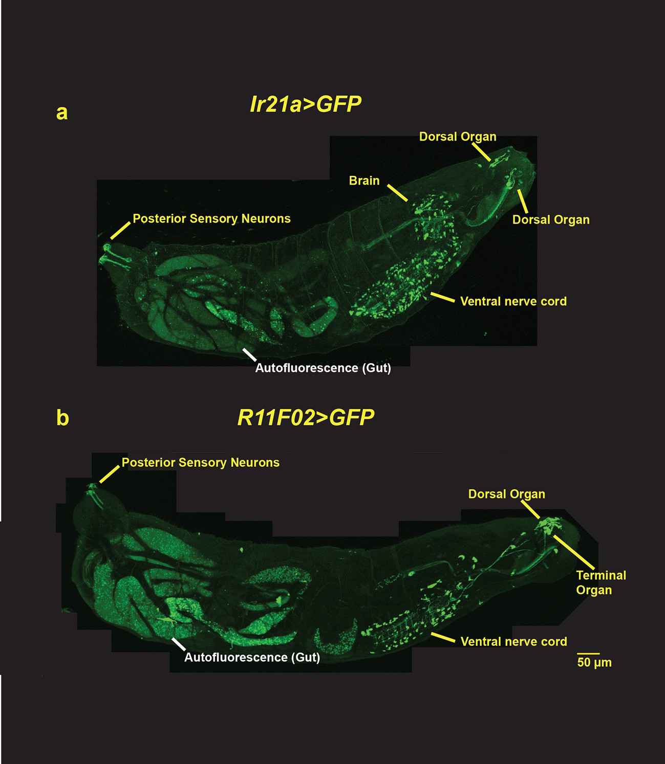
Larval-wide expression patterns of Ir21a-Gal4 and R11F02-Gal4.
(a) Ir21a-Gal4; UAS-GFP expression. (b) R11F02-Gal4; UAS-GFP expression. In addition to expression in the Dorsal Organ, both Gal4s exhibit expression in ~100 cells in the brain and ventral ganglion, neurons along the larval body wall and in the tail. R11F02-Gal4 is also expressed by sensory neurons in the Terminal Organ.
Figure 1—figure supplement 2
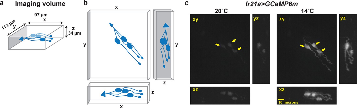
Calcium-imaging data are obtained as a three-dimensional imaging stack.
(a) Dimensions of imaging volume. DOCCs depicted in blue. (b) Maximum intensity projections used for visualizing fluorescence intensity. (c) Representative image of maximum intensity projections of Ir21a>GCaMP6m-labeled DOCCs. DOCC cell bodies remain within imaging field throughout.
Figure 2 with 1 supplement
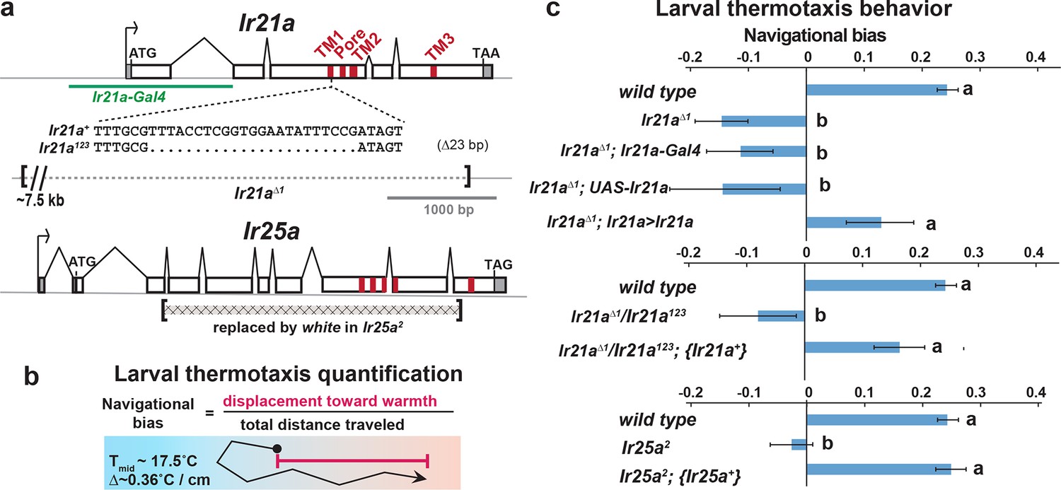
Larval cool avoidance requires Ir21a and Ir25a.
(a) Sequence alterations in Ir21a and Ir25a alleles. Ir21a regulatory sequences present in Ir21a-Gal4 are denoted in green and regions encoding transmembrane domains (TMs) and pore region in red. Additional details provided in Figure 2—figure supplement 1. (b) Thermotaxis is quantified as navigational bias. Cool avoidance behavior was assessed by tracking larval trajectories on a ~0.36˚C/cm gradient extending from ~13.5˚C to ~21.5˚C, with a midpoint of ~17.5˚C. (c) Cool avoidance requires Ir21a and Ir25a. Ir21a>Ir21a denotes a wild type Ir21a transcript expressed under Ir21a-Gal4 control. {Ir21a+} and {Ir25a+} denote wild type genomic rescue transgenes. Letters denote statistically distinct categories (alpha=0.05; Tukey HSD). wild type, n=836 animals. Ir21a∆1, n=74. Ir21a∆1;Ir21a-Gal4, n=48. Ir21a∆1;UAS-Ir21a, n=10. Ir21a∆1;Ir21a>Ir21a, n= 88. Ir21a∆1/ Ir21a123, n=71; Ir21a∆1/ Ir21a123; {Ir21a+} n=70; Ir25a2, n =100. Ir25a2; {Ir25a+} n= 247. Additional mutant analyses provided in Figure 2—figure supplement 1.
Figure 2—figure supplement 1
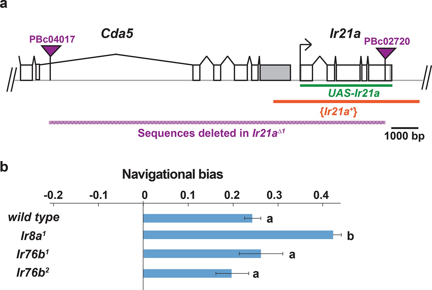
Structure of Ir21a locus and analysis of thermotaxis in Ir8a and Ir76b mutants.
(a) Cda5/Ir21a genomic region, denoting positions of the FRT-containing transposon insertions used to generate Ir21a∆1 (PBc04017 and PBc02720), the sequences deleted in Ir21a∆1, the Ir21a sequences present in the UAS-Ir21a rescue construct and the sequences present in the {Ir21+} genomic rescue construct. Untranslated regions are in gray. (b) Larval thermotaxis of Ir8a and Ir76b mutants quantified as navigational bias. Neither Ir8a nor Ir76b is required for cool avoidance; Ir8a mutants show enhanced cool avoidance compared to wild type. Letters denote statistically distinct categories (alpha=0.05; Tukey HSD). wild type, n=836 animals. Ir8a, n=166; Ir76b1, n=96, Ir76b2, n= 100.
Figure 3
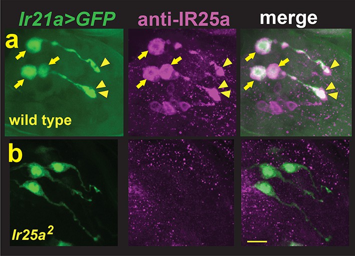
DOCCs express IR25a.
(a) Left panel, Ir21a>GFP-labeled DOCCs. Middle panel, IR25a protein expression in dorsal organ. Right panel, Ir21a>GFP-labeled DOCCs express IR25a protein. Arrows denote DOCC cell bodies and arrowheads DOCC dendritic bulbs. (b) IR25a immunostaining is not detected in Ir25a2 null mutants. Scale bar, 10 microns.
Figure 4 with 1 supplement
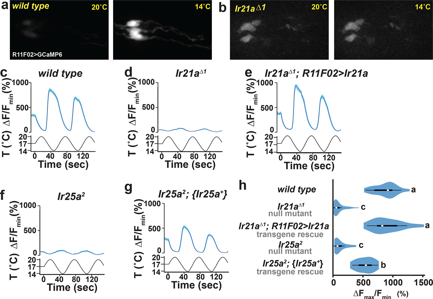
DOCC cool responses require Ir21a and Ir25a.
DOCC responses monitored using R11F02>GCaMP6m. DOCCs exhibit robust cool-responsive increases in fluorescence (a,c), which are dramatically reduced in Ir21a (b,d) and Ir25a (f) mutants. (e) Ir21a transcript expression under R11F02-Gal4 control rescues the Ir21a mutant defect. (g) Introduction of an Ir25a genomic rescue transgene rescues the Ir25a mutant defect. (h) Ratio of fluorescence at 14˚C versus 20˚C depicted using a violin plot. Letters denote statistically distinct categories, p<0.0001, Steel-Dwass test. Scale bars, 10 microns. Traces, average +/- SEM. wild type, n=33 cells (from 11 animals). Ir21a∆1, n= 58 (14). Ir21a∆1; R11F02>Ir21a, n=32 (9). Ir25a2, n=43 (16). Ir25a2; {Ir25a+}, n=30 (10). Analyses of brv1 and brv2 mutants provided in Figure 4—figure supplement 1.
Figure 4—figure supplement 1
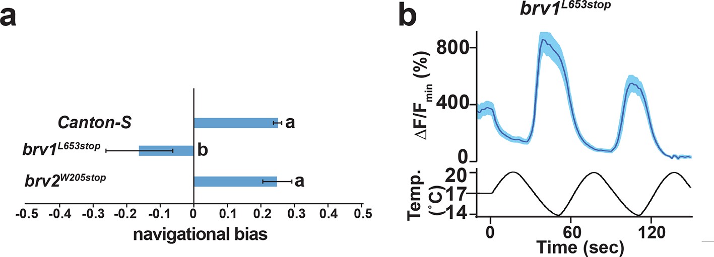
Analysis of putative null mutants of brv1 and brv2.
(a) brv1 but not brv2 mutants exhibit defects in larval cool avoidance. Thermotaxis quantified as navigational bias. Letters denote statistically distinct categories (alpha=0.05; Tukey HSD). wild type, n=836 animals. brv1L653stop, n =43. brv2W205stop, n =99. b) Ir21a>GCaMP6m-labelled DOCCs respond to cooling in brv1L653stop mutants. n= 35 cells (from 6 animals).
Figure 5 with 1 supplement

IR21a expression confers cool-sensitivity upon warmth-responsive Hot Cell neurons.
(a,b) Temperature responses of wild type (a) or N-syb>Ir21a-expressing (b) thermoreceptors in the arista, monitored with N-syb>GCaMP6m. Cell bodies of warmth-responsive Hot Cells outlined in red and cool-responsive Cold Cells in blue. Arrows highlight Hot Cells at 14˚C. Traces of Hot Cell and Cold Cell responses shown at right. Scale bar, 10 microns. (c-e) Fluorescence of Hot Cells in response to sinusoidal 14˚C to 30˚C temperature stimulus, quantified as percent ∆F/Fmin. Dotted lines denote temperature minima. Traces, average +/- SEM. (f) Difference between ∆F/Fmin at 14˚C vs 20˚C (average +/- SEM). Responses of N-syb>Ir21a cells were statistically distinct from both wild type and Ir25a2;N-syb>Ir21a (p<0.01, Steel-Dwass test; letters denote statistically distinct groups). wild type, n= 16 cells (from 8 animals). N-syb>Ir21a, n= 16 (10). Ir25a2; N-syb>Ir21a, n= 20 (10). Analysis of endogenous IR25a expression in the Hot Cells and of the consequences of Hot Cell-specific misexpression of IR21a provided in Figure 5—figure supplement 1.
Figure 5—figure supplement 1
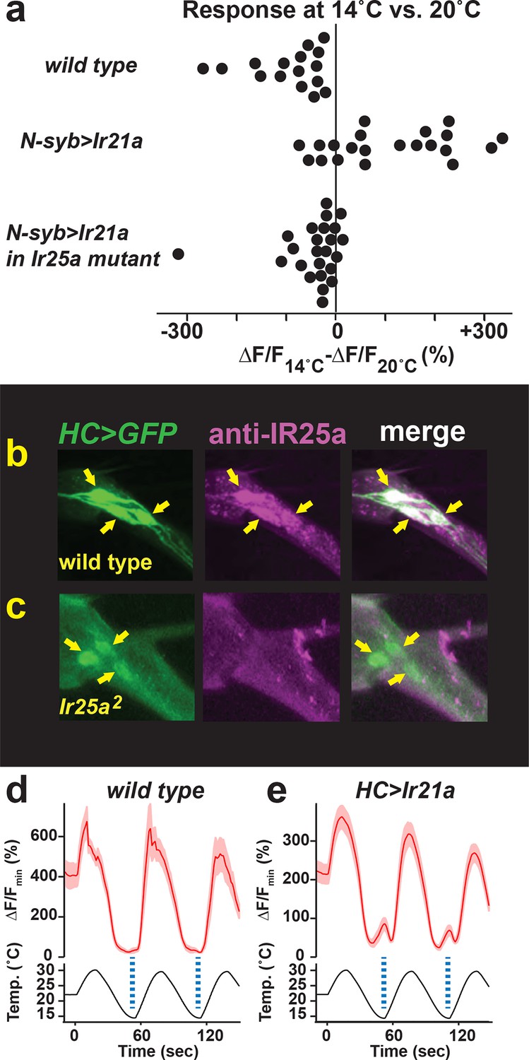
Hot Cell neurons express IR25a protein, and IR21a confers cool-sensitivity upon the Hot Cell neurons.
(a) Difference between ∆F/Fmin at 14˚C vs 20˚C for each cell imaged in Figure 5. (b, c) Left panel, HC>GFP-labeled Hot Cell neurons. Middle panel, IR25a immunostaining. Right panel, HC>GFP and IR25a co-expression. Arrows indicate Hot Cell neuron cell bodies. Specific IR25a immunostaining is absent in Ir25a null mutants (b). (d, e) Temperature responses of wild type (d), HC>Ir21a-expressing (e) thermoreceptors in the adult arista, monitored using HC>GCaMP6m. Dotted lines denote temperature minima. Traces, average +/- SEM. wild type, n=4 cells (2 animals). HC>IR21a n=25 (9).
Figure 6

Hot Cell-specific expression of IR21a confers cool-sensitivity upon Gr28b mutant Hot Cell neurons.
(a-c) Temperature responses of wild type (a), Gr28b mutant (b), and HC>Ir21a-expressing Gr28b mutant (c) thermoreceptors in the adult arista, monitored using HC>GCaMP6m. Dotted lines denote temperature minima. Traces, mean +/- SEM. wild type, n=11 cells (3 animals). Gr28bMi n=9 (3). HC>IR21a; Gr28bMi n=11 (3). (d) Cool responses (∆F/F14˚C - ∆F/F30˚C) of HC>IR21a; Gr28bMicells were distinct from both wild type and Gr28bMi (p<0.01, Steel-Dwass test, letters denote statistically distinct groups).
Download links
A two-part list of links to download the article, or parts of the article, in various formats.
Downloads (link to download the article as PDF)
Open citations (links to open the citations from this article in various online reference manager services)
Cite this article (links to download the citations from this article in formats compatible with various reference manager tools)
The Ionotropic Receptors IR21a and IR25a mediate cool sensing in Drosophila
eLife 5:e13254.
https://doi.org/10.7554/eLife.13254


