Endothelial cell-derived CD95 ligand serves as a chemokine in induction of neutrophil slow rolling and adhesion
- German Cancer Research Center, Germany
- University of Münster, Germany
Figures
Figure 1 with 3 supplements
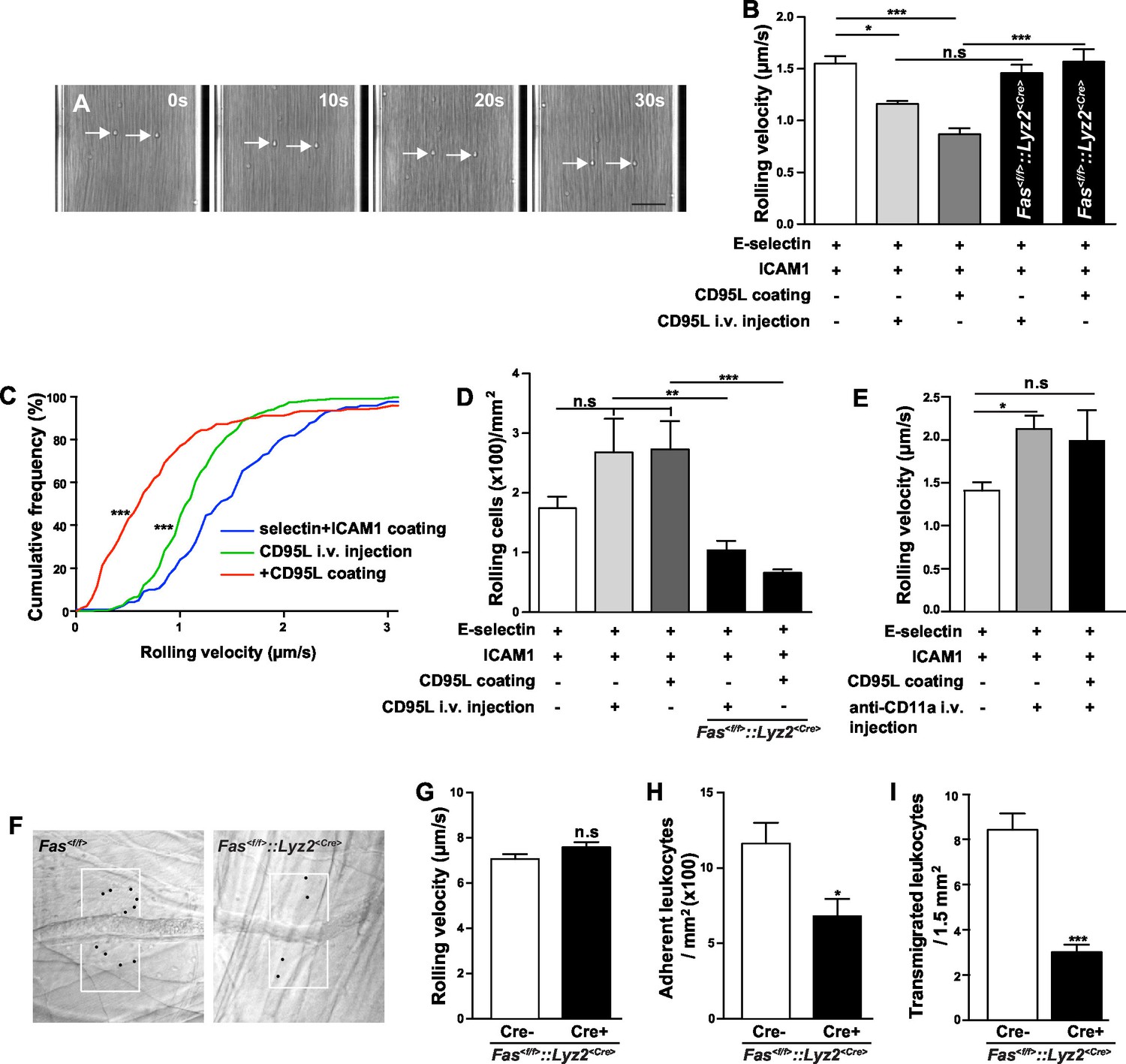
CD95 signaling in myeloid cells is involved in mediating slow rolling, adhesion and transmigration.
(A) Representative time lapse pictures of neutrophil slow rolling in flow chamber. Arrows indicate the rolling cells. Scale bar: 50 μm. (B) Rolling velocity of WT or Fas<f/f>::Lyz2<Cre> neutrophils in flow chambers upon the stimulation of immobilized CD95L or soluble CD95L. Data are presented as mean ± SEM, n=3–4. (C) Cumulative histogram shows the velocity of rolling neutrophils in flow chambers coated with E-selectin/ICAM1, E-selectin/ICAM1/CD95L or E-selectin/ICAM1+soluble CD95L stimulation. (D) Number of WT or Fas<f/f>::Lyz2<Cre> rolling cells in flow chambers upon the stimulation of immobilized CD95L or soluble CD95L. Data are presented as mean ± SEM, n=3–4. (E) Rolling velocity of neutrophils in flow chambers coated with E-selectin/ICAM1 in the presence of immobilized CD95L or anti-CD11a antibody. Data are presented as mean ± SEM, n=3. (F) Representative reflected light oblique transillumination pictures of postcapillary venules of Fas<f/f> and Fas<f/f>::Lyz2<Cre> mice 2 hr after TNF-α application. Demarcations on each side of the venule determine the areas in which extravasated leukocytes were counted. (G–I) Rolling velocity of leukocytes (G) and numbers of adherent leukocytes (H) in the inflamed cremaster muscle venules and numbers of transmigrated leukocytes (I) in inflamed cremaster muscle of Fas<f/f> and Fas<f/f>::Lyz2<Cre> mice. Data are presented as mean ± SEM, n=6. Statistical significance was evaluated by one-way ANOVA followed by Bonferroni multiple comparison post hoc test in (B, C, D, E) (F=13.44, p<0.0001 in B, F=37.37, p<0.0001 in C, F=10.21, p<0.0001 in D, F=4.40, p=0.0135 in E) and two-tailed unpaired Student's t test in (G–I), *p<0.05, **p<0.01, ***p<0.001, n.s not significant.
Figure 1—figure supplement 1
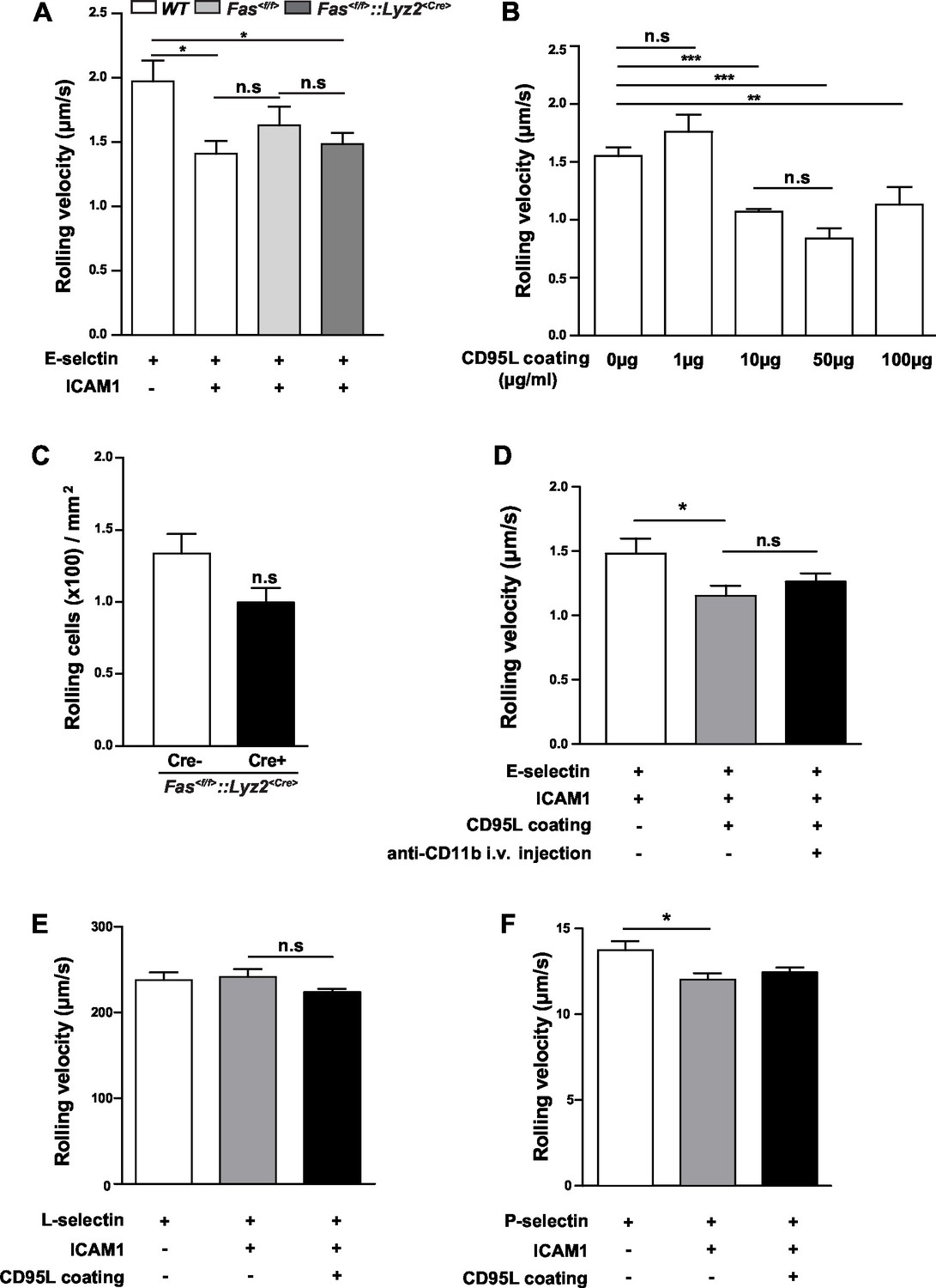
Rolling velocity of WT or Fas<f/f>::Lyz2<Cre> neutrophils in different conditions.
(A) Rolling velocity of neutrophils from WT, Fas<f/f> and Fas<f/f>::Lyz2<Cre> mice in flow chambers coated with E-selectin or E-selectin /ICAM1. n=3. (B) Rolling velocity of neutrophils in flow chambers coated with E-selectin/ICAM1 and different concentration of CD95L. n=3. (C) Number of Fas<f/f>::Lyz2<Cre±> rolling cells in flow chambers coated with E-selectin and ICAM1., n=3. (D) Rolling velocity of neutrophils in flow chambers coated with E-selectin/ICAM1 in the presence of immobilized CD95L or anti-CD11b antibody. n=3–4. (E, F) Rolling velocity of neutrophils in flow chambers coated with L-selectin/ICAM1/CD95L (E) or P-selectin/ICAM1/CD95L (F). n=3–4. Data are presented as mean ± SEM. One-way ANOVA followed by Bonferroni multiple comparison post hoc test in (A, B) (F=3.462, p=0.0161 in A, F=16.23, p<0.0001 in B) and two-tailed unpaired Student's t test in (C), *p<0.05, **p<0.01, ***p<0.001, n.s not significant.
Figure 1—figure supplement 2

TNFRs surface expression level of neutrophils from Fas<f/f> and Fas<f/f>::Lyz2<Cre> mice in homeostasis and inflamed conditions.
(A–B) TNFR1 and TNFR2 surface expression level of neutrophils from Fas<f/f> and Fas<f/f>::Lyz2<Cre> mice in homeostasis. n=6. (C–D) TNFR1 and TNFR2 surface expression level of neutrophils from Fas<f/f> and Fas<f/f>::Lyz2<Cre> mice at 6 hr post CLP. n=6. Data are presented as mean ± SEM, Two-tailed unpaired Student's t test in, *p<0.05.
Figure 1—figure supplement 3

CD95L i.v. injection or deletion of CD95 in myeloid cells doesn’t influence the integrin level in neutrophils.
(A) Flow cytometry plot of blood neutrophils. (B–D) Mice were i.v. injected with saline or CD95L (10 μg). One hour later, blood samples were stained with antibodies of neutrophil markers and integrin subunits and analyzed by flow cytometry. Neutrophils expression levels of integrin αL (B), integrin αM (C) and integrin β2 (D) are presented as mean ± SEM, n=3. (E) Scheme of CD95 deletion in myeloid cells of Fas<f/f>::Lyz2<Cre> mouse line. (F) Blood samples of Fas<f/f> and Fas<f/f>::Lyz2<Cre> mice were stained with antibodies of neutrophil markers and CD95 levels in neutrophils were analyzed by flow cytometry. n=3. (G–J) Blood samples of Fas<f/f> and Fas<f/f>::Lyz2<Cre>mice were stained with antibodies of neutrophil markers and integrin subunits and analyzed by flow cytometry. Neutrophils expression levels of integrin αL (G), integrin αM (H), integrin β2 (I) and CD44 (J) are presented as mean ± SEM, n.s., not significant, n=3. (K–L) Ratio of neutrophils (CD11b+Ly6G+), monocytes (CD11b+CD115+), T cells (CD3) and B cells (CD19) among CD45+ cells in blood of Fas<f/f> and Fas<f/f>::Lyz2<Cre>mice. n=3. (M) Absolute number of neutrophils in blood of Fas<f/f> and Fas<f/f>::Lyz2<Cre>mice. n=6. Data are presented as mean ± SEM, Two-tailed unpaired Student's t test in (C, F, H, I, K, M), *p<0.05, ***p<0.001, n.s not significant.
Figure 2 with 2 supplements

Endothelial cells-derived CD95L is necessary for neutrophil recruitment during inflammation.
(A) Scheme of inducible CD95L deletion in endothelial cells of Fasl<f/f>::Cdh5<CreERT2> mouse line. (B) Representative reflected light oblique transillumination pictures of postcapillary venules of Fasl<f/f> and Fasl<f/f>::Cdh5<CreERT2> mice 2 hr after TNF-α application. Demarcations on each side of the venule determine the areas in which extravasated leukocytes were counted. (C–F) Rolling velocity of leukocytes (C), rolling flux fraction (D) and numbers of adherent leukocytes (E) in inflamed cremaster muscle venules and numbers of transmigrated leukocytes in inflamed cremaster muscle (F) of Fasl<f/f> and Fasl<f/f>::Cdh5<CreERT2> mice. Data are presented as mean ± SEM, n=6. (G) Injection schedule of tamoxifen and thioglycollate is depicted. (H) Flow cytometry plot of peritoneal neutrophils at 6 hr after thioglycollate injection. (I) Influx of peritoneal neutrophils 6 hr after thioglycollate injection in Fasl<f/f> and Fasl<f/f>::Cdh5<CreERT2> mice. n=11–14. (J) Influx of peritoneal neutrophils 6 hr after thioglycollate injection in Fasl<f/f> and Fasl<f/f>::Cdh5<CreERT2> mice. Data in I–J are presented as mean ± SEM and were pooled from two independent experiments, n=16–17. Statistical significance was evaluated by two-tailed unpaired Student's t test in (C–F, I, J), *p<0.05, **p<0.01, ***p<0.001, n.s not significant.
Figure 2—figure supplement 1
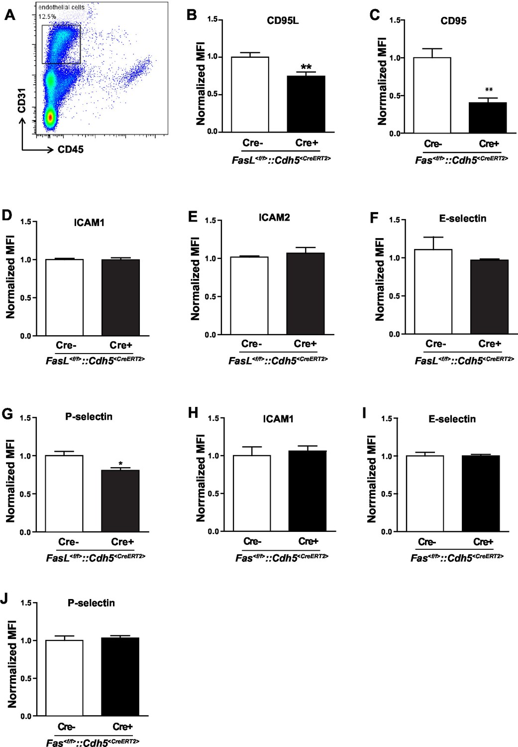
Induced-deletion of CD95L or CD95 has no influence on ICAM and E-selectin level in endothelial cells.
(A) Flow cytometry plot of liver endothelial cells. (B–C) Endothelial cells were dissociated from the liver of Fasl<f/f> and Fasl<f/f>::Cdh5<CreERT2> mice (B) or Fas<f/f> and Fas<f/f>::Cdh5<CreERT2> mice (C) and stained with antibodies of endothelial cell markers and CD95L or CD95. CD95L or CD95 level was analyzed by flow cytometry. (D–G) Endothelial cells of Fasl<f/f> and Fasl<f/f>::Cdh5<CreERT2> mice were stained with antibodies of endothelial cell markers and subtypes of ICAM and selectin. ICAM1 (D), ICAM2 (E), E-selectin (F) and P-selectin (G) levels were analyzed by flow cytometry. (H–J) Endothelial cells of Fas<f/f> and Fas<f/f>::Cdh5<CreERT2> mice were stained with antibodies of endothelial cell markers, ICAM and selectin. ICAM1 (H), E-selectin (I) and P-selectin (J) levels were analyzed by flow cytometry. Data are presented as mean ± SEM in (B–J) and evaluated by two-tailed unpaired Student's t test in (B, C, G), *p<0.05, **p<0.01, n=4.
Figure 2—figure supplement 2

Characterization of Fasl<f/f>::Cdh5<CreERT2> mice.
(A, B) Percentage of blood neutrophils, monocytes, T cells, B cells, dendritic cells and natural killer cells among CD45+ blood cells in Fasl<f/f> and Fasl<f/f>::Cdh5<CreERT2> mice before tamoxifen induction (A) or 10 days after tamoxifen induction (B). n=6. (C) Absolute number of neutrophils in blood of Fasl<f/f> and Fasl<f/f>::Cdh5<CreERT2> mice. n=6. Data are presented as mean ± SEM, n.s. not significant.
Figure 3
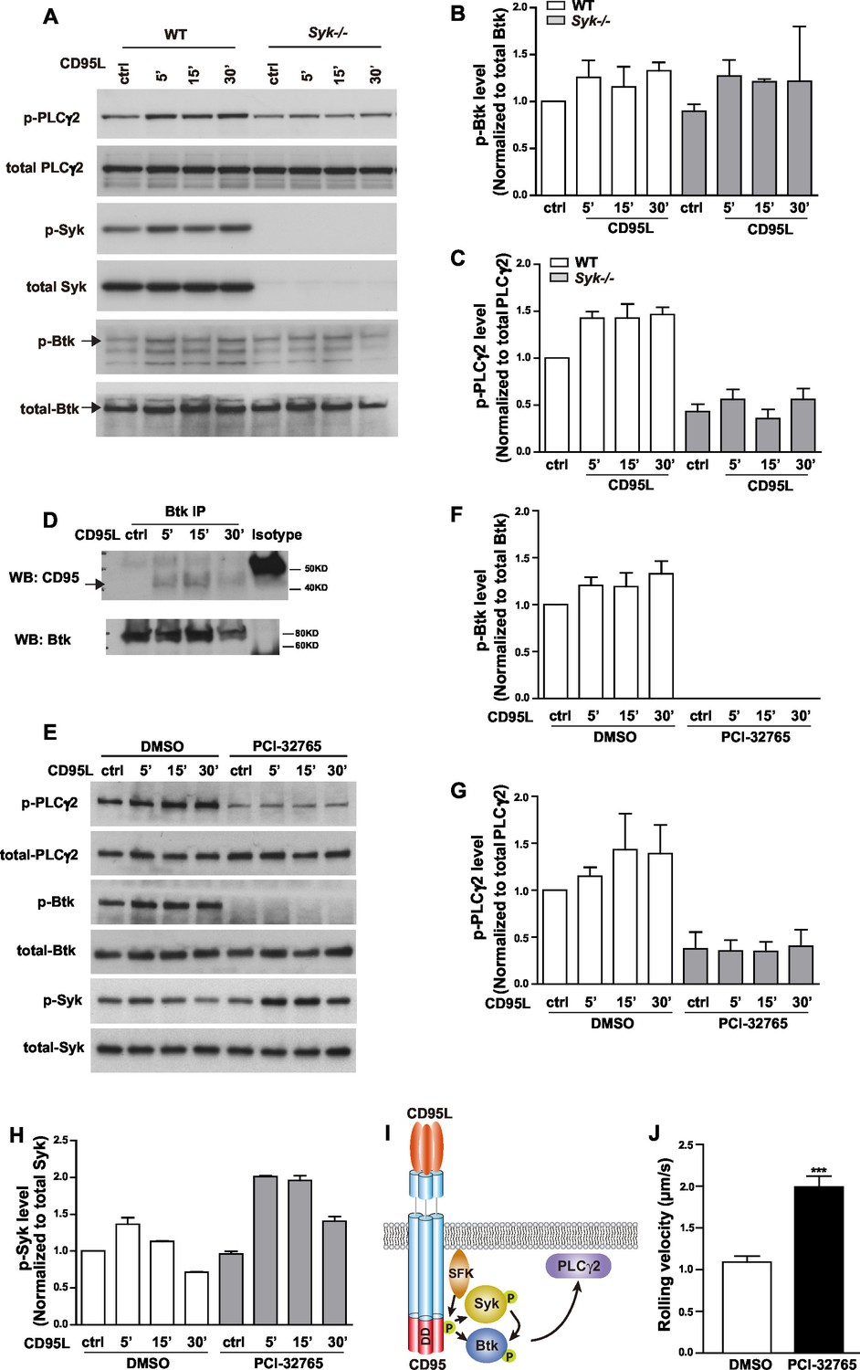
CD95L stimulation induces phosphorylation of PLCγ2 via activating Syk and Btk in myeloid cells.
(A) Macrophages cultured from WT or Syk-/- embryonic liver cells were treated with CD95L (40ng/ml). Lysates were prepared at the indicated time points and immunoblotted for the indicated proteins. (B–C) Quantification analysis of PLCγ2 and Btk phosphorylation level in (A) from three independent experiments. Data are presented as mean ± SEM, n=3. (D) Macrophages cultured from bone marrow cells were treated with CD95L (40 ng/ml). Lysates were prepared at the indicated time and immunoprecipitated with anti-Btk followed by immunoblotting with CD95 and Btk antibody. (E) Macrophages cultured from bone marrow cells were treated with DMSO or Btk inhibitor PCI-32765 (1 µM) one hour prior to CD95L stimulation (40 ng/ml). Lysates were prepared at the indicated time points and immunoblotted for the indicated proteins. (F–H) Quantification analysis of Btk, PLCγ2 and Syk phosphorylation level in (E) from three independent experiments. Data are presented as mean ± SEM, n=3. (I) Scheme of CD95L stimulation-induced PLCγ2 activation. (J) Rolling velocity of neutrophils from DMSO or Btk inhibitor pre-treated mice in a flow chamber coated with E-selectin/ICAM1/CD95L. Data are presented as mean ± SEM, two-tailed unpaired Student's t test, ***p<0.001, n=3.
Figure 4 with 1 supplement
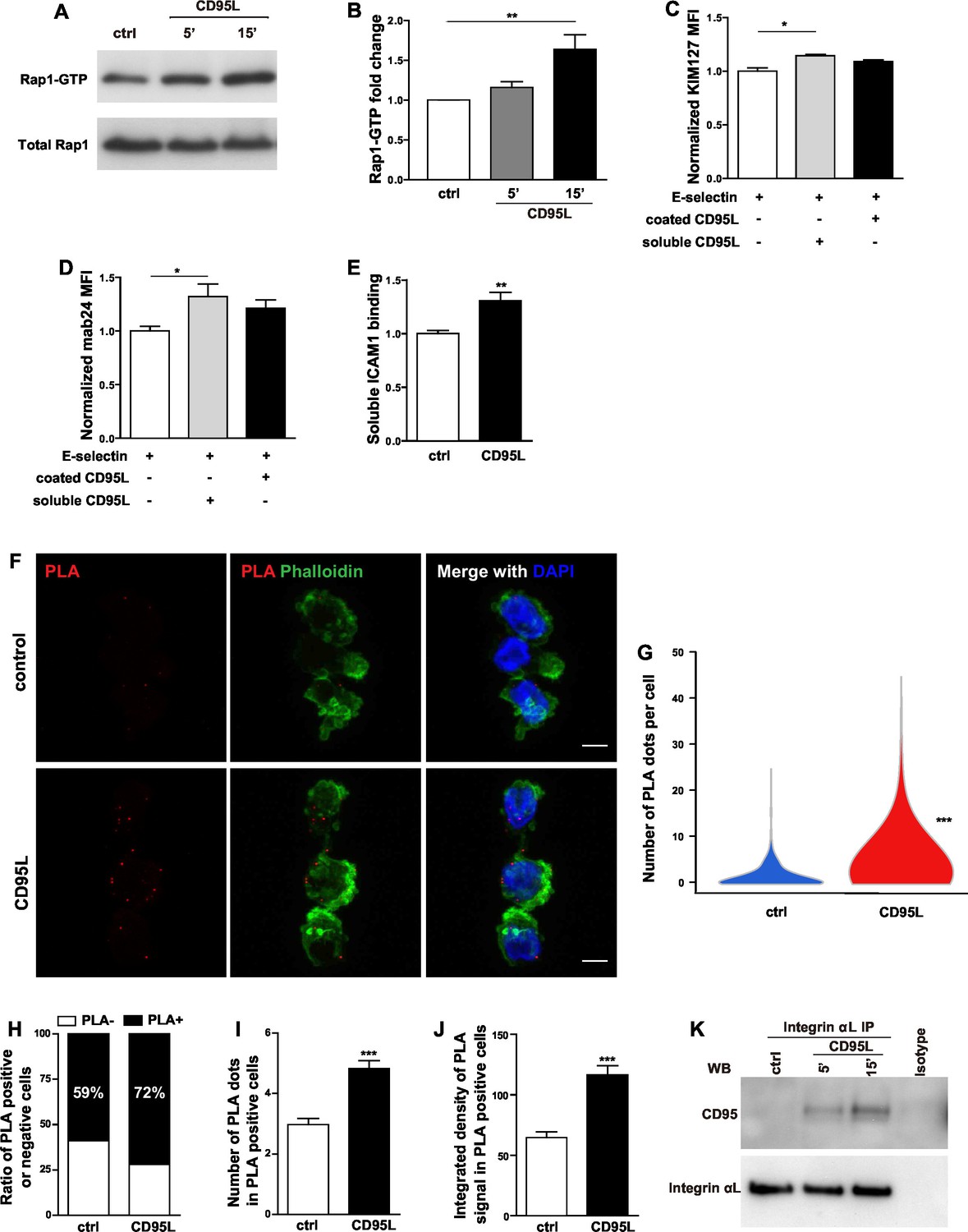
CD95L stimulation induces integrin activation and recruitment of integrin to CD95.
(A) Bone marrow-derived murine neutrophils were treated with CD95L (40 ng/ml). Lysates were prepared at the indicated time points and GST-RalGDS-RBD peptide affinity-precipitated for Rap1 immunoblotting. (B) Quantification analysis of Rap1-GTP activation in (A) from three independent experiments. Data are presented as mean ± SEM, n=3. (C–D) U937 cells were perfused through human E-selectin coated flow chamber in the presence of soluble or immobilized CD95L. The binding of KIM127 (C) or mAb24 (D) were analyzed by flow cytometry and presented as mean ± SEM, n=3. (E) Bone marrow-derived murine neutrophils were treated with coated CD95L for 10 min. The binding of soluble ICAM1 was analyzed by flow cytometry and data presented as mean ± SEM, n=3. (F) Representative pictures show PLA of integrin αL and CD95 in control or CD95L-treated dHL60 cells. Red, PLA; green, Phalloidin; blue, DAPI. Scale bar: 10 µm. (G) The number of PLA signal in each control or CD95L-treated dHL60 cell. Data are presented as violin plot, 383 control cells and 630 CD95L-treated dHL60 cells from 8 random fields were evaluated. (H) Ratio of PLA negative and positive cells in control or CD95L-treated dHL60. Data are presented as stacked bar. (I–J) Number of PLA signal (I) and integrated density (J) of PLA signal in PLA positive cells. Data are presented as mean ± SEM, n=226–453. (K) Bone marrow-derived macrophages were treated with CD95L (40 ng/ml). Lysates were prepared at the indicated time and immunoprecipitated with anti-CD11a followed by immunoblotting for CD95 and CD11a antibody. Statistical significance was evaluated by one-way ANOVA followed by Bonferroni multiple comparison post hoc test in (B–D) and two-tailed unpaired Student's t test in (E, G, I, J), *p<0.05, **p<0.01, ***p<0.001, n.s not significant.
Figure 4—figure supplement 1
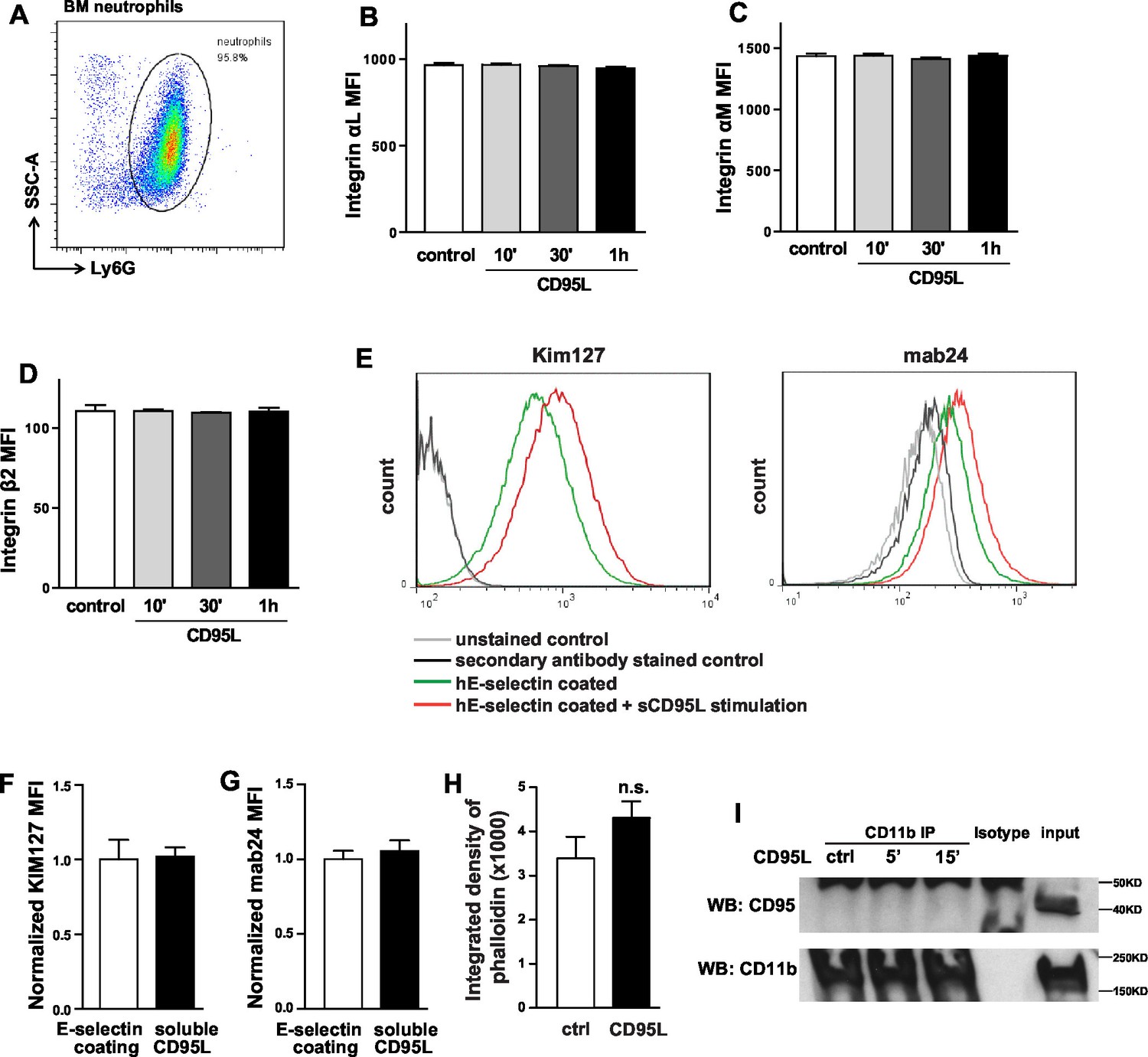
CD95L treatment doesn’t influence the integrin level of neutrophils in vitro.
(A) Flow cytometry plot of percoll isolated-bone marrow neutrophils. (B–D) Bone marrow-derived neutrophils were treated with CD95L (40 ng/ml) and fixed at the indicated time points. Fixed neutrophils were stained with antibodies of neutrophil markers and integrin subunits and analyzed by flow cytometry. Neutrophils expression levels of integrin αL (B), integrin αM (C) and integrin β2 (D) are presented as mean ± SEM, n=3. (E) FACS histogram plots show KIM127 and mAb24 reporter antibodies binding on hE-seletin/CD95L stimulated U937 cells. (F–G) CD95L ligand-treated or non-treated U937 cells were perfused through human E-selectin coated or non-coated flow chamber. The binding of KIM127 (E) or mAb24 (F) were analyzed by flow cytometry and presented as mean ± SEM, n=3. (H) Integrated density of phalloidin in control or CD95L-treated dHL60 cells is presented as mean ± SEM. (I) Bone marrow-derived macrophages were treated with CD95L (40 ng/ml). Lysates were prepared at the indicated time and immunoprecipitated with anti-CD11b followed by immunoblotting for CD95 and CD11b antibody.
Figure 5
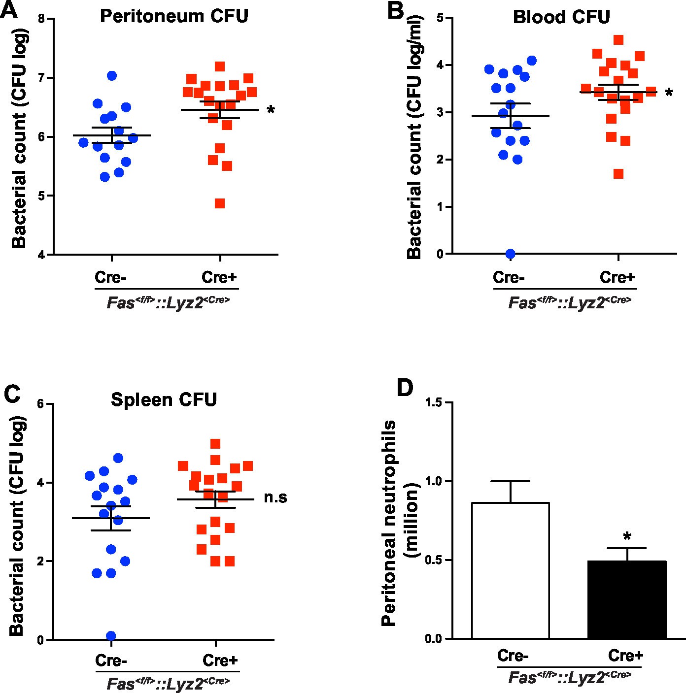
CD95 in myeloid cells is required for bacterial clearance.
(A–C) Bacterial counts of peritoneal lavage fluid (A), blood (B) and spleen (C) from Fas<f/f> or Fas<f/f>::Lyz2<Cre> mice 6 hr after CLP. Data are pooled from three independent experiments and presented as dot plot with mean ± SEM, n=16–19. (D) Infiltrating peritoneal neutrophils 6 hr after CLP in Fas<f/f> or Fas<f/f>::Lyz2<Cre> mice. Data are pooled from three independent experiments and presented as mean ± SEM, n=14–18. Significance between groups was evaluated by running a linear mixed model for the log-CFU with the random covariable of time point and the fixed covariable of gender. *p<0.05, n.s not significant,
Author response image 1

Download links
A two-part list of links to download the article, or parts of the article, in various formats.
Downloads (link to download the article as PDF)
Open citations (links to open the citations from this article in various online reference manager services)
Cite this article (links to download the citations from this article in formats compatible with various reference manager tools)
Endothelial cell-derived CD95 ligand serves as a chemokine in induction of neutrophil slow rolling and adhesion
eLife 5:e18542.
https://doi.org/10.7554/eLife.18542


