Gating of reafference in the external cuneate nucleus during self-generated movements in wake but not sleep
- The University of Iowa, United States
Figures
Figure 1

Forelimb twitches, but not wake movements, trigger neural activity in forelimb sensorimotor cortex.
(A) Representative data depicting sleep and wake behavior, MUA, LFP, and forelimb and nuchal EMG during spontaneous sleep-wake cycling. Red tick marks denote forelimb twitches and red horizontal bars denote forelimb wake movements as scored by the experimenter. (B) Mean (+SEM) rate of spindle burst (n = 6 pups) and unit activity (n = 8 units) during periods of wake and active sleep (AS). The mean rate of spindle bursts and unit activity was significantly higher during active sleep than during wake. * significant difference from active sleep, p<0.01. (C) Waveform average and event correlation for LFP power and unit activity, respectively, in relation to forelimb twitches (2413 and 2943 twitches, respectively). The blue dashed lines denote upper and lower acceptance bands (p<0.01). LFP, Local field potential; MUA, Multiunit activity.
Figure 2
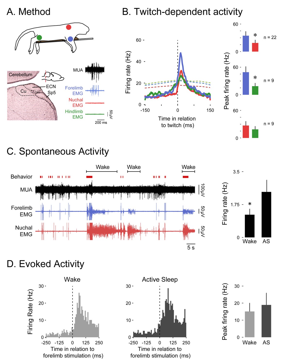
The ECN exhibits wake-dependent inhibition of sensory reafference.
(A) Top: For all ECN recordings, P8-10 rats were instrumented with forelimb (blue) and nuchal (red) EMGs (n = 22). A subset of these rats also had a hindlimb (green) EMG (n = 9). The torso was supported by a platform and the limbs dangled freely. Bottom left: Coronal brain section depicting the anatomical location of the ECN in the hindbrain (inset; red dashed line depicts AP position of the coronal section). Bottom right: Sample record of a burst of ECN reafference in response to forelimb twitches. (B) Left: Event correlations for unit activity in relation to forelimb (blue, n = 8146 twitches), nuchal (red, n = 9603 twitches), and hindlimb (green, n = 1243 twitches) twitches. The colored dashed lines denote upper acceptance bands (p<0.01) for the event correlations. Right: Pairwise comparisons of mean (+SEM) peaks in unit activity (Hz) in response to forelimb, nuchal, and hindlimb twitches. Comparisons are between forelimb and nuchal muscles (top), forelimb and hindlimb muscles (middle), and nuchal and hindlimb muscles (bottom). (C) Left: Representative data depicting sleep and wake behavior, MUA, and forelimb and nuchal EMG during spontaneous sleep-wake cycling. Red tick marks denote forelimb twitches, and red horizontal bars denote forelimb wake movements as scored by the experimenter. Right: Mean (+SEM) unit activity (n = 16) during wake and active sleep (AS) periods. * significant difference from active sleep, p<0.05. (D) Left: Event correlations for evoked unit activity in response to forelimb stimulations performed during active sleep (n = 188 stimulations) and wake (n = 238 stimulations) across 6 ECN units in six pups. Right: Mean (+SEM) peak unit activity (n = 6) derived from event correlations during wake and active sleep. ECN, External cuneate nucleus; MUA, Multiunit activity.
Figure 3
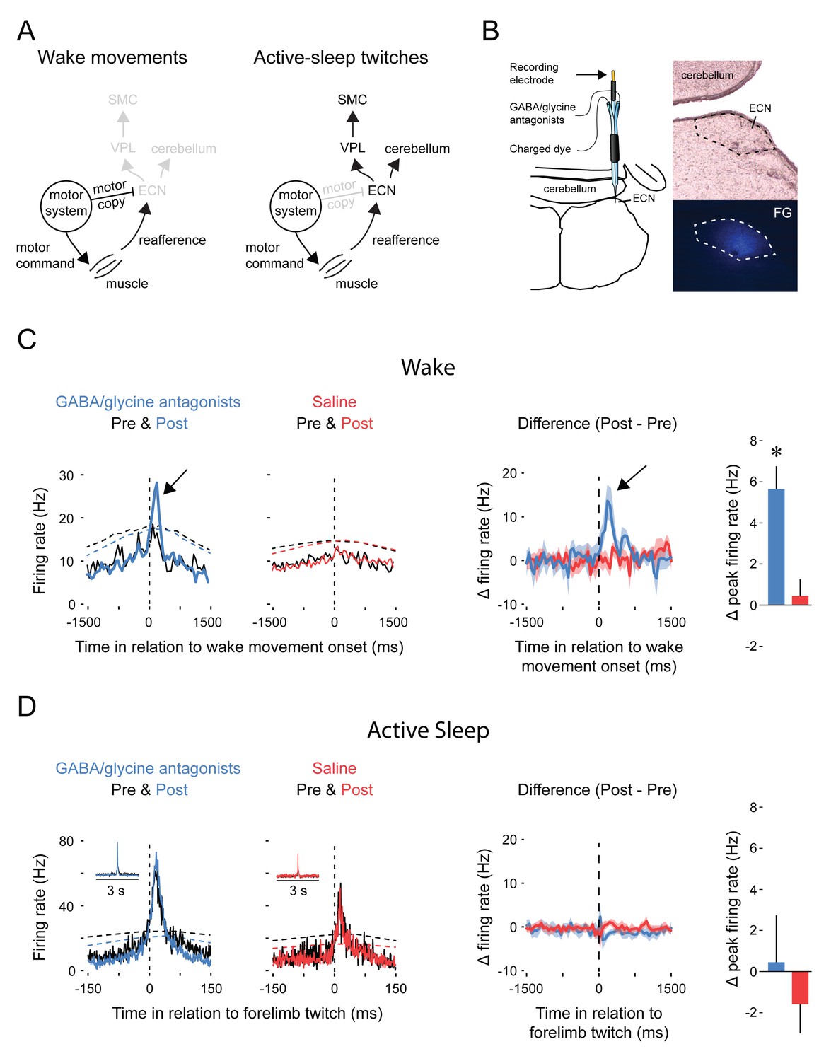
Pharmacological blockade of GABAA and glycine receptors in the ECN specifically unmasks reafference from self-generated wake movements.
(A) Proposed circuitry depicting how reafference arising from self-generated movements is modulated at the level of the ECN. Left: During wake, ECN inputs arising from motor areas convey a corollary discharge (i.e., motor copy) signal that gates expected reafference, resulting in decreased activity in the ECN and downstream sensory areas. Right: During active sleep, the motor copy is absent or inhibited and, therefore, ECN inputs arising from motor areas do not gate reafference, resulting in activity in the ECN and downstream sensory areas. VPL: ventral posterolateral nucleus of thalamus; SMC: primary sensorimotor cortex. (B) ECN recordings were performed using multibarrel electrodes filled either with a GABAA antagonist (10 mM bicuculline methiodide) and a glycine antagonist (10 mM strychnine hydrochloride) or saline. For all animals, a separate barrel of the electrode was filled with fluorogold (FG) to mark the location of the recording site and estimate the spread of the drug (image at bottom right). (C) Left: Event correlations for unit activity in relation to the onset of forelimb wake movements in animals in the GABA/glycine (blue) or saline (red) groups before (Pre) and after (Post) infusion. Data are pooled across all pups. The dashed lines denote upper acceptance bands (p<0.01) for the event correlations. Right: Event correlations depicting changes in unit activity between the pre-infusion and post-infusion periods for the GABA/glycine (blue) and saline (red) groups. Color-coded shaded regions denote +SEM. Histograms depict mean (+SEM) peak changes in unit activity. n = 5 per group. * significant difference from saline, p<0.05. (D) Same as in (C) but during active sleep; event correlations are triggered on forelimb twitches. ECN, External cuneate nucleus.
Figure 4
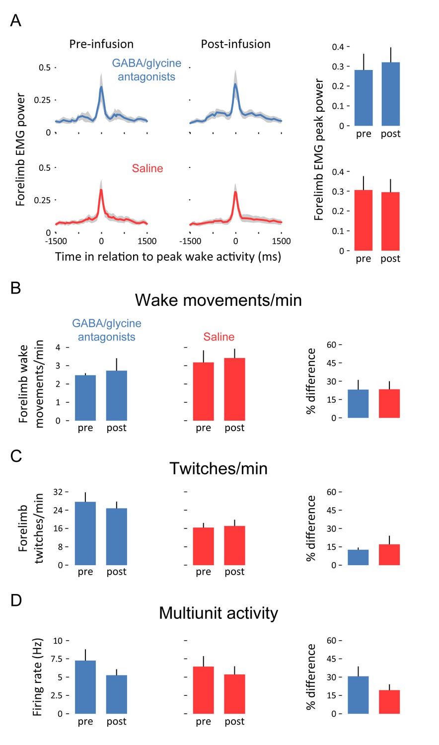
Pharmacological blockade of GABAA and glycine receptors in the ECN does not affect forelimb motor activity or tonic ECN unit activity.
(A) Waveform averages (3-s time windows) depicting forelimb EMG power in relation to peak forelimb wake activity before and after infusion of GABAA and glycine receptor antagonists (blue) or saline (red). Shaded regions denote +SEM. At right, mean (+SEM) forelimb EMG peak power derived from waveform averages. (B) Top row: Mean (+SEM) forelimb wake movements/min before and after infusion of GABAA and glycine receptor antagonists (blue) or saline (red). At right, mean (+SEM) percent difference (pre vs. post) in forelimb wake movements/min for both experimental groups. (C) and (D): Same as in (B) but for twitches/min and unit activity (in Hz), respectively. None of the differences in this figure are significant. ECN, External cuneate nucleus.
Download links
A two-part list of links to download the article, or parts of the article, in various formats.
Downloads (link to download the article as PDF)
Open citations (links to open the citations from this article in various online reference manager services)
Cite this article (links to download the citations from this article in formats compatible with various reference manager tools)
Gating of reafference in the external cuneate nucleus during self-generated movements in wake but not sleep
eLife 5:e18749.
https://doi.org/10.7554/eLife.18749


