Neural circuitry coordinating male copulation
- University of Oxford, United Kingdom
- University of Sheffield, United Kingdom
- National Institute of Mental Health, United States
- McGill University, Canada
Figures
Figure 1 with 1 supplement
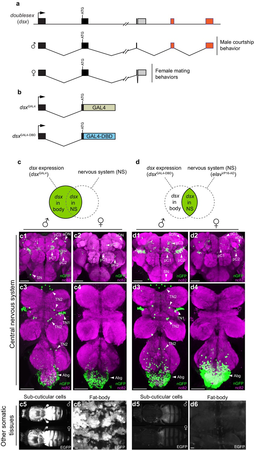
Spatial restriction of GFP expression to dsx neurons using novel dsx Split-GAL4 allele.
(a) Schematic of doublesex (dsx) gene and male and female predicted transcripts. Arrows indicate transcriptional start sites. Colored boxes depict non-sex-specific (black) and sex-specific (red: male and grey: female) exons. (b) Schematic of dsxGAL4 and dsxGAL4-DBD knock-in alleles. (c) GFP expression in five day-old males and females driven by dsxGAL4. (c1–4) dsxGAL4 driving UAS-nuclear GFP (nGFP) in (c1) adult male brain and (c3) VNC and (c2) adult female brain and (c4) VNC. (c5–6) Epifluorescence images of dsxGAL4 driving UAS-2XEGFP (EGFP) in (c5) adult male and female whole-fly preparations revealing EGFP expression in sub- and peri-cuticular cells and (c6) adult male filleted dorsal abdominal wall revealing EGFP expression in the adult fat body. (d) GFP expression in five day-old males and females driven by dsxGAL4-DBD combined with pan-neuronal elavVP16-AD hemidriver. (d1–4) dsxGAL4-DBD/elavVP16-AD (referred to as dsx/elav in text) driving UAS-nGFP in (d1) adult male brain and (d3) VNC and (d2) adult female brain and (d4) VNC. Epifluorescence images of dsxGAL4DBD/elavVP16-AD driving UAS-2XEGFP in (d5) adult male and female whole-fly preparations revealing no EGFP expression in sub- and peri-cuticular cells and (d6) adult male filleted dorsal abdominal wall revealing no EGFP expression in the adult fat body. nGFP realized with anti-GFP antibody (green) and neuropil counterstained with nc82 (magenta). EGFP realized with anti-GFP antibody (white). (c1–4) and (d1–d4) views are ventral, with anterior up. Scale bar = 50 μm.
Figure 1—figure supplement 1
dsx-expressing neurons specify male and female sexual behaviors.
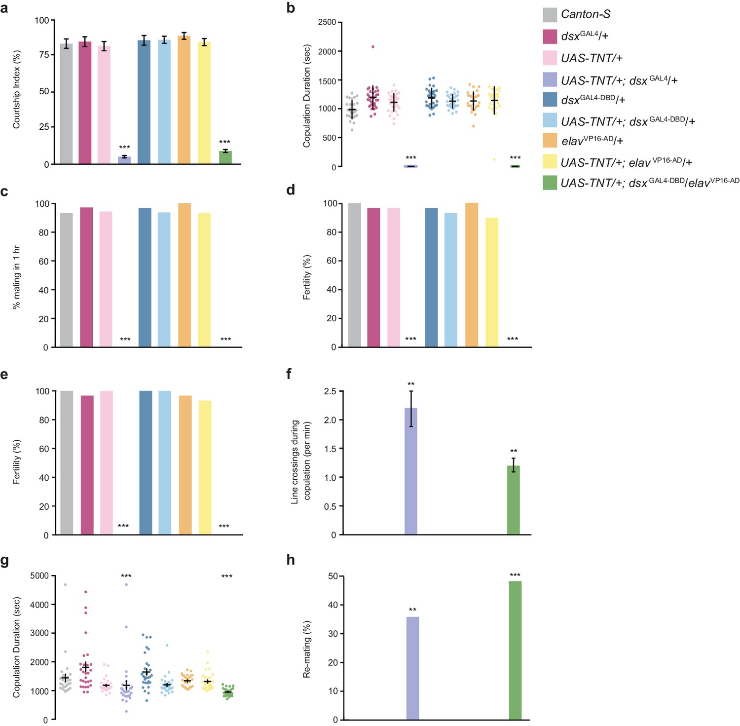
(a-d) Effects of silencing dsx/elav neurons on male courtship and copulatory behaviors. (a) Courtship index (mean ± S.E.M.; n = 30). (b) Copulation duration (mean ± S.E.M. n = 30). (c) Percentage of male matings in 1 hr (n = 30). (d) Male fertility (n = 30). Genotypes indicate males. (e–h) Effects of silencing dsx/elav neurons on female courtship behaviors. (e) Female fertility (n = 30). (f) Line crossings during copulation (mean ± S.E.M.; n = 30). (g) Copulation duration (mean ± S.E.M.; n = 30). (h) Percentage of females that re-mate with the same male in 2 hr (n = 30). Genotypes indicate females. (a–h) **p<0.001, ***p<0.0001 by Fisher exact test (a,b,f,g) or Kruskal-Wallis and Dunn’s test (c,d,e,h).
Figure 2 with 1 supplement
Sexually dimorphic dsx/glutamatergic neurons control genital coupling during copulation.
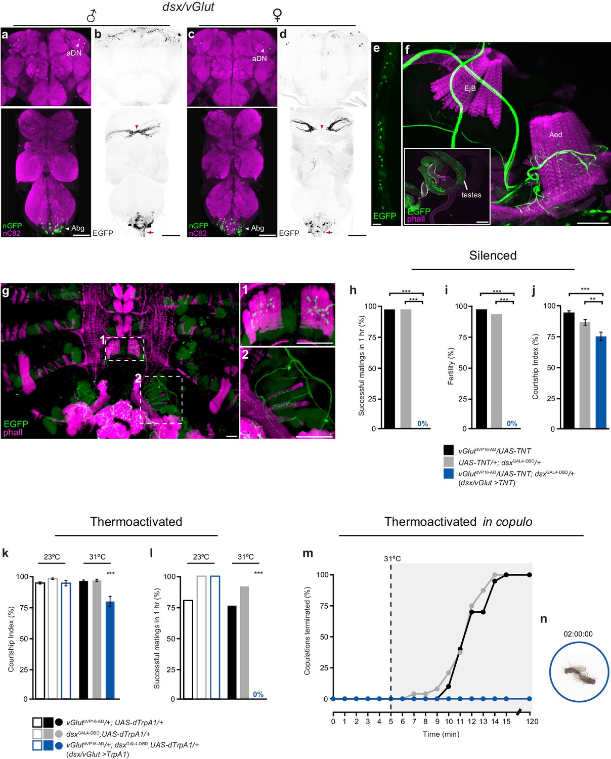
(a-d) Sexually dimorphic expression of dsx/vGlut neurons in the brain (top) and VNC (bottom) of adult males (a,b) and females (c,d). (a,c) dsx/vGlut cell bodies visualized by vGlutdVP16-AD/dsxGAL4-DBD driving UAS-nGFP in (a) male and (c) female CNSs. nGFP stained with anti-GFP (green); neuropil counterstained with anti-nC82 (magenta). (b,d) dsx/vGlut projection patterns visualized by vGlutdVP16-AD/dsxGAL4-DBD driving UAS-2XEGFP in (b) male and (d) female CNSs. EGFP stained with anti-GFP (black); sexually dimorphic midline crossing (red arrowhead) and neurons of the Abg and their descending projections (red arrow) are shown. (e) dsx/vGlut driven EGFP expression in the T1 tarsi of the male foreleg. Projections from these neurons form the male-specific contralateral commissural bridge in the mesothoracic gangion of the male VNC (red arrowheads in bottom panels of b,d). (f,g) dsx/vGlut driven EGFP expression in the (f) internal reproductive system and (g) abdomen reveals motor neuron arborizations onto (f) muscles of the aedeagus and (g) dorsal and ventral muscles of the sixth abdominal segment. (g1-2) Higher magnification of ventral (g1) and dorsal (g2) longitudinal muscles of sixth abdominal segment showing dsx/vGlut motor neuron innervations and synaptic termini. EGFP stained with anti-GFP (green). Internal reproductive system and abdominal muscles counterstained with the F-actin specific antibody Phalloidin (phall; magenta). Detail of internal genitalia: testes, ejaculatory bulb (EjB), and aedeagus (Aed) indicated. Scale bar = 50 μm. (h-j) Effects of silencing dsx/vGlut neurons on male copulatory and courtship behaviors. (h) Percentage of successful matings in 1 hr (n = 24–30). (i) Male fertility (n = 30). (j) Courtship index (mean ± S.E.M.; n = 24–30). Genotypes indicate males. See also Video 1. (k,l) Effects of thermoactivating dsx/vGlut neurons on male courtship and copulatory behaviors. (k) Courtship index (mean ± S.E.M.; n = 20–30). (l) Percentage of successful matings in 1 hr (n = 20–30). Statistical comparisons of the experimental genotype at 31°C in (k-l) were made against the same genotype at 23°C and all control genotypes at 31°C. Genotypes indicate males. (m) Effects of thermoactivating male dsx/vGlut neurons 5 min into copulation. Percentage of copulations terminated over a 2 hr period is graphed (n = 22–24). (n) Video still showing ‘stuck’ dsx/vGlut>TrpA1 male at the end of the 2 hr observation period. See also Video 2. (h-l) **p<0.001, ***p<0.0001 by Fisher exact test (h,i,k) or Kruskal-Wallis and Dunn’s test (j,l).
Figure 2—figure supplement 1
Characterisation of dsx/glutamatergic neurons in the adult male CNS.
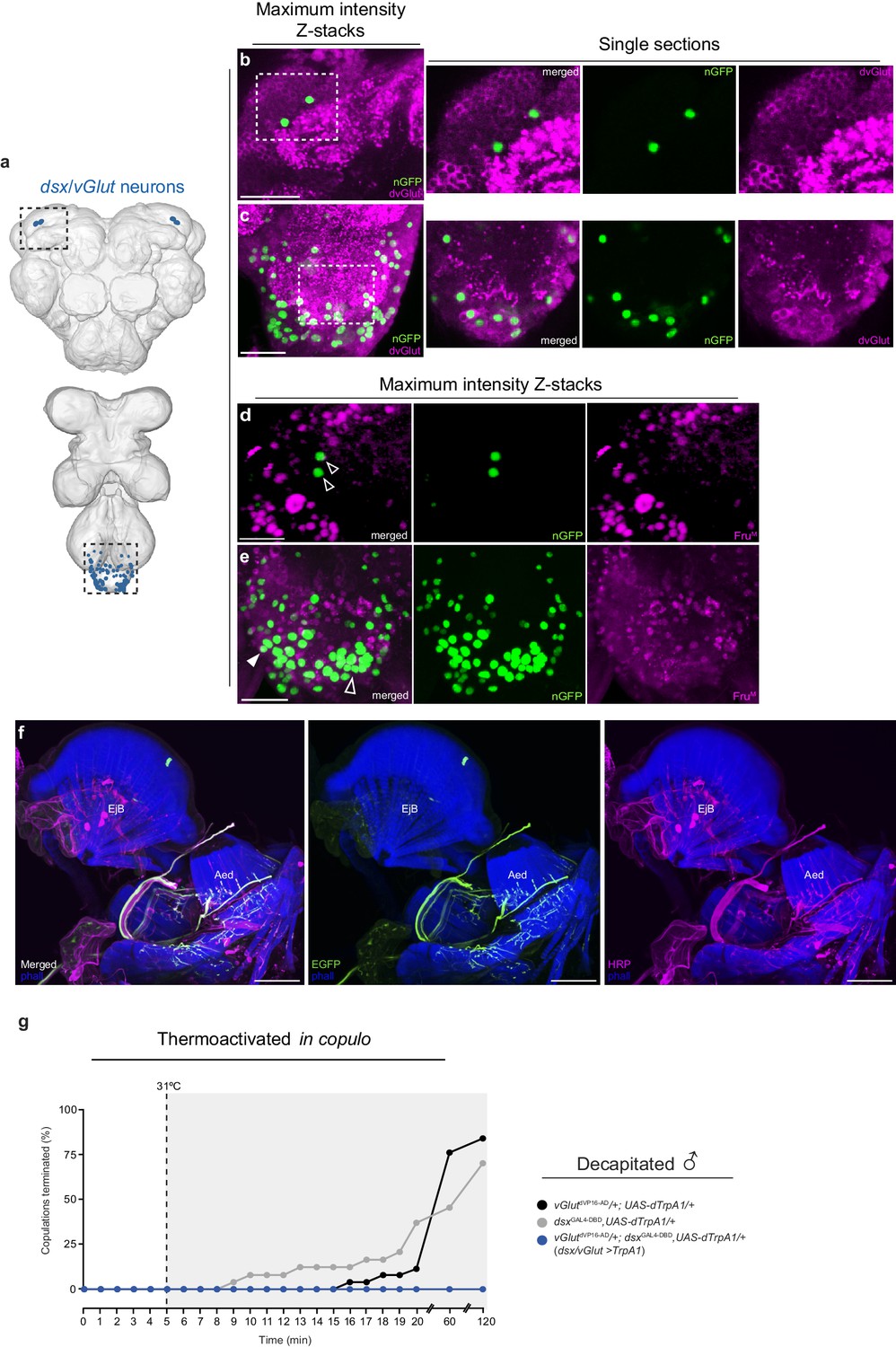
(a) Schematic representation of dsx/vGlut neurons in the adult male CNS. Black dotted boxes depict regions shown in b-e. (b-c) Co-localization of nGFP and anti-dvGlut in dsx/vGlut neurons in the (b) brain and (c) VNC of vGlutVP16-AD/ UAS-pStinger; dsxGAL4-DBD/+ males. Scale bar = 10 μm. Single section view of marked regions in b and c (white dotted box) showing overlap between nGFP and dvGlut antibody at higher optical magnification shown on right of panels b and c. nGFP stained with anti-GFP (green); vGlut stained with anti-dvGlut (magenta). (d-e) Co-localization between nGFP and anti-FruM in dsx/vGlut neurons in the (d) brain and (e) VNC of vGlutVP16-AD/ UAS-pStinger; dsxGAL4-DBD/+ males. Solid arrowheads = co-localization; Empty arrowheads = no co-localization. nGFP stained with anti-GFP (green); FruM stained with anti-FruM (magenta). Scale bar = 10 μm. (f) Co-localisation of dsx/vGlut driven EGFP and anti-HRP in male internal reproductive system. All neuronal innervations are revealed with anti-HRP. Expression of dsx/vGlut>EGFP and anti-HRP shown separately in right panels. EGFP stained with anti-GFP (green). Internal reproductive system and abdominal muscles counterstained with the F-actin specific antibody Phalloidin (phall; blue). Detail of internal genitalia: ejaculatory bulb (EjB), and aedeagus (Aed) indicated. Scale bar = 50 μm. (g) Effects of thermoactivating dsx/vGlut neurons in decapitated males 5 min into copulation. Percentage of copulations terminated over a 2 hr period is graphed (n = 22–26).
Figure 3 with 2 supplements
Sexually dimorphic dsx/GABAergic neurons control genital uncoupling during copulation.
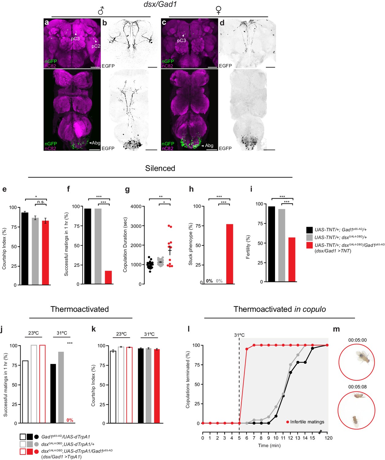
(a-d) Sexually dimorphic expression of intersected dsx/Gad1 neurons in the brain (top) and VNC (bottom) of adult males (a,b) and females (c,d). (a,c) dsx/Gad1 cell bodies visualized by dsxGAL4-DBD/Gad1p65-AD driving UAS-nGFP in (a) male and (c) female CNSs. nGFP stained with anti-GFP (green); neuropil counterstained with anti-nC82 (magenta). (b,d) dsx/Gad1 projection patterns visualized by dsxGAL4-DBD/Gad1p65-AD driving UAS-2XEGFP in (b) male and (d) female CNSs. EGFP stained with anti-GFP (black). Scale bar = 50 μm. (e-i) Effects of silencing dsx/Gad1 neurons on male copulatory and courtship behaviors. (e) Courtship index (mean ± S.E.M.; n = 24–30). (f) Percentage of successful matings in 1 hr (n = 24–30). (g) Copulation duration (n = 12–24). (h) Percentage of males displaying ‘stuck’ phenotype (n = 12–24). (i) Male fertility (n = 30). Genotypes indicate males. See also Video 3. (j,k) Effects of thermoactivating dsx/Gad1 neurons on male courtship and copulatory behaviors. (j) Percentage of successful mating’s in 1 hr (n = 20–30). (k) Courtship index (mean ± S.E.M.; n = 20–30). Statistical comparisons of the experimental genotype at 31°C in (j,k) were made against the same genotype at 23°C and all control genotypes at 31°C. Genotypes indicate males. (l) Effects of thermoactivating male dsx/Gad1 neurons 5 min into copulation. Percentage of copulations terminated over a 2 hr period is graphed (n = 20–24). (m) Video stills showing that activation of dsx/Gad1 >TrpA1 neurons in copulo results in an almost immediate termination of copulation. Top panel shows dsx/Gad1 >TrpA1 male and wild type female mating 5 min into copulation at the point of shifting the temperature to 31°C. Bottom panel shows the same mating pair 8 s later, at which time the male has terminated copulation. See also Video 4. (e-k) n.s. = not significant, *p<0.05, *p<0.05, **p<0.001, ***p<0.0001 by Fisher exact test (f,h-j) or Kruskal-Wallis and Dunn’s test (e,g,k).
Figure 3—figure supplement 1
Characterisation of dsx/GABAergic neurons in the adult male CNS.
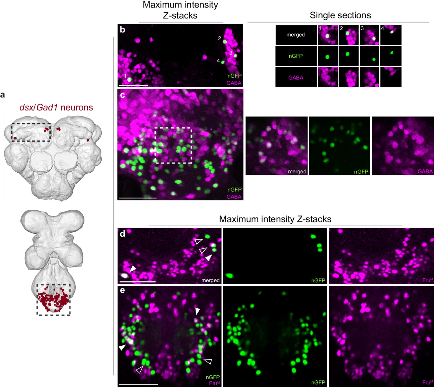
(a) Schematic representation of dsx/Gad1 neurons in the adult male CNS. Black dotted boxes depict regions shown in b-e. (b-c) Co-localization of nGFP and anti-GABA in dsx/Gad1 neurons in the (b) brain and (c) VNC of UAS-pStinger/+; dsxGAL4-DBD/Gad1p65-AD males. Scale bar = 10 μm. Single section view of marked regions in b and c (numbers and white dotted box) showing overlap between nGFP and GABA antibody at higher optical magnification shown to the right of panels b and c. nGFP stained with anti-GFP (green); GABA stained with anti-GABA (magenta). (d-e) Co-localization between nGFP and anti-FruM in dsx/Gad1 neurons in the (d) brain and (e) VNC of UAS-pStinger/+; dsxGAL4-DBD/Gad1p65-AD males. Solid arrowheads = co-localization; Empty arrowheads = no co-localization. nGFP stained with anti-GFP (green); FruM stained with anti-FruM (magenta). Scale bar = 10 μm.
Figure 3—figure supplement 2
dsx/GABAergic neurons in the male brain do not specify male copulatory behaviors, but instead mediate male courtship.
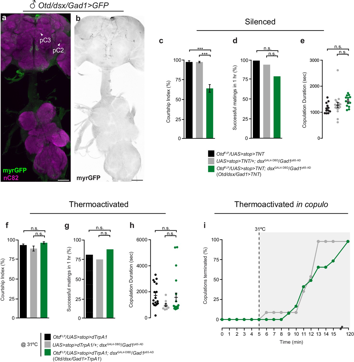
(a-b) Brain-restricted GFP expression in dsx/Gad1 neurons of the adult male CNS. (a) Expression of myrGFP in dsx/Gad1 neurons in the brain and not the VNC of OtdFLP/UAS-myrGFP; dsxGAL4-DBD/Gad1p65-AD males. myrGFP stained with anti-GFP (green); neuropil is counterstained with anti-nC82 (magenta). (b) Projections of a 10 μm subset of (a) showing skeleton expression of myrGFP in dsx/Gad1-brain neurons. myrGFP shown in black. Scale bar = 50 μm. (c-e) Effects of silencing dsx/Gad1-brain neurons on male courtship and copulatory behaviors. (c) Courtship index (mean ± S.E.M.; n = 18–24). (d) Percentage of successful matings in 1 hr (n = 18–24). (e) Copulation duration in seconds (mean ± S.E.M.; n = 18–24). Genotypes indicate males. (f-h) Effects of thermoactivating dsx/Gad1-brain neurons on male courtship and copulatory behaviors. (f) Courtship index (mean ± S.E.M.; n = 12–24). (g) Percentage of successful mating’s in 1 hr (n = 12–24). (h) Copulation duration in seconds (mean ± S.E.M.; n = 12–24). (i) Effects of thermoactivating male dsx/Gad1-brain neurons 5 min into copulation. Percentage of copulations terminated over a 2 hr period is graphed (n = 12–24). (c) ***p<0.001, (d-h) n.s. = not significant by Fisher exact test (d and g) or Kruskal-Wallis and Dunn’s test (c,e,f,h).
Figure 4 with 1 supplement
dsx/glutamatergic neurons are poised for dsx/GABAergic inhibition.
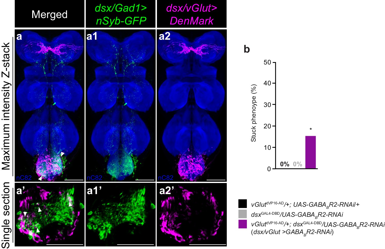
(a–a2) Overlay of expression of (a1) dsx/Gad1 presynaptic boutons and dsx/vGlut dendrites on standardized template VNC. (a1) dsx/Gad1 presynaptic boutons visualized by dsxGAL4-DBD/Gad1p65-AD driving UAS-nSyb::GFP in male VNC stained with anti-GFP (green). (a2) dsx/vGlut dendrites visualized by vGlutdVP16-AD/dsxGAL4-DBD driving UAS-DenMark in male VNC stained with anti-DsRed (magenta); neuropil counterstained with anti-nC82 (blue). (a–a2) Maximum intensity z-stacks and (a’–a2’) single section images. Solid arrowheads point at regions of close proximity. Scale bar = 50 μm. See also Video 5. (b) Effects of knocking down GABAB-R2 receptor by RNAi in dsx/vGlut neurons on male copulatory behavior. Percentage of males displaying a ‘stuck’ phenotype is graphed (n = 20–30). *p<0.05 by Fisher exact test.
Figure 4—figure supplement 1
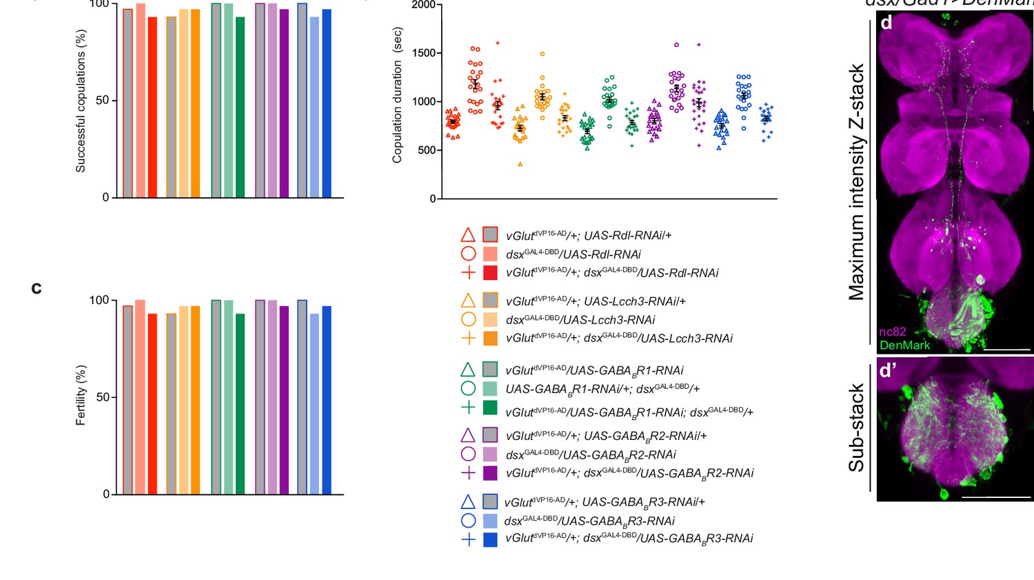
Effects of knocking down GABA receptor subunits in dsx/glutamatergic neurons on copulatory behaviors.
(a) Percentage of successful matings in 1 hr (n = 24–30). (b) Copulation duration (n = 12–24). (c) Male fertility (n = 30). Genotypes indicate males. Statistical comparisons of the experimental genotype were made against controls of the same receptor. (d) dsx/Gad1 neurons have prolific dendrites in regions of the Abg occupied by genital sensory terminals. dsx/Gad1 dendrites visualized by dsxGAL4-DBD/Gad1p65-AD driving UAS-DenMark in male VNC. Maximum intensity z-projection (d) and higher magnification image of 10 μm sub-stack (d’) is shown. DenMark stained with anti-dsRed (green); neuropil counterstained with anti-nC82 (magenta). Scale bar = 50 μm.
Figure 5
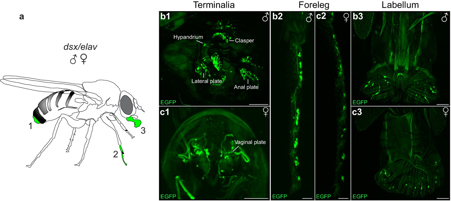
Novel patterns of dsx expression in the male and female peripheral nervous system.
(a) Cartoon of adult fly depicting regions of dsx-expression in peripheral sense organs in males and females; a1: terminalia, a2: foreleg and a3: labellum of the mouthparts. (b,c) Sexually dimorphic dsxGAL4-DBD/elavVP16-AD expression in peripheral sense organs in male and female adult flies. dsx/elav EGFP expression in bristle sensory neurons of the (b1) male clasper teeth, lateral plates, hypandrium and anal plates of the male terminalia and (c1) female vaginal plates of the female terminalia. dsx/elav EGFP expression in sensory neruons of the T1 tarsus of the foreleg in both males (b2) and females (c2). dsx/elav EGFP expression in sensory neruons of the labellum in both males (b3) and female (c3). EGFP is shown in green. Scale bar = 50 μm.
Figure 6
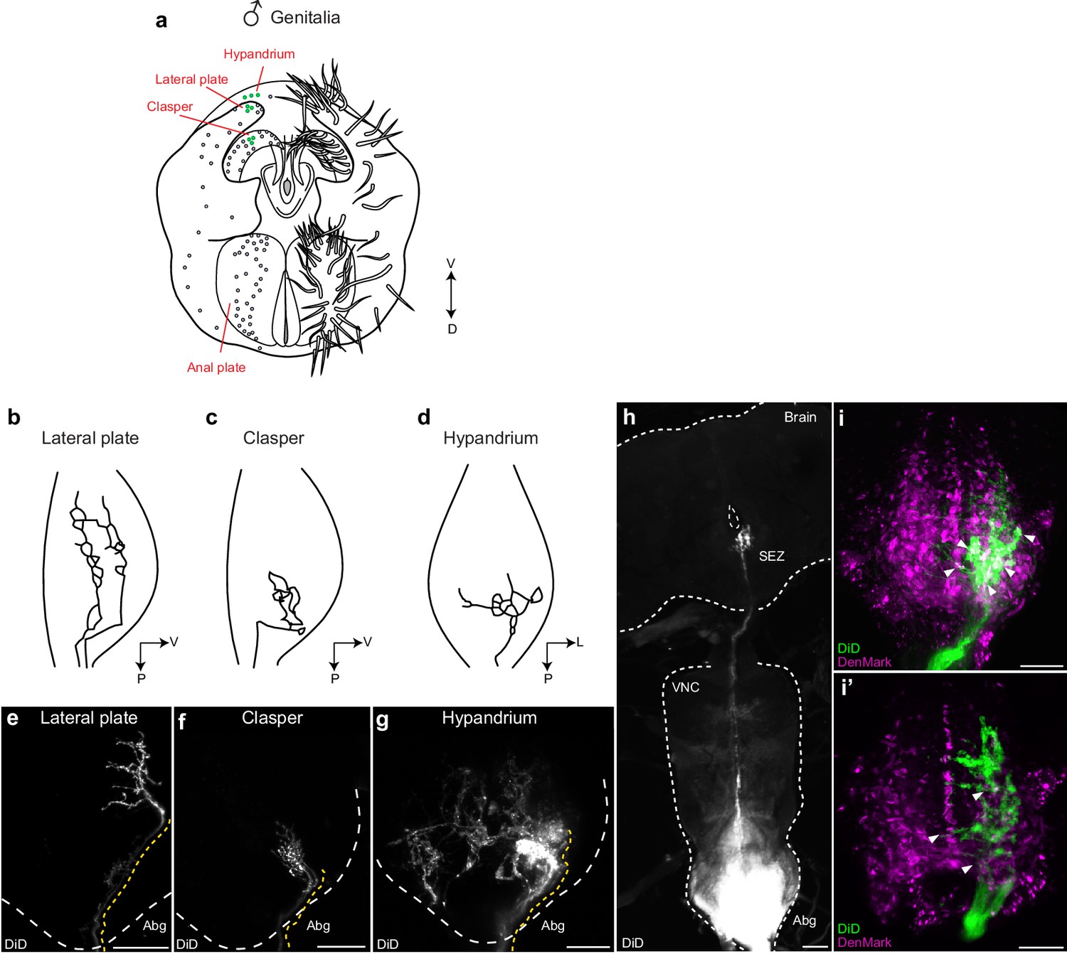
Mechanosensory neurons of the genitalia arborize onto the Abg and brain, and interdigitate with glutamatergic dsx motor neurons in the Abg.
(a) Schematic of male terminalia depicting bristles (right) and bristle topography (dots on left). Dye-filled bristle topography in shown with green dots. (b-d) Schematic of representative lateral plate (b), clasper (c), and hypandrium (d) arborizations in the abdominal ganglion (Abg) of male flies, as previously described (Taylor, 1989). (e-g) Representative images showing topographically distinctive patterns of dye-filled genital neuron arborizations in the male Abg. Maximum intensity z-projections of confocal stacks showing unilateral arborizations of (e) lateral plate, (f) clasper, and (g) hypandrium neurons in the male Abg, which reiterate previously described arborizations (Taylor, 1989). Afferent projections from lateral plate and clasper neurons occupy the same dorso-ventral area but differ in their anterior to posterior positions within the Abg, with clasper neurons ending more posteriorly than lateral plate neurons (b,c and e,f). See also Videos 6,7. Hypandrium neurons typically exhibit a unique contralateral arborization pattern within the Abg (d,g). See also Video 8. (h) A subset of clasper neurons project to the brain. Unilateral dye-fill of clasper neurons together with extended incubation (10 days) reveals single afferent axon (per hemisphere) that transverses the VNC and terminates in the subesophageal zone (SEZ) of the brain. DiD dye-filled arborizations shown in white. (e-h) DiD dye-filled arborizations shown in white. Boundaries of Abg and brain shown with dotted white line. Afferent projections of dye-filled neurons traced with dotted yellow line. D, dorsal, L, lateral, P, posterior, V, ventral. Scale bar = 25 μm. (i) Arborisations of clasper, lateral plates and hypandrium neurons interdigitate with dsx/vGlut dendrites in the adult male Abg. Neurons of all three genital structures were unilaterally dye-filled in males expressing dendritic marker (UAS-DenMark) in dsx/vGlut neurons. Maximum intensity Z-projection of Abg (i) and 10 μm sub-stack (i’) show overt interdigitation (arrowheads) between neurons of all three genital structures and dendrites of dsx/vGlut neurons in the Abg. DenMark shown in magenta; DiD shown in green. Scale bar = 25 μm.
Figure 7
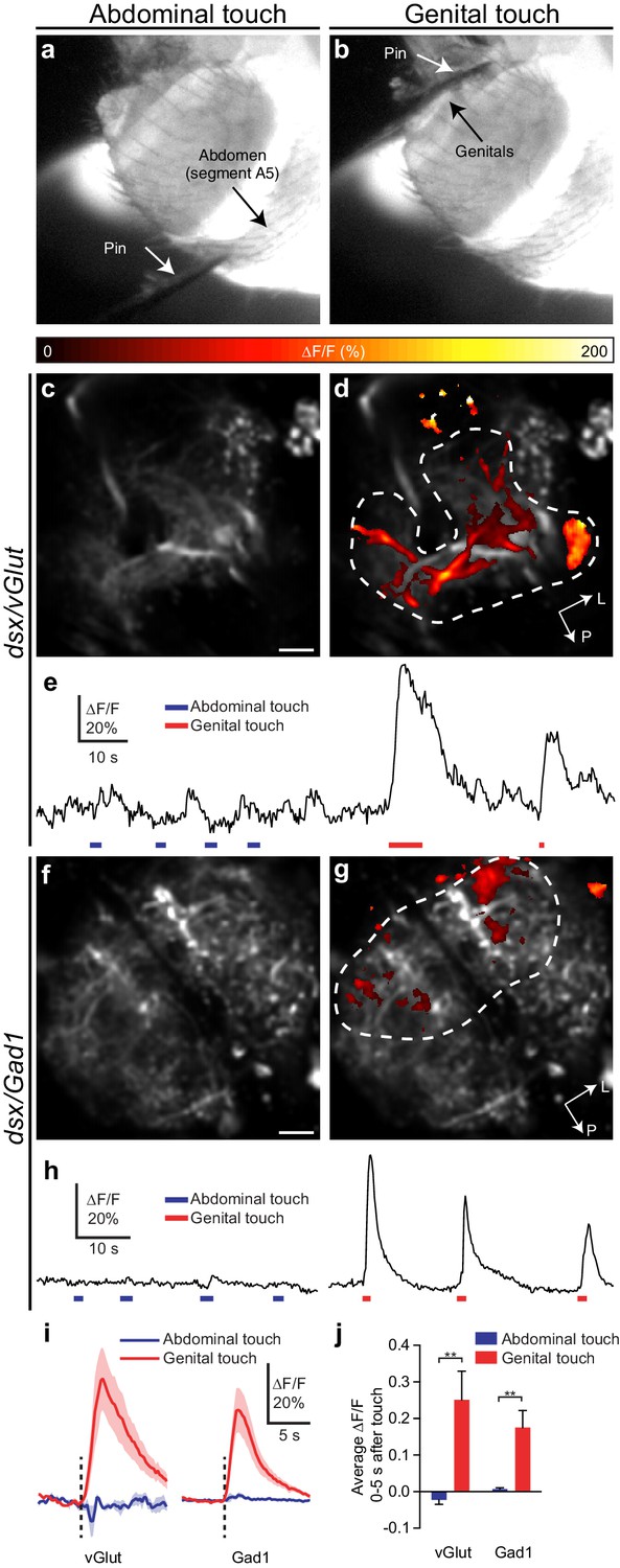
Glutamatergic and GABAergic dsx neurons of the Abg respond to mechanical stimulation of genitalia.
(a,b) Examples of pin touching a male fly’s abdomen on (a) segment A5 and (b) genitalia. The fly is illuminated at the VNC by the 910 nm two-photon laser and imaged with an infrared-sensitive camera. (c,d) dsx/vGlut>GCaMP6m neuropil in the abdominal ganglion: Pseudocolored activity maps of responses to (c) abdominal or (d) genital touch, overlaid on grayscale baseline fluorescence See also Video 9. There is no response to abdominal touch (c). Dotted outline indicates region of interest for panel E. L, lateral, P, posterior. (e) ∆F/F of the outlined region in panel D. Bars under traces represent abdominal (blue) or genital (red) touch. The two traces come from a single movie. (f,g) as with (c,d) but for dsx/Gad1>GCaMP6m neuropil. See also Video 10. (h) as with (e) but referring to the outlined region in (g). (i) Average of ∆F/F traces as in (e) and (h), aligned to touch onset. dsx/vGlut and dsx/Gad1 neurons in the abdominal ganglion respond strongly to genital touch (red) but not abdominal touch (blue). Traces: average ∆F/F (fluorescence normalized to baseline); shading, S.E.M.; vertical line, onset of touch. Only touches < 3 s long are included. n = 7 (vGlut genital), 5 (vGlut abdominal), 6 (Gad1 genital and abdominal). (j) Average ∆F/F 0–5 s after onset of touch is significantly larger for genital touch than abdominal touch. ** p<0.01, Mann-Whitney test. Scale bars, 10 µm.
Figure 8

Model of circuit organization underlying copulation in males.
(a) Musculature of male genitalia and terminalia involved in copulation. Protractor muscles shown in orange. Retractor muscles shown in blue. Muscles with no designated colour have unknown functions. D: dorsal, V: ventral. (b) dsx/vGlut motor neurons local to the Abg (blue) mediate genital coupling by controlling muscles of the phallic and periphallic organs. dsx/Gad1 Abg neurons (red), depicted as a heterogeneous population of neurons, some of which inhibit glutamatergic neurons that control copulatory muscles (bottom), and other which shorten copulation duration (stopwatch) by reducing copulation motivation by inhibiting dopaminergic (DA) neurons (top). dsx sensory neurons of the genitalia (grey) innervate the Abg and brain, and are anatomically and functionally connected to dsx/vGlut motor neurons (blue) and dsx/Gad1 inhibitory centres (red) of the Abg, likely aiding the male in adopting the correct posture to successfully achieve copulation. Brackets depict control over all encompassing neurons.
Videos
Video 1
Inhibition of dsx/vGlut neurons in males blocks genital coupling and the initiation of copulation.
This movie shows a dsx/vGlut>TNT male failing to achieve genital coupling and initiate copulation with a wild-type female. These males do however display the normal complement of courtship behaviors.
Video 2
Activation of dsx/vGlut neurons in copulating males blocks genital uncoupling and the termination of copulation.
This movie shows three dsx/vGlut>TrpA1 males that remain attached to their wild-type female mating partners via their genitals after ~2 hr of in copulo thermal activation (31°C).
Video 3
Inhibition of dsx/Gad1 neurons in males blocks genital uncoupling and the termination of copulation.
This movie shows a dsx/Gad1>TNT male displaying the distinctive ‘stuck’ behavior, whereby he has dismounted the wild-type female but remains attached via his genitals for prolonged periods of time.
Video 4
Activation of dsx/Gad1 neurons in copulating males elicits genital uncoupling and the termination of copulation.
This movie shows a dsx/vGlut>TrpA1 male and wild-type female copulating pair that have been shifted to 31°C 5 min into copulation. Thermal activation of male dsx/vGlut neurons in this manner results in the near-immediate termination of copulation by dsx/Gad1>TrpA1 males.
Video 5
dsx/Gad1 presynaptic boutons are in close proximity to dsx/vGlut dendrites.
This movie shows the 3D reconstruction of a template abdominal ganglion showing an overlay of dsx/vGlut dendrites (magenta) and dsx/Gad1 presynaptic boutons (green).
Video 6
Clasper neurons of the male genitalia innervate the abdominal ganglion.
This movie shows the 3D reconstruction of an adult male abdominal ganglion with innervations of dye-filled neurons from bristles on the clasper of the male genitalia. White: Lipophilic dye (DiD).
Video 7
Lateral plate neurons of the male genitalia innervate the abdominal ganglion.
This movie shows the 3D reconstruction of an adult male abdominal ganglion with innervations of dye-filled neurons from bristles on the lateral plate of the male genitalia. White: Lipophilic dye (DiD).
Video 8
Hypandrium neurons of the male genitalia innervate the abdominal ganglion.
This movie shows the 3D reconstruction of an adult male abdominal ganglion with innervations of dye-filled neurons from bristles on the hypandrium of the male genitalia. White: Lipophilic dye (DiD).
Video 9
dsx/vGlut Abg neurons respond to mechanical stimulation of genitalia.
The upper panel shows GCaMP6m signal in dsx/vGlut neuropil in the Abg and the lower panel shows the simultaneous view of the fly’s abdomen, illuminated by the 910 nm laser used for two-photon imaging. The minutien pin touches the genitalia at 0:05 and 0:12 depicted with ***. Movies are 5x actual speed and false colored.
Video 10
dsx/Gad1 Abg neurons respond to mechanical stimulation of genitalia.
The upper panel shows GCaMP6m signal in dsx/Gad1 neuropil in the Abg and the lower panel shows the simultaneous view of the fly’s abdomen, illuminated by the 910 nm laser used for two-photon imaging. The minutien pin touches the genitalia at 0:04 and 0:08 depicted with ***. Note that the pin approaches the genitalia but does not quite touch until 0:04. Movies are 5x actual speed and false colored.
Tables
Table 1
Cell counts for dsx-intersected neurons in male and female adult CNS. Male and female dsx/elav, dsx/vGlut and dsx/Gad1 cell counts are listed in black. Subsets of neurons that co-express FruM in males are listed in italics.
dsx neuronal clusters | dsx/elav | dsx/vGlut | dsx/Gad1 | |||
|---|---|---|---|---|---|---|
Male | Female | Male | Female | Male | Female | |
Brain | ||||||
pC1* | 52.8 ± 4.1 (12) | 8.3 ± 1.6 (12) | 0 ± 0 (12) | 0 ± 0 (12) | 0 ± 0 (12) | 0 ± 0 (12) |
pC2* | 78.3 ± 4.8 (12) | 14.2 ± 1.5 (12) | 0 ± 0 (12) | 0 ± 0 (12) | 1.0 ± 0(12) 0.9 ± 0.3 (10) | 0 ± 0 (12) |
pC3* | 13.8 ± 0.9 (12) | 8.0 ± 1.0 (12) | 0 ± 0 (12) | 0 ± 0 (12) | 3.5 ± 0.5 (12) 0.5 ± 0.5 (10) | 3.0 ± 0 (12) |
aDN* | 2.0 ± 0 (12) | 2.0 ± 0 (12) | 1.9 ± 0.3 (12)0 ± 0 (10) | 2.0 ± 0 (12) | 0 ± 0 (12) | 0 ± 0 (12) |
SN* | 1.0 ± 0 (12) | n.a. | 0 ± 0 (12) | n.a. | 0 ± 0 (12) | n.a. |
Ventral Nerve Cord | ||||||
TN1* | 23.0 ± 1.5 (12) | n.a. | 0 ± 0 (12) | n.a. | 0 ± 0 (12) | n.a. |
TN2* | 7.9 ± 0.3 (12) | n.a. | 0 ± 0 (12) | n.a. | 0 ± 0 (12) | n.a. |
Abg† | 275.0 ± 21.7 (10) | 314.8 ± 18.9 (10) | 79.8 ± 2.3 (10)7.4 ± 3.1 (10) | 101.8 ± 6.7 (10) | 151.2 ± 3.8 (10)30.0 ± 4.8 (10) | 213.1 ± 2.1 (10) |
-
*Neuronal cluster away from CNS midline. Count represents one cluster per hemisegment of the CNS.
-
†Neuronal cluster spans the CNS midline. Count given is for the entire Abg. Counts represent mean ± S.D. n’s listed in parentheses.
Download links
A two-part list of links to download the article, or parts of the article, in various formats.
Downloads (link to download the article as PDF)
Open citations (links to open the citations from this article in various online reference manager services)
Cite this article (links to download the citations from this article in formats compatible with various reference manager tools)
Neural circuitry coordinating male copulation
eLife 5:e20713.
https://doi.org/10.7554/eLife.20713


