Age-dependent diastolic heart failure in an in vivo Drosophila model
- Howard Hughes Medical Institute, University of California, San Francisco, United States
- University of California, San Francisco, United States
Figures
Figure 1 with 1 supplement
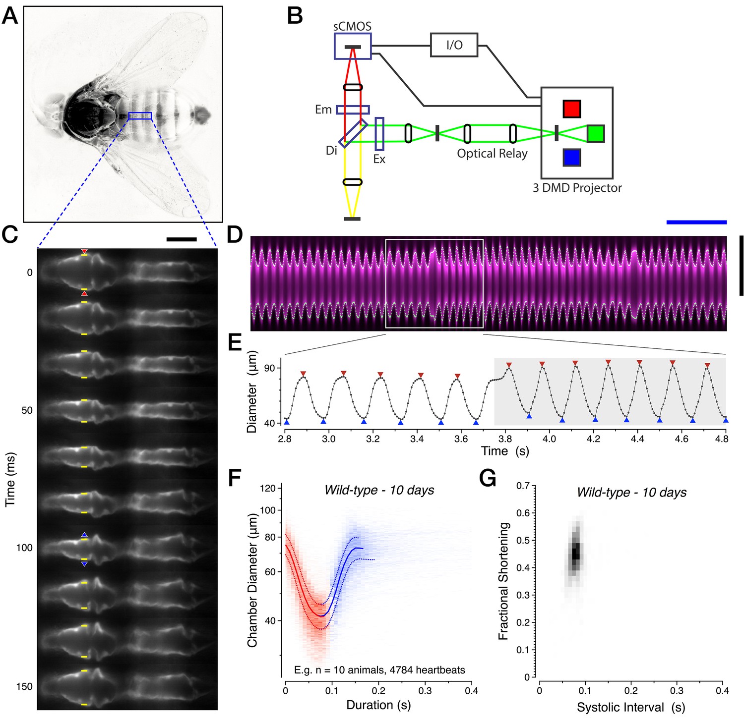
Imaging cardiac performance in intact, unanesthetized Drosophila.
(A) Full view of the intact preparation, with imaging region of interest (blue). The head and legs are freely moving, while the wings, dorsal thorax and dorsal abdomen are affixed to the coverglass with optical cement. (B) Electro-optical diagram of the imaging system. (C) Single anterograde heartbeat at half frame-rate with heart wall position (yellow), initiation of contraction (red triangles) and of relaxation (blue triangles) calls. (D) Associated YT kymograph (magenta) with heart wall detection (white dots). (E) Corresponding digitization, segmented into anterograde (white) and retrograde (grey) heartbeat epochs. The triangles denote the initiation (red) and end (blue) of contractions. (F) Two-dimensional probability map of heart chamber diameter and heartbeat duration with median +/− quartile overlay for systole (red) and diastole (blue). (G) Two-dimensional probability map of fractional shortening and systolic interval. All data in this figure are representative and from the 10-day-old w1118 wild-type dataset. See Materials and methods for all functional parameter definitions and their derivation. Scale bars: (black vertical) 75 µm, (blue horizontal) 1 s. See also Video 1.
Figure 1—figure supplement 1
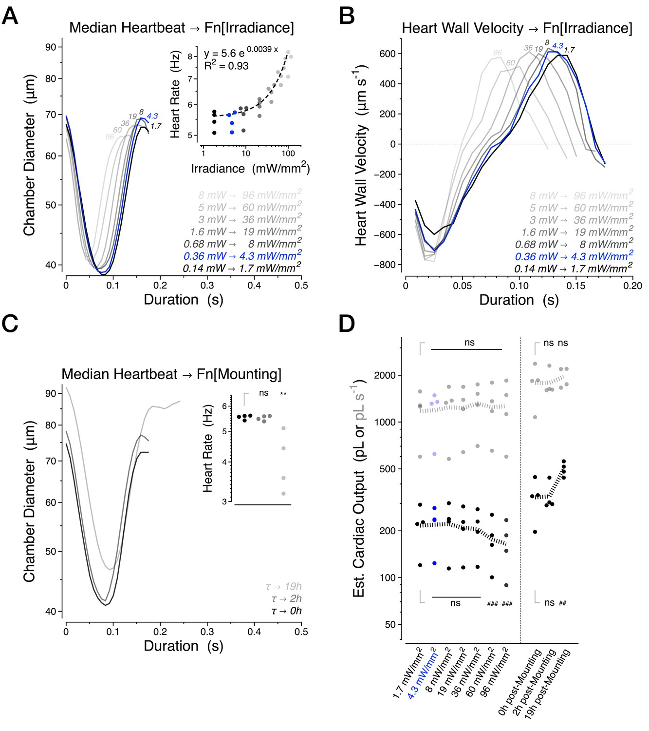
Further characterization of the intravital imaging methodology.
(A) Median heartbeat and heart rate (inset) as a function of excitation light intensity. (B) Median heart wall velocity as a function of excitation light intensity. (C) Median heartbeat and heart rate (inset) as a function of time elapsed after mounting. (D) Estimated Cardiac Output per stroke (black) and per second (grey). The banded lines in (D) represent the mean value for each condition. The blue condition highlights the irradiance level used for all experiments excepting the α-Actinin dynamics in Figure 6 which required 19 mW/mm2 of excitation light. These experiments were quantified pairwise using four 30 days old Canton S animals per condition. Statistics for panels used the paired one-way ANOVA followed by Holm-Sidak’s multiple comparisons test. ns = not significant, */#p<0.05, **/##p<0.01, ***/###p<0.001.
Figure 2 with 1 supplement
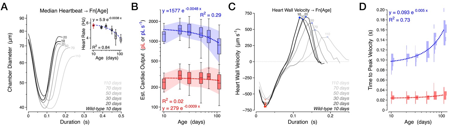
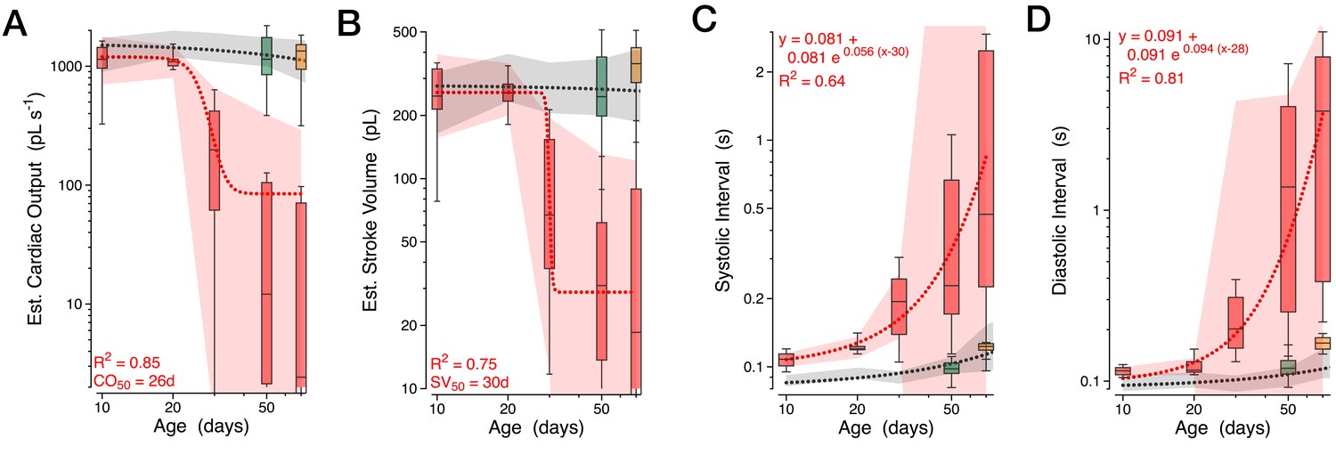
Normal aging is characterized by a diastolic decline with preserved contractility.
Various cardiac functional parameters presented by age in a combined w1118 and Canton S dataset, n = 18 to 30 animals per time-point: (A) Median heartbeat with heart rate (inset). (B) Estimated cardiac output per second (blue) and per stroke (red). (C) Median heart wall velocity with peak velocities of contraction (red dots) and relaxation (blue dots). (D) Probability histograms of the time from initiation of contraction to peak contraction velocity (red) and from the peak contraction velocity to peak relaxation velocity (blue). The shaded areas in panel B represent the mean +/− s.d., with regressions plotted as dotted lines.
-
Figure 2—source data 1
Median heartbeats for all individual animals in panel A.
Median heartbeats were calculated for individual animals (Table) and for all consolidated heartbeats for a respective age (Panel A and last column of each Table). These source data provide a representation of the observed heartbeat waveform variability between animals.
- https://doi.org/10.7554/eLife.20851.006
Figure 2—figure supplement 1
Further measures of normal aging.
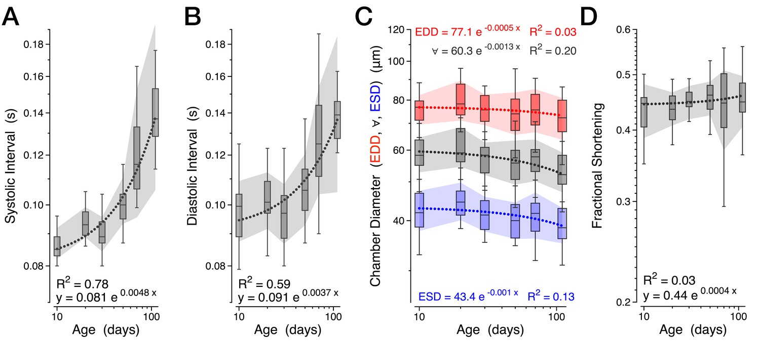
Various cardiac functional parameters presented by age in a combined w1118 and Canton S dataset, n = 18 to 30 animals per time-point: (A) Systolic interval. (B) Diastolic interval. (C) Heart chamber diameter across the cardiac cycle (∀, grey) with median end systolic diameter (ESD, blue) and end diastolic diameter (EDD, red) during aging. (D) Fractional shortening. The shaded areas are mean +/− s.d. with the dotted lines representing the best fit of each dataset (y = Aekx).
Figure 3 with 2 supplements
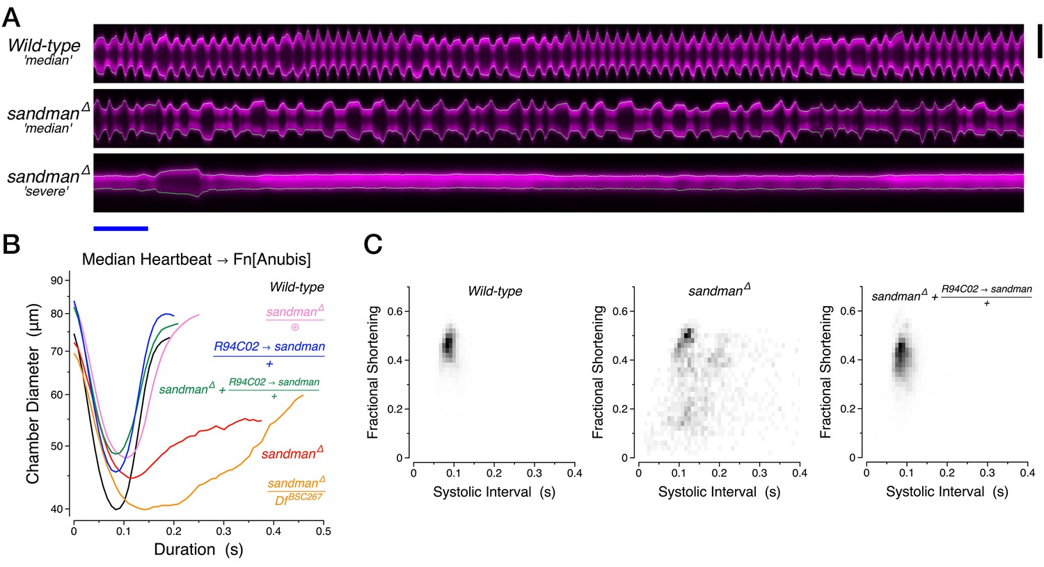
Diastolic failure in sandman mutants.
(A) Representative YT kymographs of 50-day-old animals. Scale bars: (black vertical) 75 µm, (blue horizontal) 1 s. (B) Median heartbeat per genotype at 50 days of age. ⊕, clean excision of the mutagenic piggyBac insertion e00867. (C) Two-dimensional probability map of fractional shortening and systolic interval at 50 days of age. See also Video 2.
-
Figure 3—source data 1
Median heartbeats for all individual animals in panel B.
Median heartbeats were calculated for individual animals (Table) and for all consolidated heartbeats for a respective genotype (Panel B and last column of each Table). This source data provides a representation of the observed heartbeat waveform variability between animals.
- https://doi.org/10.7554/eLife.20851.009
Figure 3—figure supplement 1

sandman and galene genetic loci.
(A,B) Genomic maps of the sandman and galene loci, adapted from the UCSC genome browser (https://genome.ucsc.edu), with a sequence conservation index across a panel of 15 insects. The piggybac insertions used to generate the deletion of sandman are detailed as is the dsRNA sequence used to knockdown galene. The gene CG8712 encodes a protein of unknown function and is reported to be predominantly expressed in the sex organs (http://flybase.org/reports/FBgn0033258.html). (C) Heart-specific RT-PCR for sandman and galene transcripts.
Figure 3—figure supplement 2
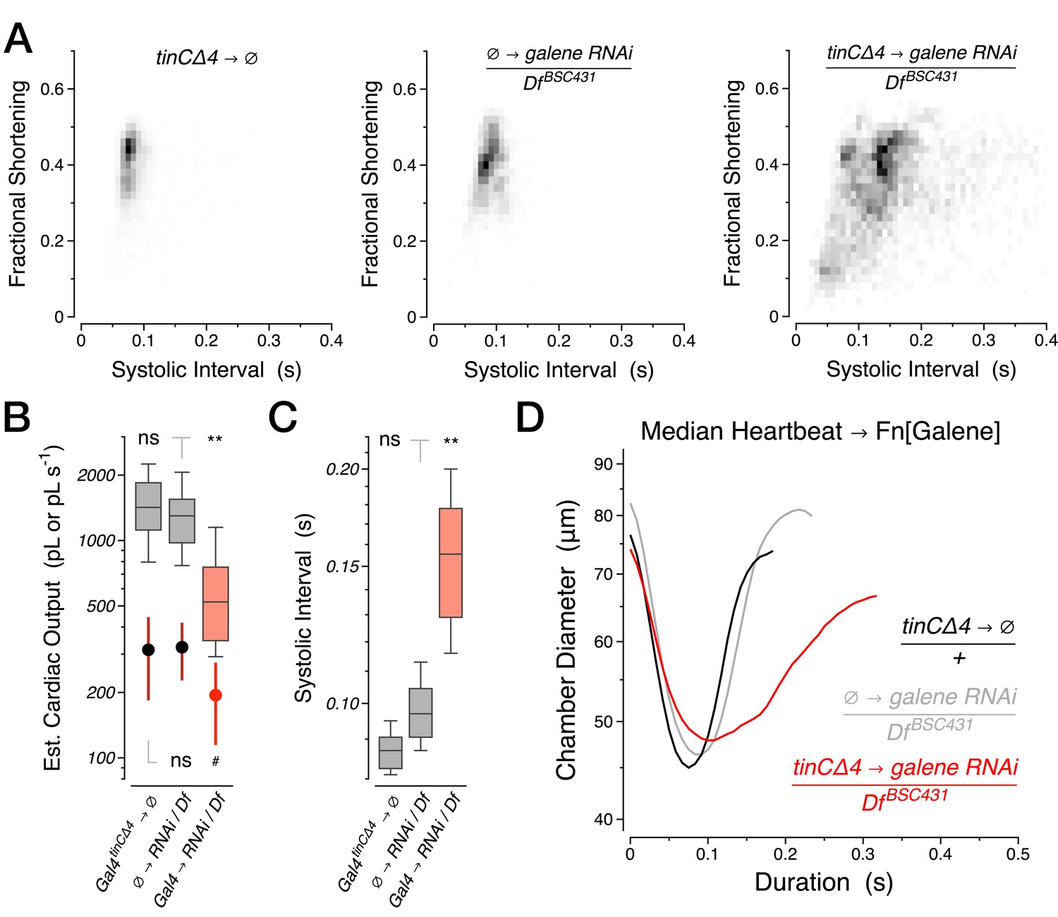
RNAi knockdown of galene from birth through 40 days of age.
(A) Two-dimensional probability map of fractional shortening and systolic interval. (B) Estimated cardiac output per second (boxplot) and per stroke (filled circles, mean +/− s.d.). (C) Systolic interval. (D) Median heartbeat. Df, Deficiency BSC431 covering the galene locus. ∅, experimental controls lacking one or the other element of the Gal4-UAS system.
Figure 4 with 3 supplements
Progressive heart failure in sandman mutants.
(A–D) Estimated cardiac output per second (A) and per stroke (B) were well fit by a Boltzmann sigmoidal regression and the systolic (C) and diastolic (D) intervals were well fit by single exponential growth regression curves for wild-type (grey), sandman (red), cardiomyocyte rescue of sandman using R94C02::Gal4 (green) and the clean excision (pink) at specified ages. n = 7 to 27 animals per genotype and age. The shaded areas represent the mean +/− s.d., with the regressions plotted as dashed lines.
Figure 4—figure supplement 1
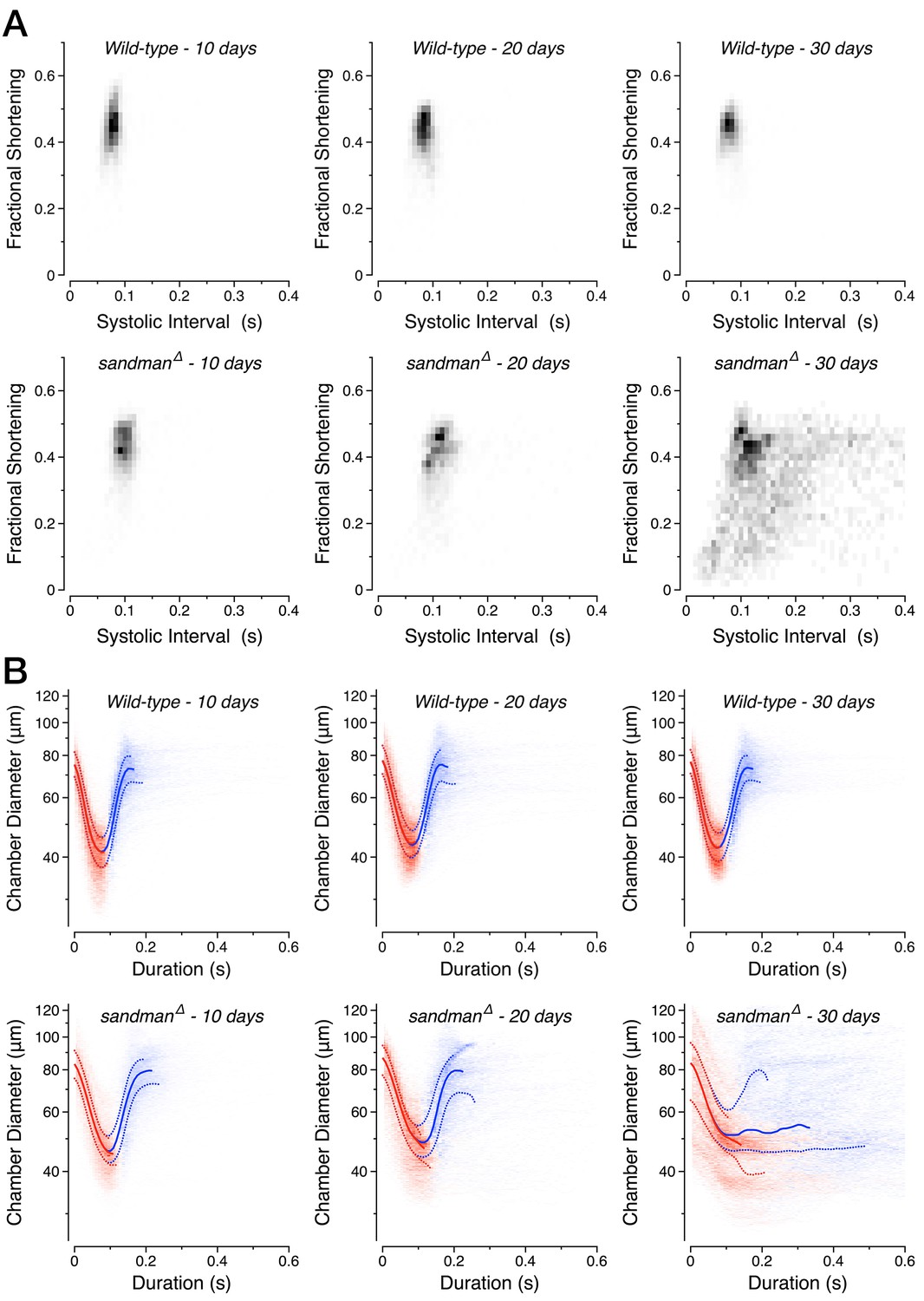
Progressive loss of diastole in sandman mutants.
(A) Two-dimensional probability map of fractional shortening and systolic interval in wild-type and sandman mutant animals at various ages. (B) Two-dimensional probability map of chamber diameter and heartbeat duration with median +/− quartile overlay for systole (red) and diastole (blue). n = 9 to 14 animals per genotype/age.
Figure 4—figure supplement 2
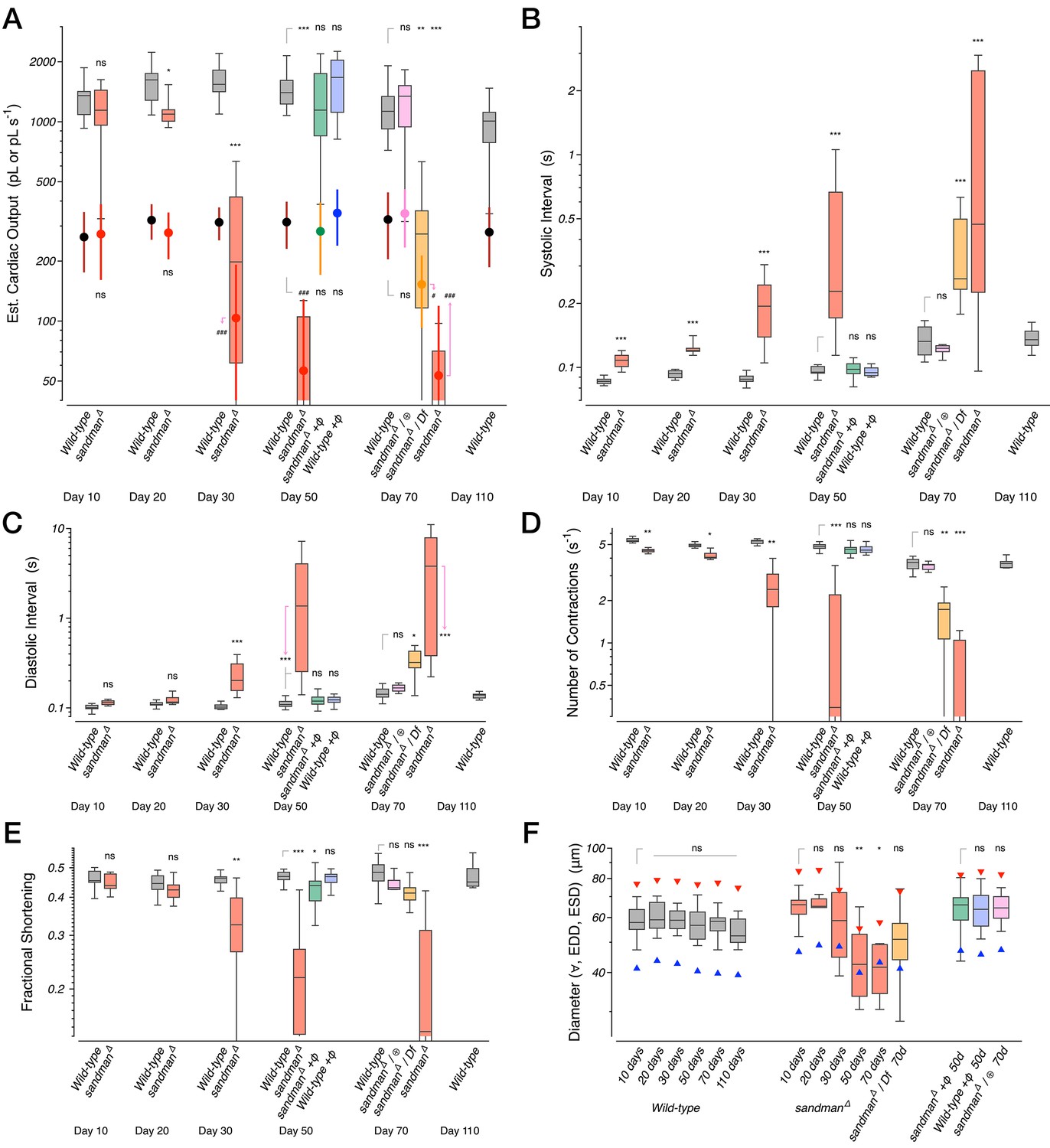
Additional cardiac functional parameters for sandman mutants.
Various functional parameters as a function of genotype and age: (A) Estimated cardiac output per second (boxplot) and per stroke (filled circles, mean +/− s.d.). (B) Systolic interval. (C) Diastolic Interval. (D) Number of contractions per second. (E) Fractional shortening. (F) Heart chamber diameter across the cardiac cycle (boxplot) with median end systolic diameter (ESD, blue triangle) and end diastolic diameter (EDD, red triangle). n = 7 to 27 animals per genotype/age. Kruskal-Wallis one-way ANOVA followed by Dunn’s multiple comparisons test. ns = not significant, *p<0.05, **p<0.01, ***p<0.001.
Figure 4—figure supplement 3
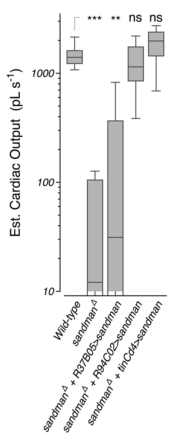
Transgenic rescue in sandman mutants at 50 days of age.
Estimated cardiac output per second (boxplot) in wild-type animals, sandman mutants, and sandman mutants expressing the sandman cDNA in body wall muscles but not the heart (R37B05), or in the heart (R94C02 and tinCΔ4). n = 12 to 27 animals per genotype. Kruskal-Wallis one-way ANOVA followed by Dunn’s multiple comparisons test. ns = not significant, *p<0.05, **p<0.01, ***p<0.001.
Figure 5 with 1 supplement

sandman and galene jointly encode a potassium channel.
(A) Representative whole-cell currents in physiological K+ and Na+ gradients from Sandman (n = 5), Galene (n = 6), and co-transfection of both (n = 11) during voltage steps (below). (B) Normalized whole-cell currents from voltage ramps in various bath solutions. The dotted line plots the I/V curve for a hypothetical ion channel with no rectification in symmetric K+. The inset plots the observed reversal potential compared to a potassium-selective conductance (dashed line) at various [K+] in/out ratios. The internal pipet solution is (in mM) 150 K+, 5 Na+, 3 Mg2+, 161 Cl−, 10 HEPES, pH 7.4 (in mM). The bath solution [K+] and [Na+] or [NMDG+] are as indicated (in mM), excepting the ‘Divalent-free’ solution which substitutes 2 mM EDTA for the divalent cations. n = 9 cells. All pooled data represent the mean +/− s.d. All voltage potentials are relative to ground.
-
Figure 5—source data 1
Normalized current-voltage data for panel B.
- https://doi.org/10.7554/eLife.20851.018
Figure 5—figure supplement 1
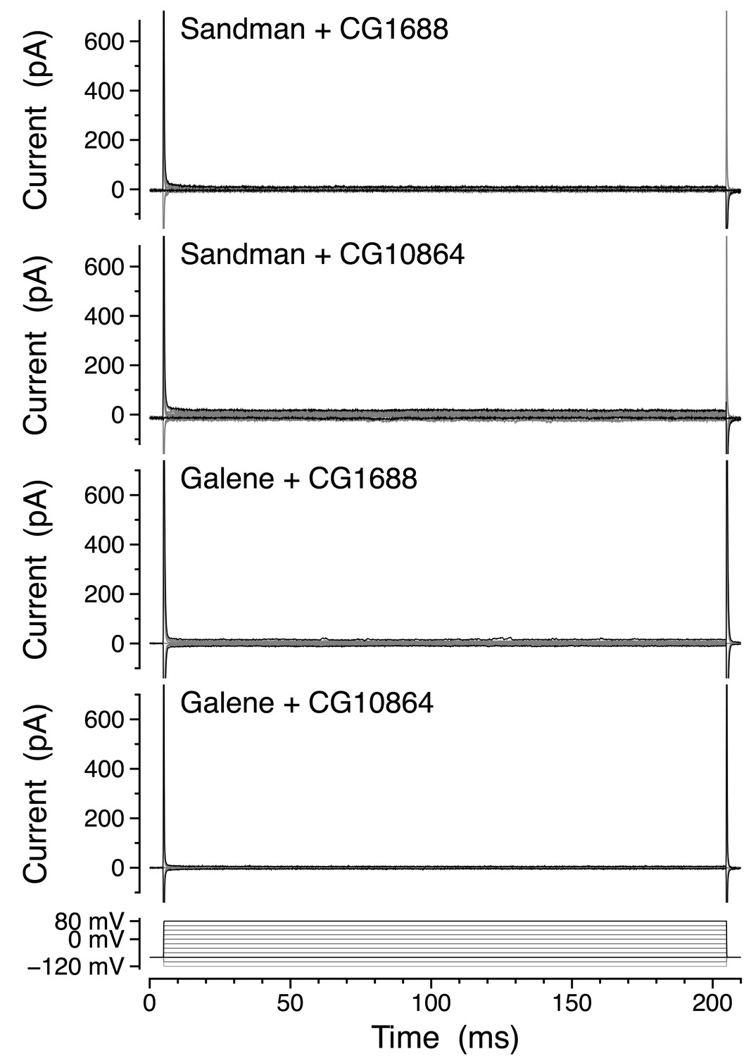
Sandman and Galene do not form functional heteromeric channels with the closely related K2P subunits CG1688 or CG10864.
Representative whole-cell currents in physiological K+ and Na+ gradients from co-transfection of Sandman + CG1688 (n = 5), Sandman + CG10864 (n = 6), Galene + CG1688 (n = 7), and Galene + CG10864 (n = 5) during voltage steps (below).
Figure 6
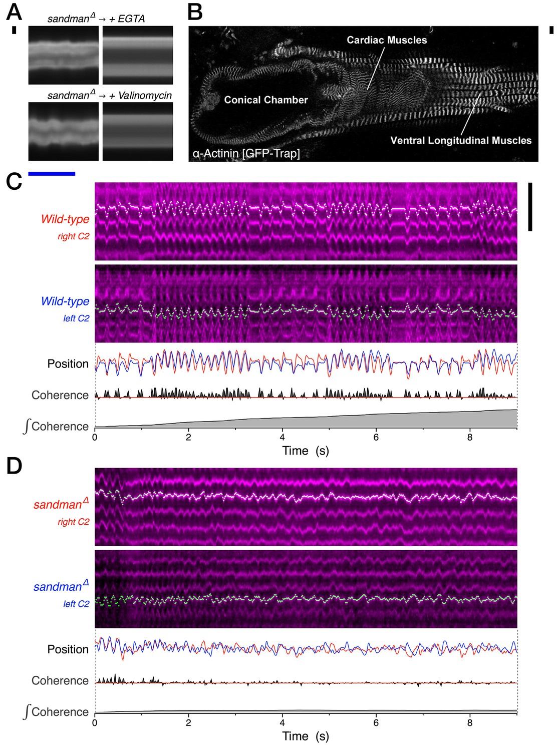
In vivo pharmacology and sarcomere dynamics implicate dyssynchronous and regenerative Ca2+ in maintaining persistent systole.
(A) Representative heart kymographs from 110-day-old sandman males before and acutely after intra-abdominal injection of the Ca2+ chelator EGTA or the potassium ionophore valinomycin. n = 3. (B) Micrograph of a dissected adult Drosophila expressing a GFP trap of the z-line protein α-actinin [CC01961]. (C–D), Representative kymographs (magenta) of second chamber right and left cardiomyocyte sarcomere dynamics from intact 30-day-old animals, as visualized intravitally using the α-actinin GFP-trap. Automated detection of one z-line for each cardiomyocyte (green/white), quantified as relative position over time (upper trace, left cardiomyocyte signal inverted), with net coherence between z-lines (middle trace) and integral coherence (bottom trace). Scale bars: (black vertical) 10 µm, (blue horizontal) 1 s. n = 9 for wild-type and four for sandman. See also Video 3.
Author response image 1

Videos
Video 1
Heartbeat visualization, digitization and segmentation.
One-third speed video of the 10 day adult female displayed in Figure 1C–E, with heart wall position calls (yellow) and the attending transformation into heart chamber diameter as a function of time in a 1 s streaming window. The initiation and end of each contraction are specified by a red and blue triangle, respectively. Note the periodic reversal in the direction of heartbeat peristalsis.
Video 2
50-day wild-type and sandman heartbeat visualization.
One-third speed videos of 50-day adult wild-type (upper video) and sandman (lower two videos) females, with heart wall position calls (yellow) and the attending transformation into heart chamber diameter as a function of time in a 1 s streaming window. The initiation and end of each contraction are specified by a red and blue triangle, respectively.
Video 3
30 day wild-type and sandman sarcomere dynamics.
Real-time videos of 30-day adult wild-type (upper video) and sandman (lower video) females, visualizing the z-lines of the dorsal aspect of the cardiomyocyte pair just posterior to the ostial valves of the second chamber using a protein trap of α-actinin [CC01961].
Tables
Table 1
DNA constructs.
| Plasmid ID | Plasmid name | Insert source | 5’ Primer | 3’ Primer | Destination Vector | Restriction Subcloning | Comments |
|---|---|---|---|---|---|---|---|
| pMK1 | 10xUAS-IVS-Syn21-tdTomato-p10 | pDEST HemmarR (Addgene #31222) | ataaggtaccAACTTAAAAAAAAAAATCAAAATGGTGAGCAAGGGCGAG | atattctagaTTACTTGTACAGCTCGTCCATGCC | pJFRC81 (Addgene #36432) | KpnI - XbaI | Intermediate plasmid - for fly transgenesis (Han et al., 2011; Pfeiffer et al., 2012) |
| pMK3 | R94C02::tdTomato | Janelia Farms, amplified from Drosophila genome | tactagtACTTTTCCGCGCCGTCTG | atatgctagcGGAAACAGACGCAAAGACTGAC | pMK1 (this paper) | HindIII - NheI | Cardiomyocyte enhancer expressing tdTomato for fly transgenesis - 5’ primer was phosphorylated to facilitate blunt ligation using Klenow fragment |
| pMK17 | pAc5.1B_GFP-CG8713 | BDGP cDNA RE21922 in clone #UFO03925 | aaaagcggccgcATGTCCTCCCGACGC | atattctagaTTAGGAGGTGCGGCAC | pAc5.1B_GFP (Addgene #21181) | NotI - XbaI | Intermediate plasmid - for S2 cell expression (Stapleton et al., 2002) |
| pMK18 | pAc5.1B_GFP-CG10864 | PCR from Drosophila genome (single exon) | aaaagcggccgcATGGCCAGCAAATTTCAGAG | atattctagaCTAGTAGTAATCATCCTCGTAC | pAc5.1B_GFP (Addgene #21181) | NotI - XbaI | Intermediate plasmid - for S2 cell expression |
| pMK21 | pAc5.1B_GFP-CG1688 | BDGP cDNA GH04802 in clone #UFO05944 | aaaagcggccgcATGTCCGACGTTGAGCAG | atattctagaTTATCCATCCGCGCGG | pAc5.1B_GFP (Addgene #21181) | NotI - XbaI | Intermediate plasmid - for S2 cell expression |
| pMK22 | pACU_GFP-CG8713 | pAc5.1B_GFP- CG8713 (this paper) | KpnI - ApaI insert from source vector | KpnI - ApaI insert from source vector | pACU (Addgene #58373) | KpnI - ApaI | UAS::GFP-CG8713 rescue construct - for fly transgenesis |
| pMK23 | peGFP_C1-CG8713 | pAc5.1B_GFP- CG8713 (this paper) | EcoRI - ApaI insert from source vector | EcoRI - ApaI insert from source vector | peGFP_C1 (Clontech) | EcoRI - ApaI | For expression in mammalian heterologous cells |
| pMK24 | pAC5.1B_eGFP-CG9194 | BDGP cDNA FI03418 in clone #UFO11253 | aaaaagcggccgcATGTCGGGTAGGCGGGCCCA | gcagcctctagaCTAATCCTCATCCTGCTCGTCGTCATCATCC | pAc5.1B_GFP (Addgene #21181) | NotI - XbaI | Intermediate plasmid - for S2 cell expression |
| pMK25 | peGFP_C1-CG9194 | pAC5.1B_eGFP- CG9194 (this paper) | EcoR1 - SacII insert from source vector | EcoR1 - SacII insert from source vector | peGFP_C1 (Clontech) | EcoR1 - SacII | For expression in mammalian heterologous cells |
| pMK29 | peGFP_C1-CG1688 | pAC5.1B_eGFP- CG1688 (this paper) | tataGCTAGCggtaccaacatggtgagcaagg | tatacgggccctctagaTTATCCATCC | peGFP_C1 (Clontech) | NheI - ApaI | For expression in mammalian heterologous cells |
| pMK30 | peGFP_C1- CG10864 | pAC5.1B_eGFP- CG10864 (this paper) | EcoR1 - ApaI insert from source vector | EcoR1 - ApaI insert from source vector | peGFP_C1 (Clontech) | EcoR1 - ApaI | For expression in mammalian heterologous cells |
Table 2
Drosophila genomic aberrations and transgenic insertions.
| Chromosomal Element | Location | Source | Description |
|---|---|---|---|
| R94C02::tdTomato | Chr. II (attP40) | This paper | R94C02 enhancer expressing tdTomato in cardiomyocytes and a subset of other muscles within the adult fly. Integrated into the attP40 docking site. We used this transgenic for all heart wall imaging experiments. |
| PBac{RB}CG8713 [e00867] | Chr. II | Exelixis via Bloomington | piggyBac{RB} insertion proximal to the 1st splice acceptor of the CG8713 mRNA. This insertion has a strong heart function phenotype. |
| PBac{RB}CG8712 [e00152] | Chr. II | Exelixis via Bloomington | piggyBac{RB} insertion into the CG8712 coding sequence. This insertion does not have a discernible heart phenotype. |
| PBac{RB}CG8713_CG8712 [e00867_e00152 deletion, mW+] | Chr. II | This paper | FLP-FRT mediated deletion of the genomic DNA between e00867 and e00152, which comprises the entire CG8713 coding sequence and most of the CG8712 coding sequence. RB(+) to RB(+) recombination reconstitutes a single PBac{RB} after the intervening sequence is deleted. |
| [e00867_e00152 deletion] | Chr. II | This paper | Clean excision of PBac{RB}CG8713_CG8712 [e00867_e00152 deletion, mW+]. This chromosome harbors the deletion without any residual pBac[RB] sequence. |
| CyO, P{Tub-PBac\T} | Chr. II | Bloomington | Source of piggyBac transposase activity for generating clean excisions, which were identified by the loss of mini-white eye pigmentation. |
| e00867 [clean excision] | Chr. II | This paper | Clean excision of PBac{RB}CG8713 [e00867] which reverts the observed heart phenotype. |
| UAS::GFP-CG8713 | Chr. III (vk00005) | This paper | nsertion of plasmid pMK22 into the attP docking site vk00005. Used to demonstrate cardiomyocyte rescue of CG8713 (anubis). |
| R94C02::Gal4 | Chr. III (attP2) | Bloomington | In a screen for new heart-specific Gal4s, we found R94C02::Gal4 as a complement to tin.CΔ4::Gal4. It is expressed in cardiomyocytes and a subset of other muscles. (Pfeiffer et al., 2008) |
| tin.CΔ4::Gal4 | Chr. III | Manfred Frasch | tin.CΔ4 is expressed in the heart and a subset of other muscles within the fly. (Lo and Frasch, 2001) |
| R37B05::Gal4 | Chr. III (attP2) | Bloomington | In a screen for new heart-specific Gal4s, we found R37B05::Gal4 to be expressed in bodywall muslces but not the heart. |
| Df(2R)BSC267 | Chr. II | Bloomington | A molecularly defined deletion spanning the CG8713 (sandman) locus. |
| Df(3L)BSC431 | Chr. III | Bloomington | A molecularly defined deletion spanning the CG9194 (galene) locus. |
| P{KK110628}VIE-260B | Chr. II | VDRC #v108758 | UAS::CG9194 dsRNA for tissue-specific knockdown of galene. UP-TORR does not predict any off-target effects. |
| P{w[+mC]=PTT-GC Actn [CC01961] | Chr. X | Bloomington, this paper | GFP translational fusion of α-Actinin, which is localized to the z-lines of all muscles. We recombined away the yellow and white alleles. |
| Canton S | N/A | Jan Lab | Wild-type stock |
| w [1118] | Chr. X | Jan Lab | Wild-type stock, outcrossed to Canton S 6x |
| M{vas-int.Dm}ZH-2A; +; PBac{y[+]-attP-9A}VK00005 | Chr. X and III | Bloomington | Used for phiC31 mediated integration. |
| P{nos-phiC31\int.NLS}X; P{CaryP}attP40 | Chr. X and II | Bloomington | Used for phiC31 mediated integration. |
Table 3
Intravital imaging sample sizes.
| Genotype and age | n | Genotype and age | n |
|---|---|---|---|
| w1118 10 days | 10 | w1118 + R94C02 > sandman cDNA 50 days | 12 |
| w1118 20 days | 12 | sandmanΔ + R94C02 > sandman cDNA 50 days | 27 |
| w1118 30 days | 13 | sandmanΔ + tinCΔ4 > sandman cDNA 50 days | 12 |
| w1118 50 days | 17 | sandmanΔ + R37B05 > sandman cDNA 50 days | 13 |
| w1118 70 days | 12 | sandmanΔ / BSC267[Df] 50 days | 12 |
| w1118 110 days | 13 | sandmanΔ / clean excision 50 days | 13 |
| Canton S 10 days | 8 | sandmanΔ / BSC267[Df] 70 days | 15 |
| Canton S 20 days | 11 | sandmanΔ / clean excision 70 days | 7 |
| Canton S 30 days | 14 | tinCΔ4 control 40 days 25C | 15 |
| Canton S 50 days | 13 | kk110628 / BSC431[Df] control 40 days 25C | 11 |
| Canton S 70 days | 9 | tinCΔ4 > kk110628 / BSC431[Df] 40 days 25C | 11 |
| Canton S 110 days | 14 | Canton S 30 days - vary light | 4 |
| sandmanΔ 10 days | 13 | Canton S 40 days - mounting duration | 4 |
| sandmanΔ 20 days | 9 | sandmanΔ 110 days + EGTA | 3 |
| sandmanΔ 30 days | 14 | sandmanΔ 110 days + valinomycin | 3 |
| sandmanΔ 50 days | 15 | wild-type 30 days - visualize sarcomeres | 9 |
| sandmanΔ 70 days | 12 | sandmanΔ 30 days - visualize sarcomeres | 4 |
Download links
A two-part list of links to download the article, or parts of the article, in various formats.
Downloads (link to download the article as PDF)
Open citations (links to open the citations from this article in various online reference manager services)
Cite this article (links to download the citations from this article in formats compatible with various reference manager tools)
Age-dependent diastolic heart failure in an in vivo Drosophila model
eLife 6:e20851.
https://doi.org/10.7554/eLife.20851


