Release-dependent feedback inhibition by a presynaptically localized ligand-gated anion channel
- University of California, San Diego, United States
Figures
Figure 1 with 1 supplement
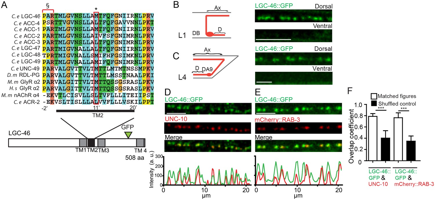
LGIC LGC-46 localizes to presynaptic terminals.
(A) Alignment of TM2 of LGC-46 with other anion channels. * marks Met314, which is mutated to Ile in LGC-46(ju825). § marks PAR motif, a signature of ionotropic anion channels. (B–F) LGC-46::GFP localizes to presynaptic terminals of cholinergic motor neurons. (B) Left panel shows a schematic of a cholinergic motor neuron in L1 animals. Right panel shows a confocal image of LGC-46::GFP from Punc-17β-LGC-46(WT)::GFP(juSi295)IV showing punctate localization in the dorsal nerve cord. (C) Left panel shows a schematic of DA9 cholinergic motor neuron in the tail of L4 animals. Right panel shows LGC-46::GFP expressed in DA9 neuron, from Pitr-1-LGC-46(WT)::GFP(juEx6843), showing punctate localization in the dorsal axon. (B, C): Ax: Axon. D: Dendrite. (D, E) LGC-46::GFP colocalizes with active zone protein UNC-10/RIM and synaptic vesicles in cholinergic motor neurons. Images of dorsal nerve cord are shown above, linescan analyses of fluorescent signal intensities below. (D) Conformal images of anti-GFP for LGC-46 (green) and anti-UNC-10 (red) from an animal carrying Punc-17β-LGC-46(WT)::GFP(juSi295)IV. (E) Presynaptic protein mCherry::RAB-3 expressed in cholinergic motor neurons overlapped with LGC-46::GFP signals. Images are from Punc-17β-LGC-46(WT)::GFP(juSi295)IV; Pacr-2-mCherry::RAB-3(juEx7053). Scale bar: 5 µm. (F) Mander’s overlap coefficient showing the extent of pixel colocalization of LGC-46 and preaynaptic marker protein signals (open bar). As negative controls, Mander’s overlap coefficient from shuffled images are shown (Filled bar). n = 10 per genotype. Data shown as mean ± SD. Statistics: one way ANOVA followed by Tukey’s post-hoc test. ***p<0.001.
Figure 1—figure supplement 1
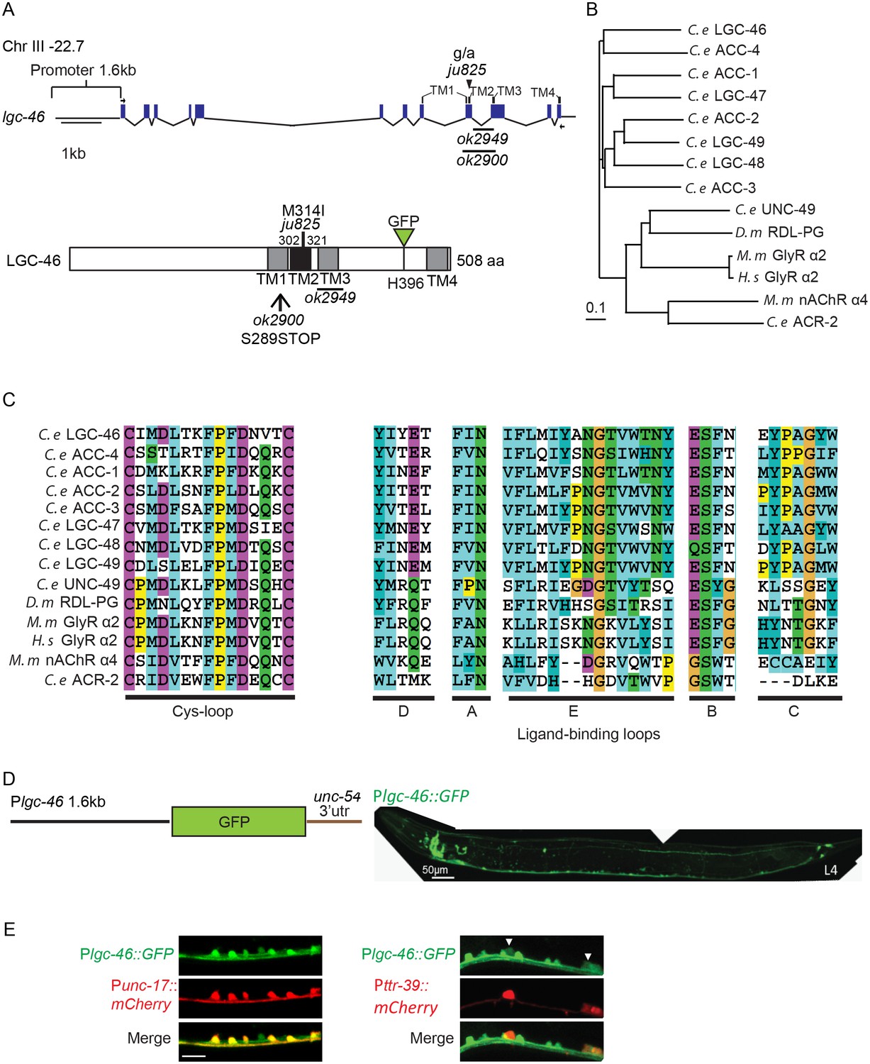
lgc-46 is expressed in the nervous system including cholinergic motor neurons.
(A) Gene and protein structure of lgc-46. The positions of the deletion alleles and the M314I mutation described in the text are noted. ok2900 removes TM2 and TM3 and generates a premature stop codon, and ok2949 generates an in-frame deletion which removes TM3. Arrows indicate the position of the primers used to amplify cDNA. Four transmembrane domains and the sites of mutations are noted in protein structure as well. For protein localization analysis, GFP was inserted at the cytoplasmic loop between TM3 and TM4. (B) Phylogenetic tree of LGICs shows that LGC-46 is a member of the ACC family of proteins from C. elegans (ACC-1 to ACC-4, LGC-46 to LGC-49). Generated by ClustalX2 using the neighbor-joining method (Larkin et al., 2007). (C) Amino acid sequence alignment of the Cys-loop region, and regions corresponding to the ligand-binding loops of nAChR. (D) (Left) Illustration of Plgc-46 transcriptional reporter. (Right) Plgc-46-GFP expression in a L4 animal is seen in the nervous system including the ventral nerve cord. (E) Expression of Plgc-46-GFP overlaps with cholinergic motor neuron-specific (Punc-17-mCherry) and GABAergic motor neuron-specific (Pttr-39-mCherry) markers.
Figure 2 with 1 supplement
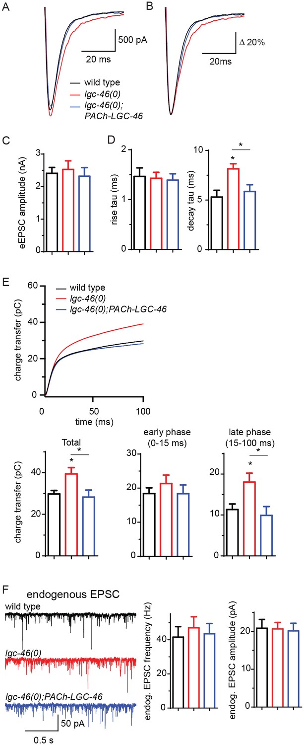
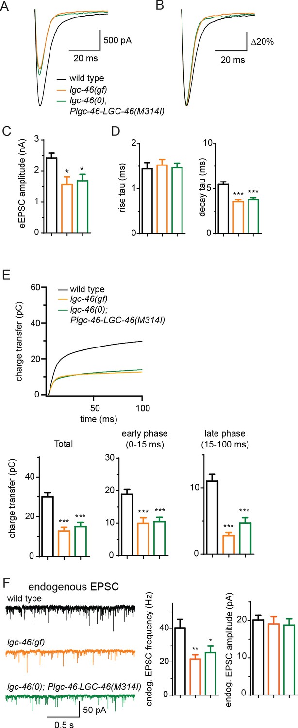
LGC-46 regulates the decay phase of eEPSCs to modulate the late phase of SV release.
(A and B) Average traces (A) and normalized average traces (B) of eEPSCs. (C) Mean amplitude of eEPSCs. (D) Rise tau and decay tau of eEPSCs. (E) Average traces of cumulative charges of eEPSCs, and charge transfers after stimulation. (F) Representative traces, mean frequency and mean amplitude of endogenous EPSCs. wild type (n = 11), lgc-46(ok2900) (n = 10), and lgc-46(ok2900); PACh-LGC-46 (n = 11). Animals were recorded at 20 degrees in 1.2 mM Ca2+ bath solutions. Statistics, one-way ANOVA, Bonferroni’s post hoc test. *p<0.05. Error bars indicate SEM.
Figure 2—figure supplement 1
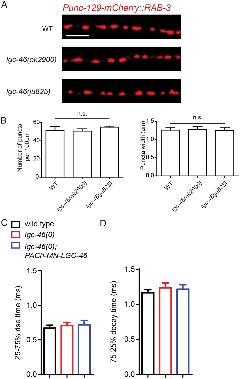
Morphology of cholinergic synapses as well as the amplitude and kinetics of endogenous EPSCs are normal in lgc-46(0).
(A) Representative images of mCherry::RAB-3 puncta in the dorsal nerve cord from each genotype. Scale bar: 5 μm. (B) Quantification of the puncta density and width show no significant differences between the wild type, lgc-46(ok2900) and lgc-46(ju825gf) backgrounds. Data shown as mean ± SEM, n = 6 animals (20 μm per animal) per genotype. Scale bar: 5 μm. (CD) 25%–75% rise time (C) and 75%–25% decay time (D) of endogenous EPSCs. n = 10 to 12 for all animals. Statistics, one way ANOVA and Bonferroni’s post hoc test. Error bars indicate SEM.
Figure 3 with 1 supplement
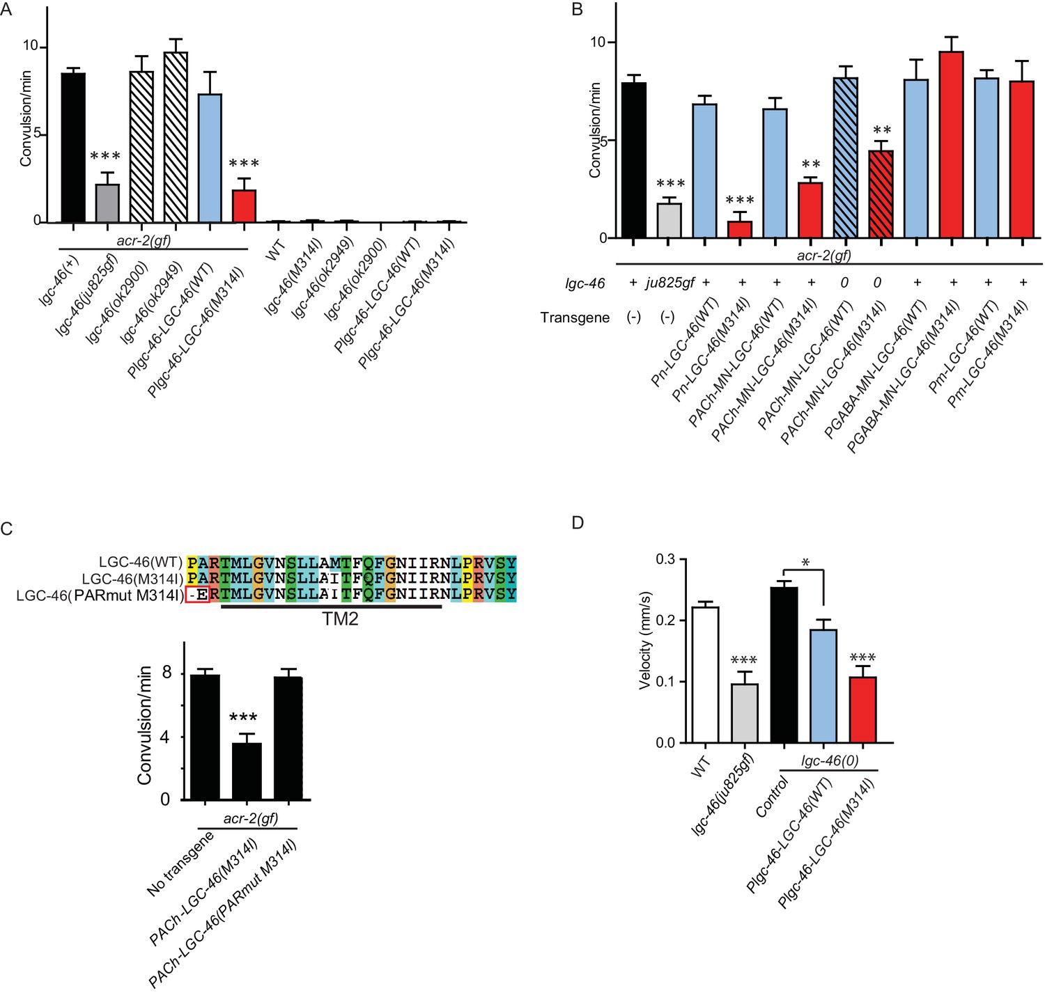
A. gain-of-function mutation LGC-46(M314I) affects excitation and inhibition imbalance in locomotor circuit.
(A) Quantification of convulsion frequencies. lgc-46(ju825), but not lgc-46(0), suppresses acr-2(gf) convulsions. Overexpression of LGC-46(M314I) under its own promoter also strongly suppresses convulsion. Data shown as mean ± SEM; n ≥ 16. (B) Overexpression of LGC-46(M314I) in cholinergic motor neurons can suppress acr-2(gf) convulsions. Data shown as mean ± SEM; n ≥ 16. Promoters used to drive expression are, Prgef-1 for neurons, Punc-17βfor cholinergic motor neurons, Punc-25 for GABAergic motor neurons, Pmyo-3 for muscles (See Supplementary file 1,2). (A and B) Statistics: one-way ANOVA followed by Dunnett’s post-hoc test. **p<0.01, ***p<0.001. (C) (Top) Amino acid sequences of LGC-46 wild type, M314I, and PAR motif mutant M314I. (Bottom) Convulsion frequencies of each genotype are shown. Cholinergic motor neuron-specific expression of LGC-46(M314I) suppresses acr-2(gf) convulsions, whereas the PAR motif mutant LGC-46(P301Δ A302E M314I) does not. Data shown as mean ± SEM; n = 24, 16, 16, respectively. Statistics: one way ANOVA followed by Dunnet’s post-hoc test. *p<0.05, ***p<0.001. (D) Off-food velocities for each genotype. Statistics: one-way ANOVA followed by Bonferroni’s post-hoc test. Data shown as mean ± SEM. **p<0.01, ***p<0.001.
Figure 3—figure supplement 1
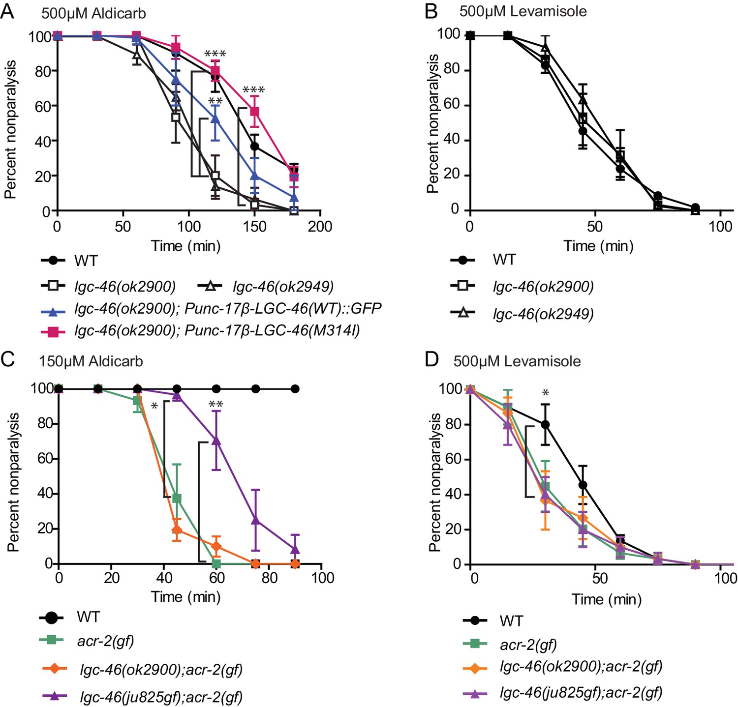
Pharmacological assays of aldicarb and levamisole.
(A) lgc-46(ok2900) is hypersensitive to aldicarb hypersensitivity, which is suppressed by cholinergic motor neuron-specific expression of functional LGC-46::GFP. (B) lgc-46(0) alleles do not affect levamisole sensitivity. (C,D) lgc-46(M314I) partially suppresses the aldicarb hypersensitivity (C) but not levamisole hypersensitivity (D) of acr-2(gf). Results are from three independent trials. n = 15 for one group per trial. Mean rate of paralysis at each time point are shown. Error bars indicate SEM. Statistics: **: p<0.01, *: p<0.05 by two-way ANOVA and Bonferroni post-hoc test.
Figure 4 with 1 supplement
LGC-46(M314I) limits synaptic transmission by shortening the decay phase of evoked release.
(A and B) Average traces (A) and normalized average traces (B) of eEPSCs. (C) Mean amplitude of eEPSCs. (D) Rise tau and decay tau of eEPSCs. (E) Average traces of cumulative charges of eEPSCs, and charge transfers after stimulation. (F) Representative traces, mean frequency and mean amplitude of endogenous EPSCs. wild type (n = 12), lgc-46(ju825gf) (n = 10), and lgc-46(0);Plgc-46-LGC-46(M314I) (n = 12). Animals were recorded at 20 degrees in 1.2 mM Ca2+ bath solutions. Statistics, one-way ANOVA, Bonferroni’s post hoc test. ***p<0.001, **p<0.01, *p<0.05. Error bars indicate SEM.
Figure 4—figure supplement 1
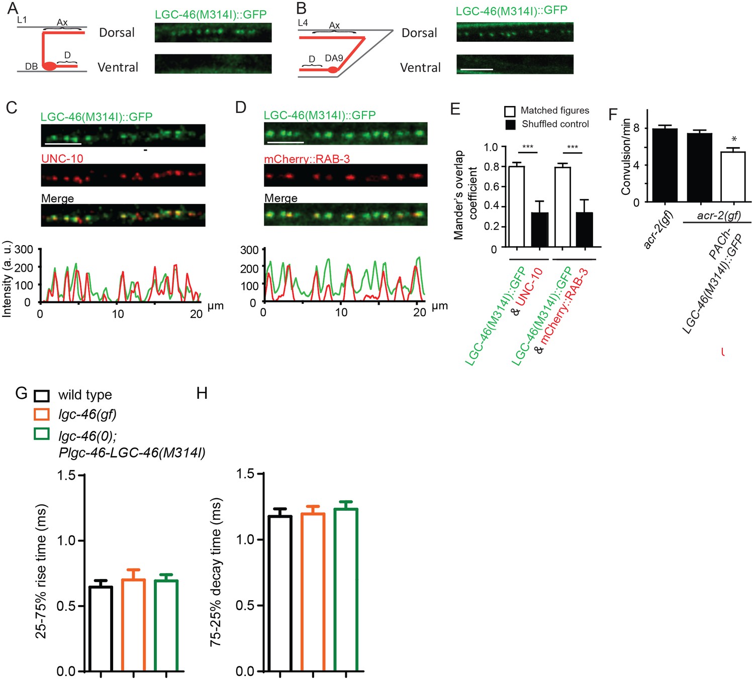
LGC-46(M314I) shows a presynaptic punctate localization pattern similar to LGC-46(WT), and amplitude and kinetics of endogenous EPSCs are normal in lgc-46(gf).
(A-D) Confocal images of animals expressing LGC-46(M314I)::GFP in cholinergic motor neurons. (A) (Left panel) Schematic of a cholinergic motor neuron in L1 animals. (Right panel) LGC-46(M314I)::GFP shows punctate localization on the dorsal process of cholinergic motor neurons of L1 animals. Images are from Punc-17β-LGC-46(M314I)(juSi282)IV. (B) (Left panel) Schematic of a cholinergic motor neuron DA9 in L4 animals. (Right panel) LGC-46(M314I)::GFP expressed in DA9 neuron shows punctate signals in the dorsal process in L4 animals, indicating their axonal localization. Images are from Pitr-1-LGC-46(M314I)::GFP(juEx6845). (C,D) Presynaptic proteins colocalize with LGC-46(M314I)::GFP in cholinergic motor neurons. Images taken from the dorsal nerve cord are shown. Lower panels show the linescan of fluorescent signal intensities. (C) Anti-GFP and anti-UNC-10 signals from an animal carrying Punc-17β-LGC-46(M314I)(juSi282)IV. (D) Presynaptic protein mCherry::RAB-3 expressed in cholinergic motor neurons overlapped with LGC-46(M314I)::GFP signals. Images are from Punc-17β-LGC-46(M314I) (juSi282)IV; Pacr-2- mCherry::RAB-3(juEx7053). (E) Mander’s overlap coefficient showing the extent of pixel colocalization of LGC-46 and preaynaptic marker protein signals (open bar). As negative controls, Mander’s overlap coefficient from shuffled images are shown (Filled bar). For each genotype, 10 images were analyzed. Data shown as mean ± SD. Statistics: one way ANOVA followed by Tukey’s post-hoc test. ***: p<0.001. (F) Single copy insertion allele Punc-17β-LGC-46(M314I)(juSi282)IV can significantly suppress the acr-2(gf) convulsion frequency. Data shown as mean ± SEM; n = 20, 16, 16, respectively. (G–H) 25%–75% rise time (G) and 75%–25% decay time (H) of endogenous EPSCs. n = 10 to 12 for all animals. Statistics, one way ANOVA and Bonferroni’s post hoc test. Error bars indicate SEM.
Figure 5 with 1 supplement
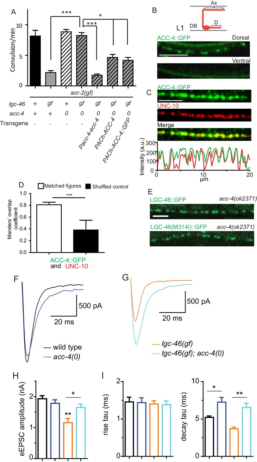
ACC-4 is required for the function of LGC-46(M314I), and localizes presynaptically.
(A) Frequency of convulsion in animals of indicated genotypes. Loss of function in acc-4 reverses the suppression effect by lgc-46(ju825) on acr-2(gf). The expression of acc-4(+) under the endogenous promoter or cholinergic motor neuron-specific promoter can rescue the effect. Data shown as mean ± SEM; n ≥ 16. Statistics: one way ANOVA followed by Dunnet’s post-hoc test. **p<0.01, ***p<0.001. (BD) ACC-4::GFP localizes to presynaptic terminals of cholinergic motor neurons, similar to LGC-46::GFP. Images are from lgc-46(gf) acc-4(0); acr-2(gf); Punc-17b-acc-4::GFP(juEx7438). (B) Upper panel shows a schematic of a cholinergic motor neuron in L1 animals. The lower panel shows a confocal image of ACC-4::GFP showing punctate localization in the dorsal nerve cord. (C) ACC-4::GFP colocalizes with active zone protein UNC-10/RIM. (D) Mander’s overlap coefficient showing the extent of pixel colocalization of ACC-4 and UNC-10 (open bar) from immunostaining. As negative controls, Mander’s overlap coefficient from shuffled images are shown (Filled bar). For each genotype, 10 images were analyzed. Data shown as mean ± SD. Statistics: Student’s t-test. ***p<0.001. (E) The punctate localization of LGC-46 is maintained in the acc-4(ok2371) null mutants. (F and G) Average traces of evoked release (eEPSC) from wild type (n = 10), acc-4(0) (n = 10), lgc-46(gf) (n = 10), and lgc-46(gf);acc-4(0) (n = 10). (H) Mean amplitude of eEPSC. (I) Rise tau and decay tau of eEPSC. Animals were recorded at 17°C in 2 mM Ca+ bath solutions. Statistics, one-way ANOVA. Bonferroni’s post hoc test for eEPSC comparisons. **p<0.01; *p<0.05. Error bars indicate SEM.
Figure 5—figure supplement 1
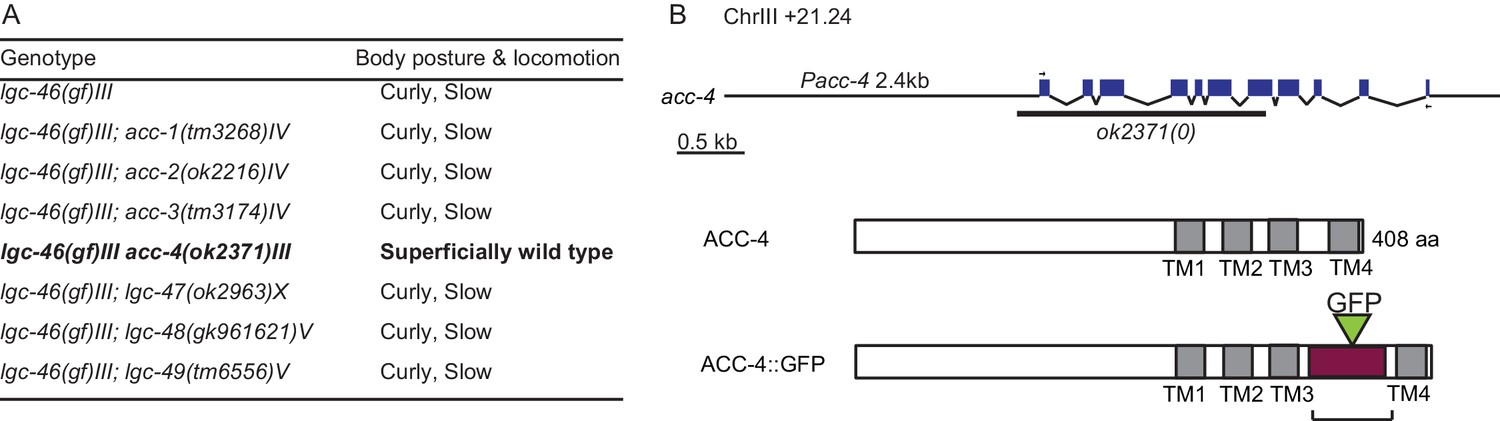
ACC-4 is required for the function of LGC-46(M314I).
(A) Locomotion observed in double mutants of lgc-46(ju825gf) and acc group genes. (B) Gene and protein structure of acc-4. Arrows indicate the position of the primers used to amplify cDNA. In ACC-4::GFP, the box filled with purple indicates the region where the GFP-tagged intracellular loop of LGC-46 was inserted.
Additional files
-
Supplementary file 1
List of strains used in the study.
- https://doi.org/10.7554/eLife.21734.012
-
Supplementary file 2
List of constructs used in the study.
- https://doi.org/10.7554/eLife.21734.013
Download links
A two-part list of links to download the article, or parts of the article, in various formats.
Downloads (link to download the article as PDF)
Open citations (links to open the citations from this article in various online reference manager services)
Cite this article (links to download the citations from this article in formats compatible with various reference manager tools)
Release-dependent feedback inhibition by a presynaptically localized ligand-gated anion channel
eLife 5:e21734.
https://doi.org/10.7554/eLife.21734


