RIPK1-RIPK3-MLKL-dependent necrosis promotes the aging of mouse male reproductive system
- National Institute of Biological Sciences, China
Figures
Figure 1 with 8 supplements
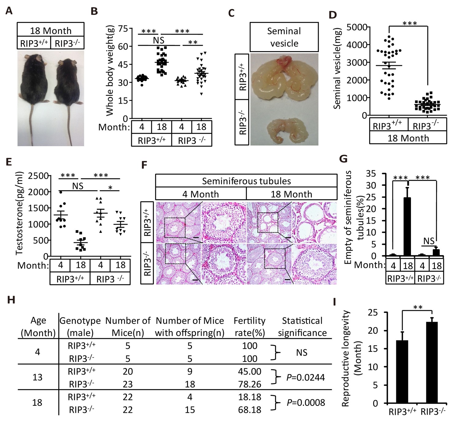
Ripk3-/- mice maintained their reproductive system function at an advanced age.
(A, B) Macroscopic features and weights of Ripk3+/+ (wild-type) and Ripk3-/- (Ripk3-knockout) male mice. Ripk3+/+ (4 Month, n = 16; 18 Month, n = 27) and Ripk3-/-(4 Month, n = 16; 18 Month, n = 27) male mice were photographed and weighed. Data represent the mean ±the standard error of the mean (s.e.m). **p<0.01, ***p<0.001. p values were determined with unpaired Student’s t-tests. NS, not significant. (C, D) Macroscopic features and weights of seminal vesicles. Mice were sacrificed at 18 months of age, and the seminal vesicles from Ripk3+/+ (n = 33) and Ripk3-/- (n = 30) mice were photographed and weighed. Data represent the mean ±s.e.m. ***p<0.001. p values were determined with unpaired Student’s t-tests. (E) Serum testosterone levels of mice assayed using ELISA. Mice were sacrificed, and the testosterone levels in serum from Ripk3+/+ (4 Month, n = 9; 18 Month, n = 9) and Ripk3-/- (4 Month, n = 9; 18 Month, n = 9) mice were measured using an ELISA kit for testosterone. Data represent the mean ±s.e.m. *p<0.05, ***p<0.001. p values were determined with unpaired Student’s t-tests. NS, not significant. (F, G) H and E of testis sections from Ripk3+/+ and Ripk3-/- mice. Ripk3+/+ (4 Months, n = 10; 18 Months, n = 10) and Ripk3-/- (4 Months, n = 10; 18 Months, n = 10) mice were sacrificed and testes were harvested and stained with H and E in (F). The number of empty seminiferous tubules was counted based on H and E staining and quantification in (G), empty seminiferous tubules were counted in five fields per testis. Scale bar, 100 μm. Data represent the mean ± S.D. ***p<0.001. p values were determined with unpaired Student’s t-tests. NS, not significant. (H) Summary of the fertility rates of Ripk3+/+ and Ripk3-/- mice. One male mice of a given age was mated with a pairs of 10-week-old wild-type female mice for 3 months; females were replaced every 2 weeks. The number of male mice with reproductive capacity was counted (see Materials and methods). p values were determined using chi-square tests. (I) Reproductive longevity. When Ripk3+/+ (n = 12) and Ripk3-/- (n = 12) male mice were 2 months old, they were continuously mated with a pairs of 10-week-old female mice until pregnancies ceased; females were replaced every 2 months. The ages of the males at which their last litter was sired was recorded (calculated as the age at birth of the litter less 21 days, see Materials and methods). Data represent the mean ± S.D. **p<0.01. p values were determined with unpaired Student’s t-tests.
-
Figure 1—source data 1
Summary of the fertility rates and mortality rates of the offspring of 4- or 18-month-old Ripk3+/+ and Ripk3-/- male mice.
- https://doi.org/10.7554/eLife.27692.003
-
Figure 1—source data 2
Summary of the fertility rates and mortality rates of the offspring of 13-month-old Ripk3+/+ and Ripk3-/- male mice.
- https://doi.org/10.7554/eLife.27692.004
Figure 1—figure supplement 1

Morphological changes in seminal vesicles and testis during aging.
(A, B) Macroscopic features and weights of seminal vesicles at different time points. Mice were sacrificed and the seminal vesicles from Ripk3+/+ and Ripk3-/- mice at different ages were photographed and weighed; each group is representative of at least twelve mice. Data represent the mean ±S.D. ***p<0.001. p values were determined with unpaired Student’s t-tests. (C) H and E of seminal vesicles sections from Ripk3+/+ and Ripk3-/- male mice. Ripk3+/+ and Ripk3-/- mice were sacrificed and seminal vesicles were harvested and stained with H and E; each group is representative of six mice. Scale bar, 50 μm. (D, E) Macroscopic features and weights of testes at different ages. Ripk3+/+ and Ripk3-/- mice of different ages were sacrificed and their testes were photographed and weighed; each group is representative of twelve mice. Data represent the mean ± S.D. **p<0.01, ***p<0.001. p values were determined with unpaired Student’s t-tests.
Figure 1—figure supplement 2
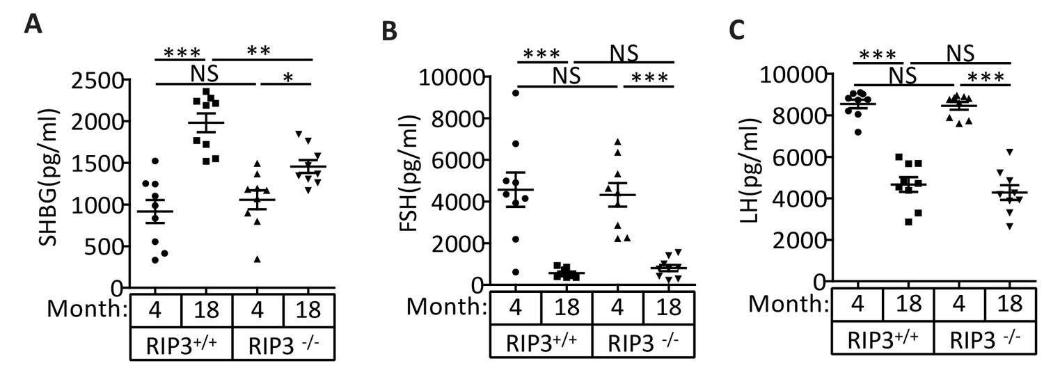
The levels of the pituitary endocrine hormones LH and FSH decline normally in Ripk3+/+ and Ripk3-/- mice.
(A) Serum SHBG levels of mice assayed using ELISA. Mice were sacrificed, and the levels of sex hormone-binding globulin (SHBG) in serum from Ripk3+/+ (4 Month, n = 10; 18 Month, n = 10) and Ripk3-/-(4 Month, n = 10; 18 Month, n = 10) mice were measured using an SHBG ELISA kit. (B) Serum FSH levels of mice assayed using ELISA. Mice were sacrificed, and the follicle-stimulating hormone (FSH) levels in serum from Ripk3+/+ (4 Month, n = 9; 18 Month, n = 9) and Ripk3-/-(4 Month, n = 9; 18 Month, n = 9) mice were measured using a FSH ELISA kit. (C) Serum LH levels of mice assayed using ELISA. Mice were sacrificed, and the luteinizing hormone (LH) levels in serum from Ripk3+/+ (4 Month, n = 9; 18 Month, n = 9) and Ripk3-/-(4 Month, n = 9; 18 Month, n = 9) mice was measured using an LH ELISA kit. All graphs present the mean ±s.e.m. **p<0.01, ***p<0.001. P values were determined with unpaired Student’s t-tests. NS, not significant.
Figure 1—figure supplement 3
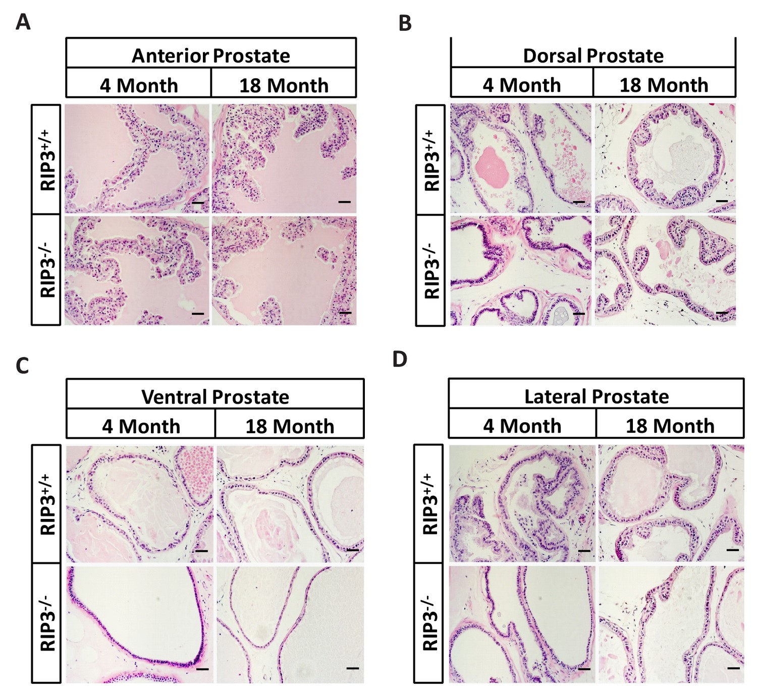
No morphological differences were apparent in the prostates of Ripk3+/+ and Ripk3-/- mice.
(A–D) H and E of Anterior/Dorsal/Ventral/Lateral prostate sections from Ripk3+/+ and Ripk3-/- mice. Mice were sacrificed, prostates from Ripk3+/+ and Ripk3-/- mice were harvested and stained with H and E; each group is representative of six mice. Scale bar, 50 μm.
Figure 1—figure supplement 4

Morphological changes in seminiferous tubules in 36-month-old mice.
(A, B) H and E of testis sections from Ripk3+/+ and Ripk3-/- mice. Mice were sacrificed, testes from Ripk3+/+ (36 Months, n = 6) and Ripk3-/- (36 Months, n = 6) mice were harvested and stained with H and E in (A). The number of empty seminiferous tubules was counted based on H and E staining and quantification in (B), empty seminiferous tubules were counted in five fields per testis. Scale bar, 200 μm. Data represent the mean ±s.e.m. ***p<0.001. p values were determined with unpaired Student’s t-tests.
Figure 1—figure supplement 5

Ripk3-/- mice have higher sperm counts than Ripk3+/+ mice at an advanced age.
(A) H and E of epididymis sections from Ripk3+/+ and Ripk3-/- mice. Mice were sacrificed and epididymides from Ripk3+/+ (4 Month, n = 6; 18 Months, n = 6) and Ripk3-/- (4 Month, n = 6; 18 Months, n = 6) mice were harvested and stained with H and E. Scale bar, 50 μm. (B) Sperm count. Mice were sacrificed, and epididymides from Ripk3+/+ and Ripk3-/- mice of at different ages were harvested. The number of sperm within the epididymis was counted using a cell counting chamber under a microscope; each group is representative of at least twelve mice. Data represent the mean ± S.D. **p<0.01, ***p<0.001. p values were determined with unpaired Student’s t-tests.
Figure 1—figure supplement 6

Histological analysis of various organs of Ripk3+/+ and Ripk3-/- mice.
(A–D) H and E of tissue sections from Ripk3+/+ and Ripk3-/- male mice. Mice of different ages were sacrificed, the organs from Ripk3+/+ and Ripk3-/- mice were harvested and stained with H and E; each group is representative of at least six mice. Scale bar, 100 μm.
Figure 1—figure supplement 7
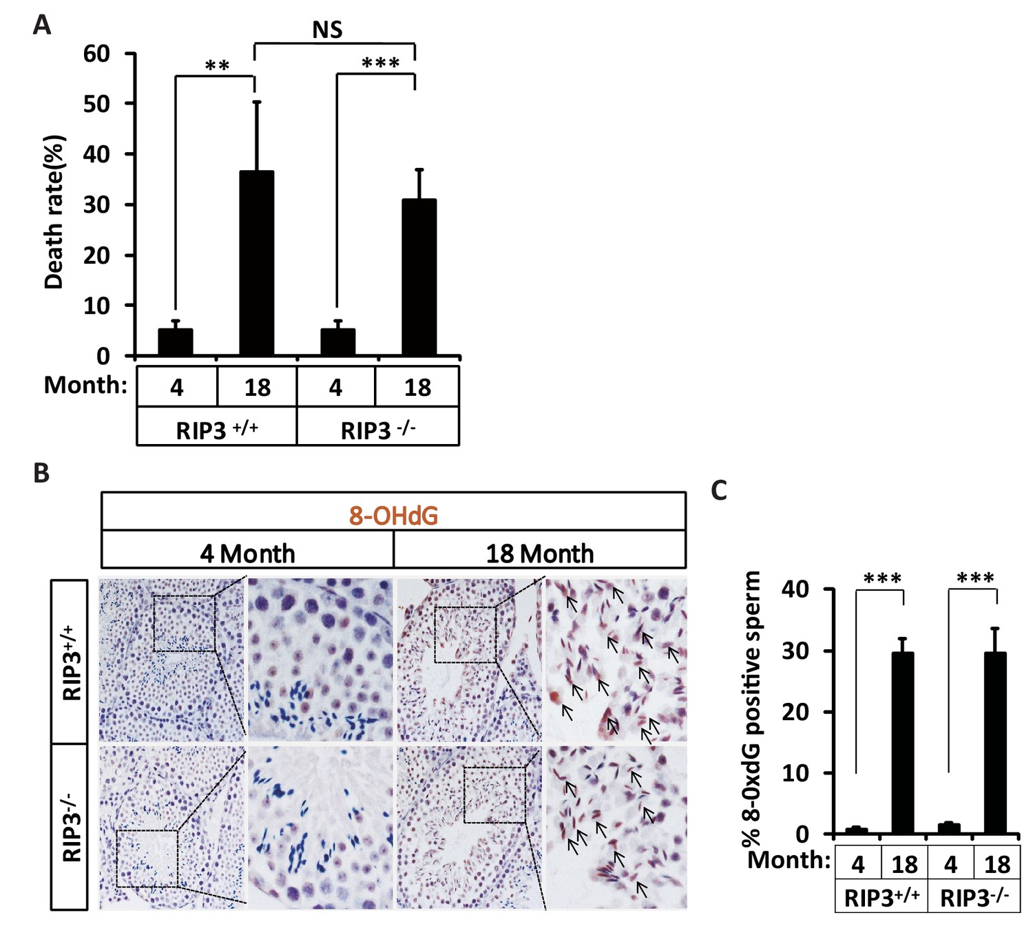
Increase of rates of birth defects and oxidative damage in sperm from aged Ripk3-/- mice.
(A) Mortality of offspring from Ripk3+/+ and Ripk3-/- male mice. One male mice of a given age was mated with a pairs of 10-week-old female wild-type mice for 3 months; females were replaced every 2 weeks. Litters were counted by date of birth of the pups; if a litter was born but did not survive, we counted the dead pups; if we were not able to count the pups, the number of pups was entered as ‘0’. Mortality of offspring from 4-month-old Ripk3+/+ (n = 5) and Ripk3-/- (n = 5) male mice; offspring from 18-month-old Ripk3+/+ (n = 4) and Ripk3-/- (n = 15) male mice were calculated. Data represent the mean ± S.D. **p<0.01, ***p<0.001. P values were determined with unpaired Student’s t-tests. NS, not significant. (B, C) IHC of Ripk3+/+ and Ripk3-/- testes with 8-OHdG antibody. Mice were sacrificed, testes from Ripk3+/+ (4 Months, n = 6; 18 Months, n = 6) and Ripk3-/-(4 Months, n = 6; 18 Months, n = 6) mice were harvested and stained with 8-OHdG antibody in (B) (black arrows for sperm with 8-OHdG staining). 8-OHdG + sperm were counted in five fields per testis and quantification in (C). Scale bar, 100 μm. Data represent the mean ± S.D. ***p<0.001. p values were determined with unpaired Student’s t-tests.
Figure 1—figure supplement 8

Weights of fat and seminal vesicles from 18-month-old Ripk3+/+ and Ripk3-/- male mice.
(A–D) Ripk3+/+ (18 Month, n = 7) and Ripk3-/-(18 Month, n = 7) male mice were weighed in (A). Mice were sacrificed and the seminal vesicles and fat from Ripk3+/+ and Ripk3-/- mice were weighed in (B, C and D). Data represent the mean ± s.e.m. ***p<0.001. p values were determined with unpaired Student’s t-tests.
Figure 2 with 1 supplement
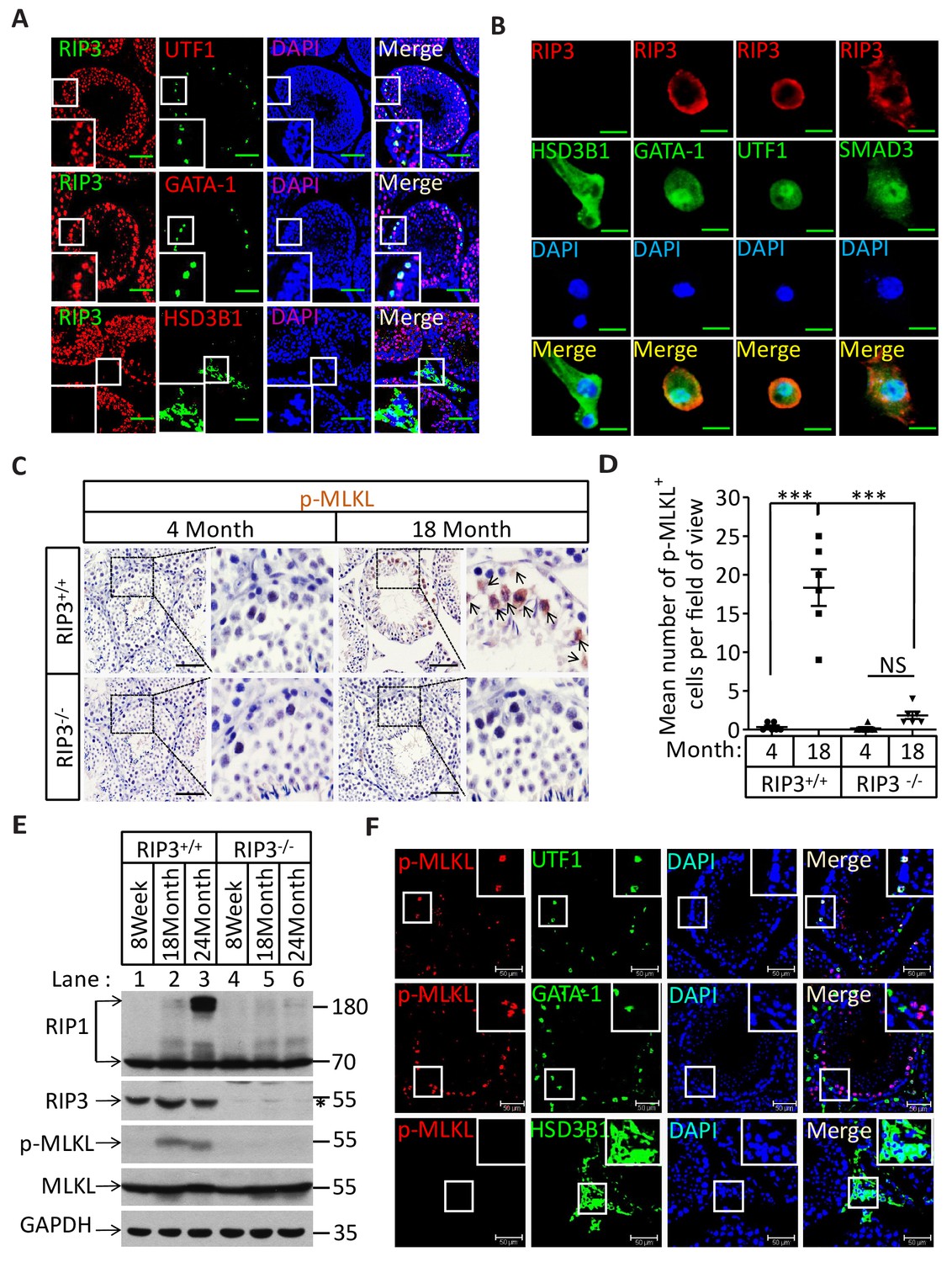
Necroptosis in seminiferous tubules of aged wild-type mice.
(A) RIPK3 expression in spermatogonia, Sertoli cells, and spermatocytes. Immunofluorescence in an 8-week-old testis with antibodies against RIPK3 (red), HSD3B1 (Leydig cells specific protein, green), GATA-1 (Sertoli cells specific protein, green), and UTF1 (spermatogonium specific protein, green). Scale bar, 100 μm. (B) RIPK3 expression in germ line cells and Sertoli cells. Primary testis cells were isolated from wild-type testes, Immunofluorescence of Leydig cells, Sertoli cells, spermatogonia, and primary spermatocytes with antibodies against RIPK3 (red), HSD3B1 (green), GATA-1 (green), UTF1 (green), and SMAD3 (primary spermatocytes specific protein, green). Counterstaining with DAPI, blue. Scale bar, 10 μm. (C, D) Immunohistochemistry (IHC) of testes from Ripk3+/+ and Ripk3-/- mice with phosphor-MLKL (p-MLKL) antibody. Ripk3+/+ (4 Months, n = 6; 18 Months, n = 6) and Ripk3-/- (4 Months, n = 6; 18 Months, n = 6) mice were sacrificed and testes were harvested and stained with p-MLKL antibody in (C) (black arrows indicate cells with p-MLKL staining). p-MLKL+ cells were counted in five fields per testis and quantification in (D). Scale bar, 100 μm. Data represent the mean ± s.e.m. ***p<0.001. p values were determined with unpaired Student’s t-tests. NS, not significant. (E) Western blot analysis of RIPK1, RIPK3, MLKL, and p-MLKL levels in the testis after perfusion, each group is representative of at least three mice. GAPDH was used as loading control. The asterisk (*) indicates non-specific bands. (F) Immunofluorescence in an 18-month-old testis with antibodies against p-MLKL (red, purple arrows indicate spermatogonium with p-MLKL staining), HSD3B1, GATA-1, and UTF1. Scale bar, 50 μm.
Figure 2—figure supplement 1
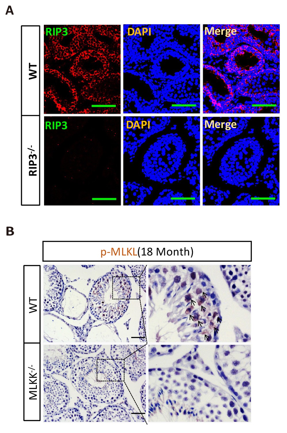
RIPK3 expression in seminiferous tubules.
(A) Immunofluorescence of testes from Ripk3+/+ and Ripk3-/- mice (2 weeks) with RIPK3 antibody. Counterstaining with DAPI, blue. Scale bar, 100 μm. (B) No p-MLKL signaling in the testes from aged Mlkl-/- mice. WT (18 Months, n = 3) and Mlkl-/- (18 Months, n = 3) mice were sacrificed and testes were harvested and stained with p-MLKL antibody (black arrows indicate cells with p-MLKL staining). Scale bar, 100 μm.
Figure 3 with 2 supplements
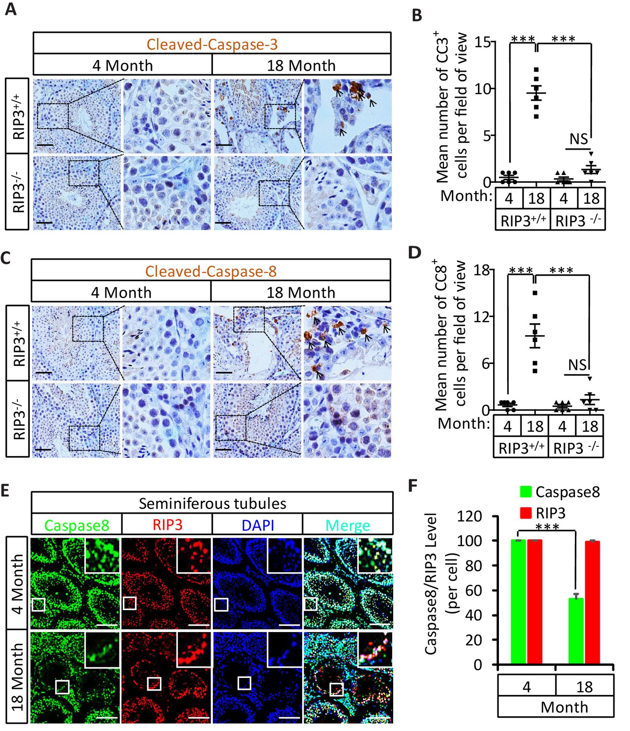
Activation of apoptosis in Leydig cells during aging.
(A, B) IHC of testis from Ripk+/+ and Ripk3-/- mice with Cleaved-Caspase-3 antibody. Mice were sacrificed, testes from Ripk3+/+ (4 Month, n = 6; 18 Month, n = 6) and Ripk3-/- (4 Month, n = 6; 18 Month, n = 6) mice were harvested and stained with Cleaved-Caspase-3 antibody in (A) (black arrows for Leydig cells with Cleaved-Caspase-3 staining). Cleaved-Caspase-3+ cells were counted in six fields per testis and quantification in (B). Scale bar, 100 μm. Data represent the mean ± s.e.m. *p<0.05, ***p<0.001. p values were determined with unpaired Student’s t-tests. (C, D) IHC of testis from Ripk+/+ and Ripk-/- mice with Cleaved-Caspase-8 antibody. Mice were sacrificed, testes from Ripk+/+ (4 Month, n = 6; 18 Month, n = 6) and Ripk3-/- (4 Month, n = 6; 18 Month, n = 6) mice were harvested and stained with Cleaved-caspase-8 antibody in (C) (black arrows for Leydig cells with Cleaved-caspase-8 staining). Cleaved-caspase-8+ cells were counted in six fields per testis and quantification in (D). Scale bar, 100 μm. Data represent the mean ± s.e.m. *p<0.05, ***p<0.001. p values were determined with unpaired Student’s t-tests. (E, F) Caspase8 levels decrease during aging in empty seminiferous tubules. Immunofluorescence of testes from 4-month-old and 18-month-old wild-type mice with caspase8 and RIPK3 antibody in (E). The caspase8 levels were quantified in (F). Counterstaining with DAPI, blue. Scale bar, 100 μm.
Figure 3—figure supplement 1
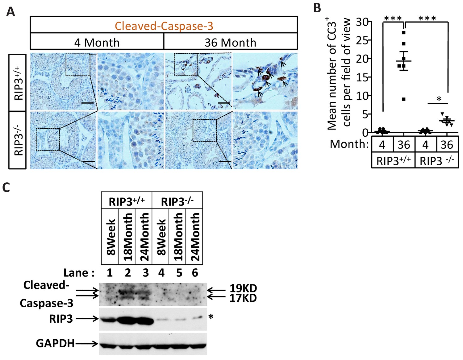
Activation of apoptosis in Leydig cells during aging.
(A, B) IHC of testis from Ripk3+/+ and Ripk3-/- mice with Cleaved-Caspase-3 antibody. Mice were sacrificed, testes from Ripk3+/+ (4 Month, n = 6; 36 Months, n = 6) and Ripk3-/- (4 Month, n = 6; 36 Months, n = 6) mice were harvested and stained with Cleaved-Caspase-3 antibody in (A) (black arrows for Leydig cells with Cleaved-Caspase-3 staining). Cleaved-Caspase-3+ cells were counted in six fields per testis and quantification in (B). Scale bar, 100 μm. Data represent the mean ± s.e.m. *p<0.05, ***p<0.001. p values were determined with unpaired Student’s t-tests. (C) Western blot analysis of RIPK3 and Cleaved-caspase-3 levels in the testis after perfusion; each group is representative of at least three mice. GAPDH was used as a loading control.
Figure 3—figure supplement 2

Caspase8 levels increase during aging in Leydig cells.
Immunofluorescence of testes from 4-month-old and 18-month-old wild-type mice with caspase8 antibody. Counterstaining with DAPI, blue. Scale bar, 100 μm.
Figure 4
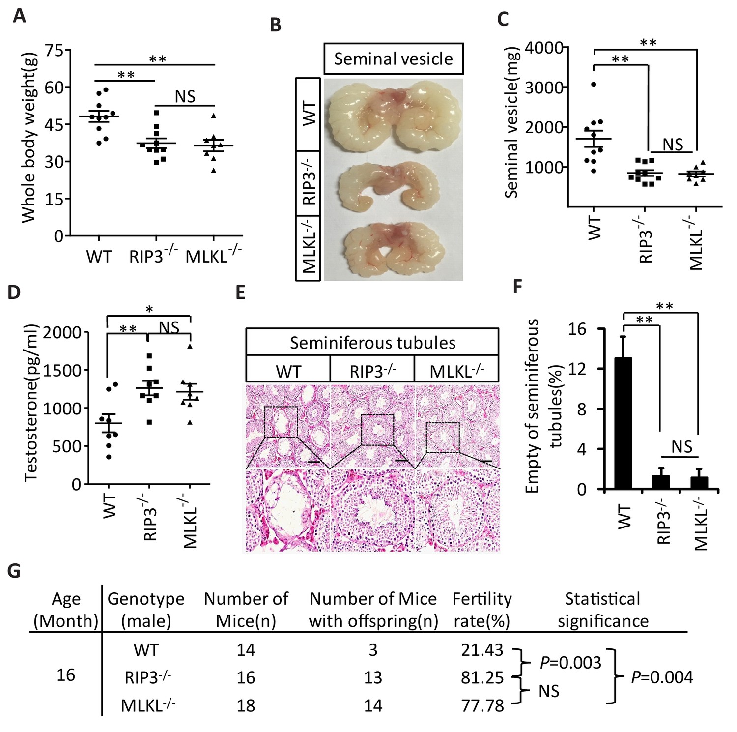
Aging of reproductive organs is delayed in Mlkl-/- mice.
(A) Weight of WT (wild-type), Ripk3-/-, and Mlkl-/- male mice. WT (15 Month, n = 10), Ripk3-/-(15 Month, n = 10) and Mlkl-/- (15 Month, n = 8) male mice were weighed. Data represent the mean ± s.e.m. **p<0.01. p values were determined with unpaired Student’s t-tests. NS, not significant. (B, C) Macroscopic features and weights of seminal vesicles. WT (15 month, n = 10), Ripk3-/- (15 Month, n = 10) and Mlkl-/-(15 Month, n = 8) male mice were sacrificed and the seminal vesicles were photographed and weighed. Data represent the mean ± s.e.m. **p<0.01. p values were determined with unpaired Student’s t-tests. NS, not significant. (D) Serum testosterone levels of mice assayed using ELISA. Mice were sacrificed and the level of testosterone in serum from WT (15 Month, n = 8), Ripk3-/-(15 Month, n = 8) and Mlkl-/-(15 Month, n = 8) mice was measured using a testosterone ELISA kit. Data represent the mean ± s.e.m. *p<0.05, **p<0.01. p values were determined with unpaired Student’s t-tests. NS, not significant. (E, F) H and E of testis sections from WT, Ripk3-/-, and Mlkl-/mice. Testes from WT (15 Months, n = 8), Ripk3-/- (15 Months, n = 8), and Mlkl-/-(15 Month, n = 8) mice were harvested and stained with H and E in (E). The number of empty seminiferous tubules was counted based on H and E staining and quantification in (F), empty seminiferous tubules were counted in five fields per testis. Scale bar, 100 μm. Data represent the mean ± S.D. **p<0.01. p values were determined with unpaired Student’s t tests. (G) Summary of the fertility rates of WT, Ripk3+/+ and Mlkl-/- mice. One male mice of a given age was mated with a pairs of 10-week-old wild-type female mice for 3 months; females were replaced every 2 weeks. The number of male mice with reproductive capacity was counted (see Materials and methods). p values were determined using chi-square tests.
Figure 5 with 1 supplement

Induction of necroptosis in testis depleted cells in seminiferous tubules.
(A–E) Testes of WT (2 Months, n = 6), Ripk3-/- (2 Months, n = 6), and Mlkl-/-(2 Month, n = 6) mice were injected with TSZ or saline (see Materials and methods); 72 hr after the injection, mice were sacrificed and the testes were harvested. The proteins were extracted from testes and were analyzed with western blotting in (A, B). GAPDH was used as a loading control. The testes were stained with p-MLKL antibody in (C) (black arrows indicate cells with p-MLKL staining). Scale bar, 100 μm. The testes were stained with H and E in (D). The number of empty seminiferous tubules was counted based on H and E staining and quantification in (E), empty seminiferous tubules were counted in five fields per testis. Scale bar, 100 μm. Data represent the mean ± S.D. ***p<0.001. p values were determined with unpaired Student’s t-tests. NS, not significant.
Figure 5—figure supplement 1

Activation of necroptosis in germ line stem cells and Sertoli cells in seminiferous tubules.
Primary testis cells were isolated from wild-type testes, immunofluorescence of Leydig cells, Sertoli cells, spermatogonia, and primary spermatocytes with antibodies against p-MLKL (red), HSD3B1 (green), GATA-1 (green), UTF1 (green), and SMAD3 (green) after stimulation with TSZ for 10 hr. Counterstaining with DAPI, blue. Scale bar, 10 μm.
Figure 6

Induction of necroptosis in testes accelerates aging of the male reproductive system.
(A–D) Testes from WT (3 Months, n = 9), Ripk3-/- (3 Months, n = 9), and Mlkl-/-(3 Month, n = 7) mice were injected with TSZ or saline (see Materials and methods) and were maintained in SPF facility for 3 months. Mice were then sacrificed and the seminal vesicles were photographed and weighed. Macroscopic features and weights of seminal vesicles form mice in (A, B). Data represent the mean ± s.e.m. ***p<0.001, p values were determined with unpaired Student’s t-tests. Testes were harvested and stained with H and E in (C). The number of empty seminiferous tubules was counted based on H and E staining and quantification in (D). Empty seminiferous tubules were counted in five fields per testis. Scale bar, 100 μm. Data represent the mean ± S.D. ***p<0.001, p values were determined with unpaired Student’s t-tests. NS, not significant. (E) Summary of the fertility rate of WT, Ripk3-/-, and Mlkl-/- male mice after injection with TSZ. Testes from WT (3 Months, n = 8), Ripk3-/- (3 Months, n = 8), and Mlkl-/-(3 Month, n = 8) male mice were injected with TSZ or saline (see Materials and methods) and mice were maintained in SPF for 3 months. One male mouse was mated with a pairs of 10-week-old female wild-type mice for 2 months; females were replaced every 2 weeks. The number of male mice with reproductive capacity was counted (see Materials and methods). p values were determined using chi-square tests.
Figure 7 with 1 supplement
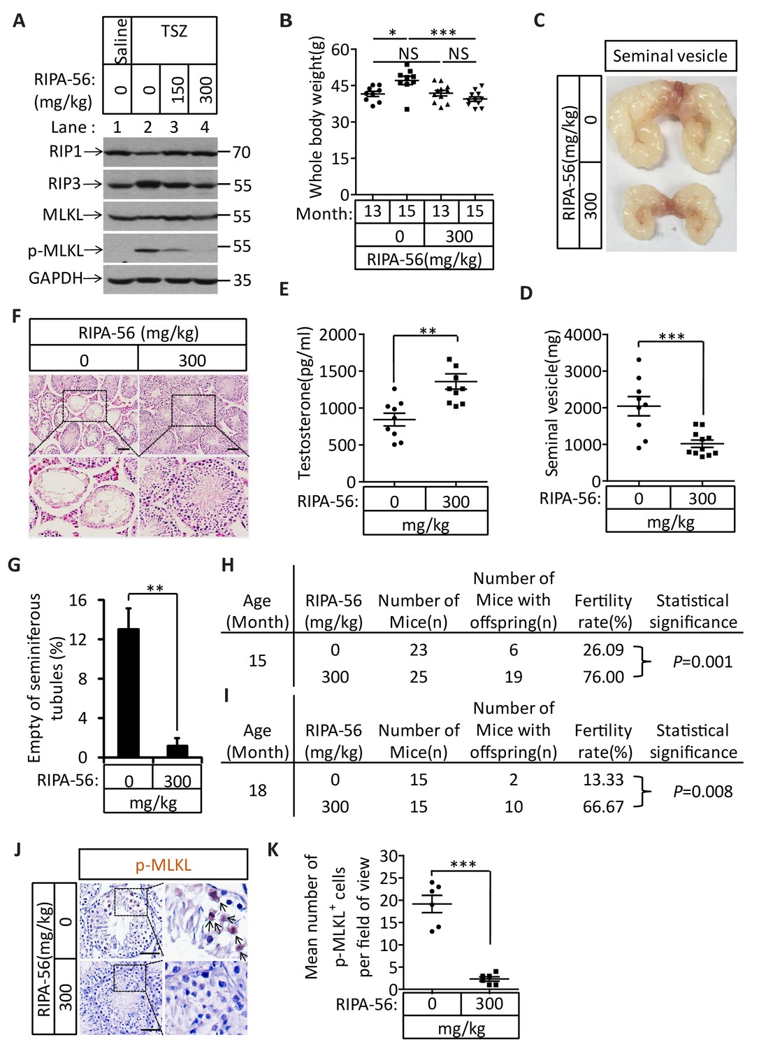
An RIPK1 inhibitor blocks aging of the male reproductive system.
(A) Western blot analysis of RIPK1, RIPK3, MLKL, and p-MLKL levels in testes after injection with TSZ. The 2-month-old wild-type male mice continuously feed with RIPA-56 (0 mg/kg, n = 6; 150 mg/kg, n = 3; 300 mg/kg, n = 3) for one week. Testes were injected with TSZ (see Materials and methods); 72 hr after the injection, mice were sacrificed and the testes were harvested. The proteins were extracted from testes and were analyzed with western blotting. GAPDH was used as a loading control. (B–H) 13-month-old wild-type male mice were feed with AIN93G (RIPA-56:0 mg/kg) or AIN93G-RIPA-56 (RIPA-56:300 mg/kg) for 2 months in SPF facility. Mice were weighed before and after feed with RIPA-56 in (B). Data represent the mean ± s.e.m. *p<0.05, ***p<0.001. p values were determined with unpaired Student’s t-tests. Mice were sacrificed and the seminal vesicles were photographed and weighed. Macroscopic features and weights of seminal vesicles form mice in (C, D). Data represent the mean ± s.e.m. ***p<0.001. p values were determined with unpaired Student’s t-tests. Testosterone levels in serum from mice were measured using a testosterone ELISA kit in (E). Data represent the mean ± s.e.m. **p<0.01. p values were determined with unpaired Student’s t-tests. The testes were harvested and stained with H and E in (F). The number of empty seminiferous tubules was counted based on H and E staining and quantification in (G). Empty seminiferous tubules were counted in five fields per testis. Scale bar, 100 μm. Data represent the mean ± S.D. **p<0.01. p values were determined with unpaired Student’s t-tests. The fertility rate of each RIPA-56-treated (0 mg/kg, n = 23; 300 mg/kg, n = 25) male mice was assessed by mating it with four 10-week-old wild-type female mice successively (see Materials and methods). The number of mice with reproductive capacity was counted in (H). p values were determined using chi-square tests. (I–K) 13-month-old wild-type male mice were feed with AIN93G (RIPA-56:0 mg/kg) or AIN93G-RIPA-56 (RIPA-56:300 mg/kg) for 5 months in SPF facility. The fertility rate of each RIPA-56-treated (0 mg/kg, n = 15; 300 mg/kg, n = 15) male mice was assessed by mating it with four 10-week-old wild-type female mice successively (see Materials and methods). The number of mice with reproductive capacity was counted in (I). p values were determined using chi-square tests. After fertility test, mice were sacrificed and testes were harvested and stained with p-MLKL antibody in (J) (black arrows indicate cells with p-MLKL staining). p-MLKL+ cells were counted in five fields per testis and quantification in (K). Scale bar, 100 μm. Data represent the mean ± s.e.m. ***p<0.001. P values were determined with unpaired Student’s t-tests.
Figure 7—figure supplement 1

TNF-α increased in the testes from aged wild-type mice.
Western blot analysis of TNF-α, RIPK1, RIPK3, MLKL, Cleaved-caspase3 and p-MLKL levels in the testis after perfusion, each group is representative of at least three mice. GAPDH was used as loading control.
Download links
A two-part list of links to download the article, or parts of the article, in various formats.
Downloads (link to download the article as PDF)
Open citations (links to open the citations from this article in various online reference manager services)
Cite this article (links to download the citations from this article in formats compatible with various reference manager tools)
RIPK1-RIPK3-MLKL-dependent necrosis promotes the aging of mouse male reproductive system
eLife 6:e27692.
https://doi.org/10.7554/eLife.27692


