Inhibition of PIP4Kγ ameliorates the pathological effects of mutant huntingtin protein
- Jan and Dan Duncan Neurological Research Institute, Texas Children's Hospital, United States
- Texas Medical Center, United States
- University of Michigan, United States
- National Center for Advancing Translational Sciences, United States
- Northwestern University, United States
- University of Cambridge, United Kingdom
Figures
Figure 1 with 3 supplements
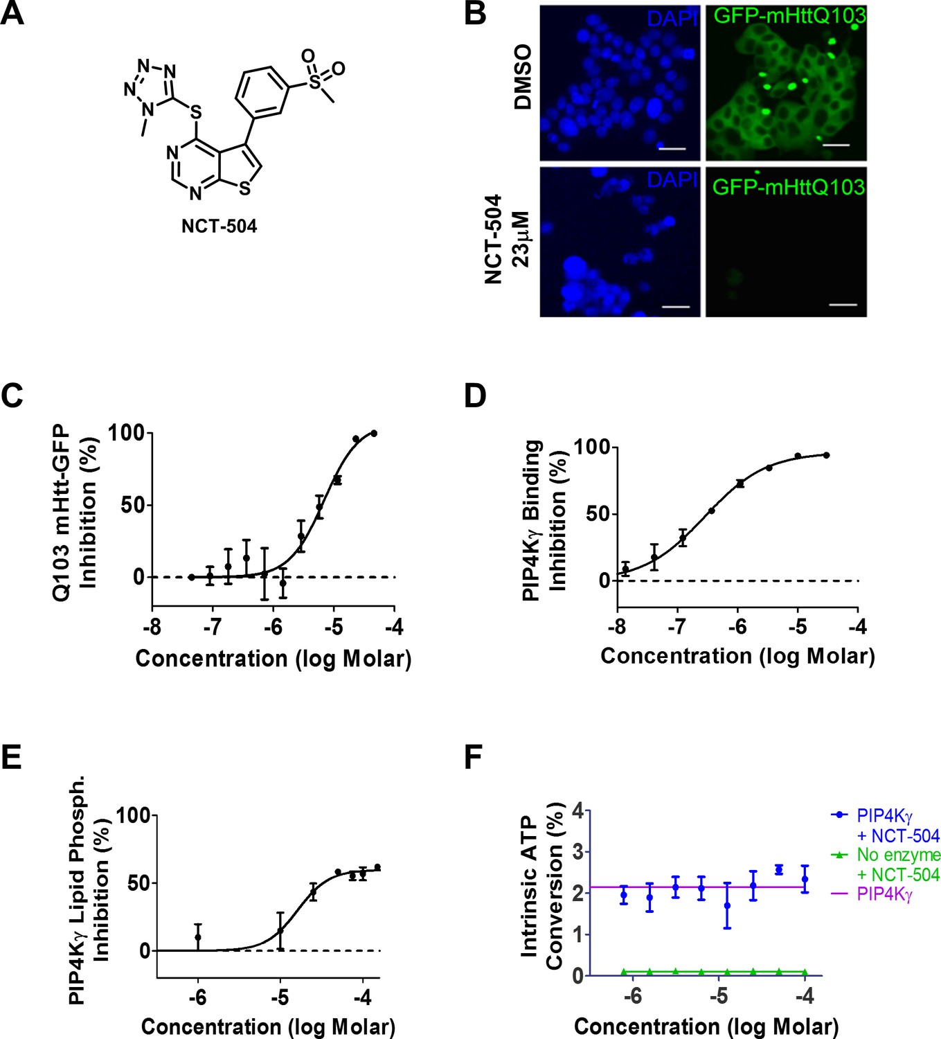
Identification of NCT-504 and its inhibition of PIP4Kγ.
(A) Structure of NCT-504. (B) NCT-504 treatment reduces Htt(exon1)-Q103 in PC12 cells. Cells with stable expression of ecdysone-inducible GFP-Htt(exon1)-Q103 (green), induced for 24 hr, and treated with DMSO (top panels) or 23 μM NCT-504 (bottom). Cells stained with DAPI (blue). Scale Bar = 50 μm. (C) Concentration-response curve of NCT-504 inhibition of cellular accumulation of GFP-Htt(exon1)-Q103 in PC12 cells. (D) NCT-504 inhibition of PIP4Kγ binding to an immobilized proprietary active site ligand (DiscoverX KINOMEscan https://www.discoverx.com/services/drug-discovery-development-services/kinase-profiling/kinomescan). (E) NCT-504 exhibits dose-dependent inhibition of phosphorylation of PI4P by full length isolated PIP4Kγ. (F) The intrinsic ATPase specific activity of full length isolated PIP4Kγ in the absence of PI5P substrate as a function of NCT-504 concentration is the same in the presence (blue) or in the absence (purple) of NCT-504.
Figure 1—figure supplement 1

NCT-504 suppresses the accumulation of HTT-exon1 aggregates.
HEK293T cells were either transfected with GFP-HTT(exon1)-Q23 or GFP-HTT(exon1)-Q74. Two hours after transfection, cells were either treated with DMSO or NCT-504 (2 μM). After 48 hr of treatment, cells were fixed and quantification of the cells transfected with GFP-HTT(exon1)-Q74 for the percentage of cells with aggregates is shown below (results from three independent experiments. Statistical significance was analyzed using paired one tailed student T test ***p<0.005. Bar = 50 μm.
Figure 1—figure supplement 2
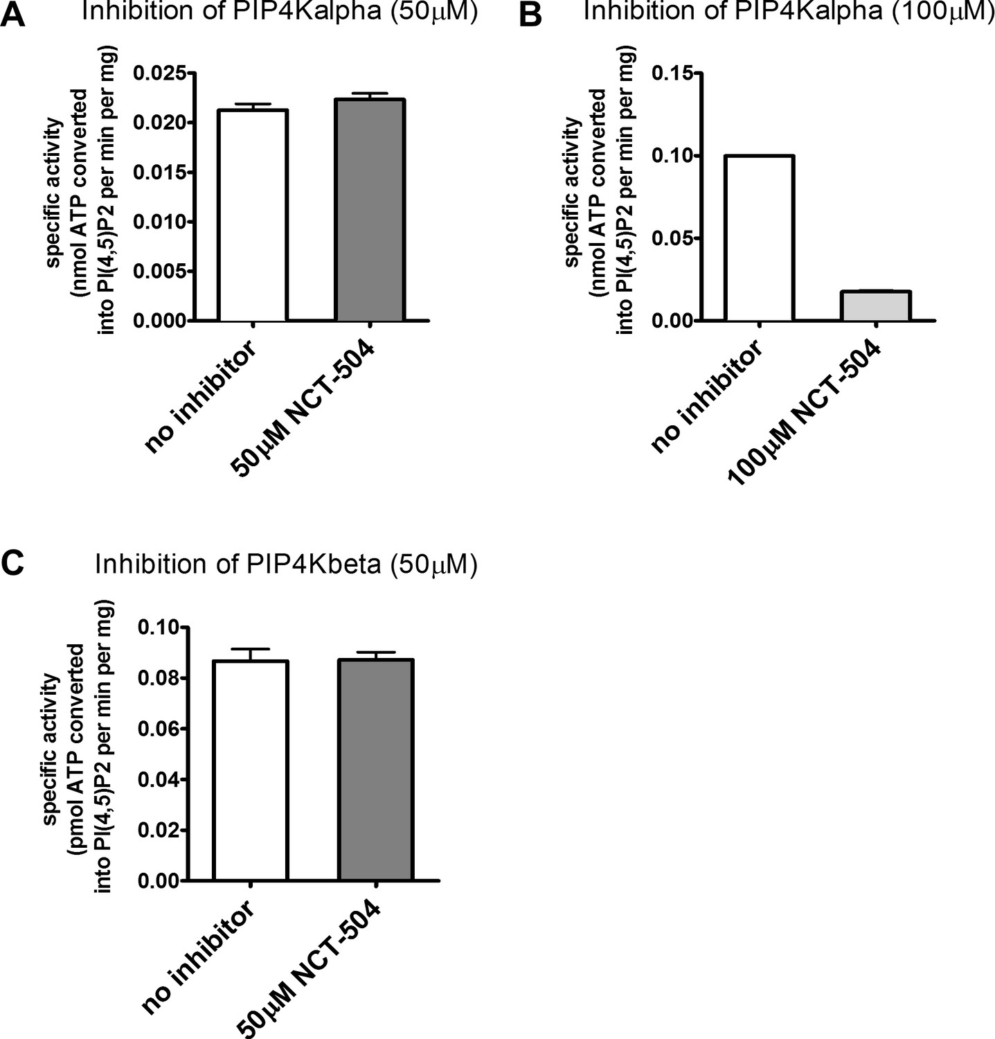
NCT-504 does not inhibit PIP4Kbeta and weakly inhibits PIP4Kalpha phosphorylation of PI5P.
Results from three independent experiments are shown for inhibition of phosphorylation of PI5P. (A) PIP4Kalpha with 50 μM NCT-504, (B) 100 μM NCT-504, and (C) PIP4Kbeta with 100 μM NCT-504.
Figure 1—figure supplement 3
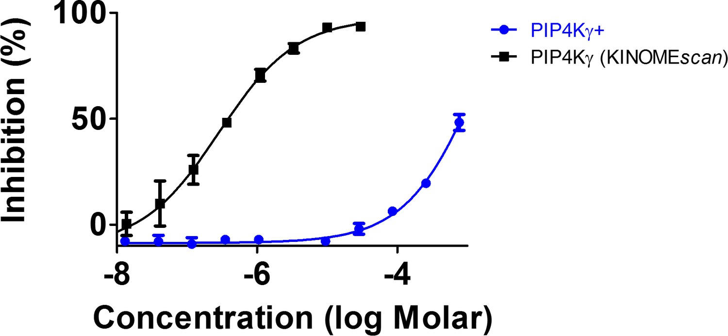
A PIP4Kγ+ G-loop mutant is resistant to inhibition by NCT-504, consistent with NCT-504 functioning as an allosteric inhibitor.
PIP4Kγ+ contains mutations in the G-loop and additional mutations that increase the low intrinsic ATP turnover exhibited by PIP4Kγ in the presence of PI5P. The PIP4Kγ+ mutant kinase is described as PI5P4Kγ G3A + B in reference (Clarke and Irvine, 2013). NCT-504 was almost inactive (potency >500 μM) against PIP4Kγ+ (blue). In comparison, activity against the PIP4Kγ construct at DiscoverRx KINOMEscan assay) results are shown in black. N = 3 for each concentration tested. The data is presented as % inhibition of kinase binding to a proprietary active site immobilized ligand by a compound that binds to the kinase active site directly (sterically) or indirectly (allosterically). (https://www.discoverx.com/technologies-platforms/competitive-binding-technology/kinomescan-technology-platform).
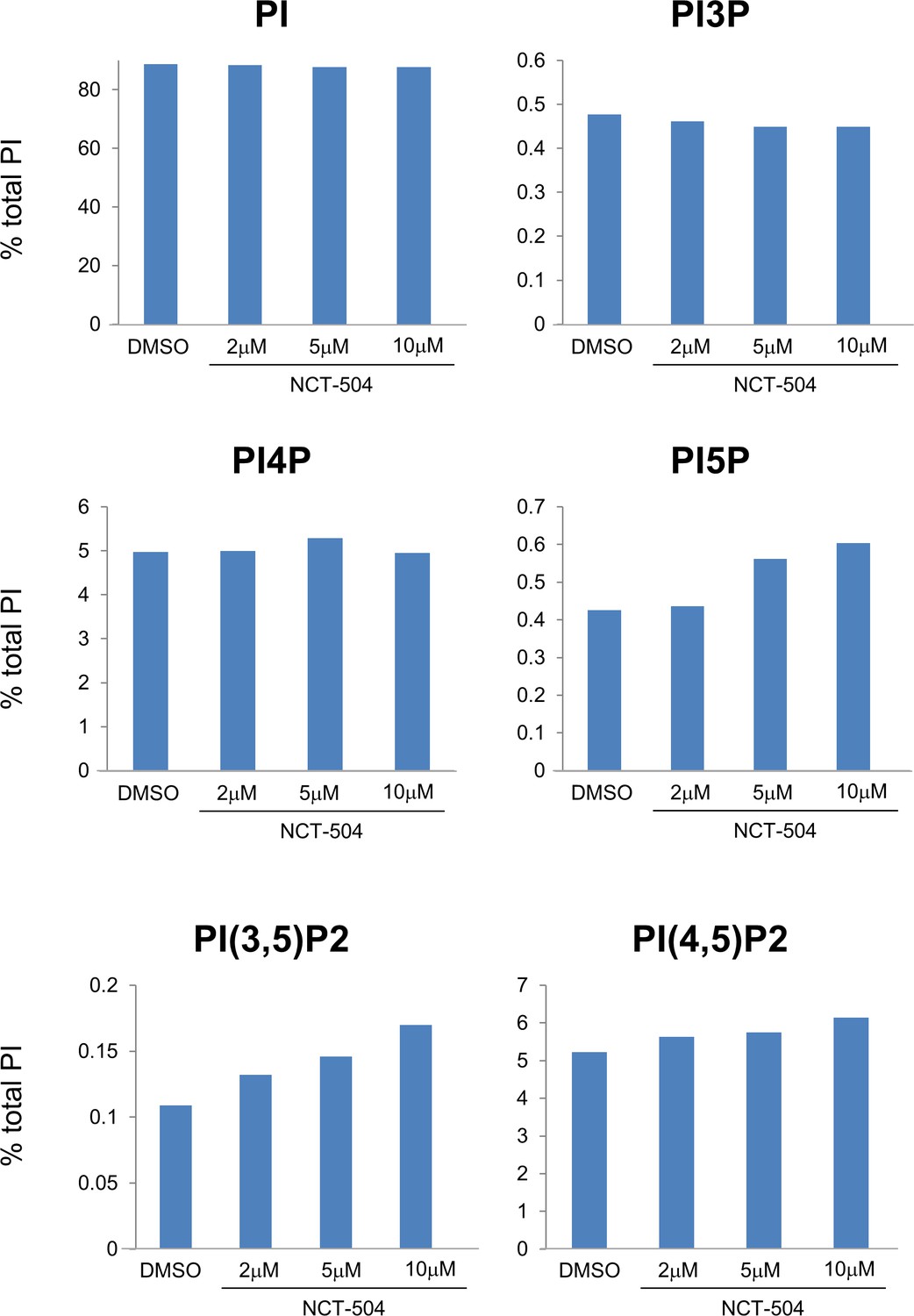
Figure 2 with 3 supplements

Pharmacologic and genetic inhibition of PIP4Kγ elevates the levels of PI(3,5)P2, PI3P and PI5P in MEFs.
(A–F) Pharmacologic (NCT-504 10 μM, 12 hr) and genetic (shRNA) inhibition of PIP4Kγ leads to increased levels of PI5P (D), PI(3,5)P2 (E) and PI3P (B), with no significant change in the levels of phosphatidylinositol (A), PI4P (C) or PI(4,5)P2 (F). However, there was a modest reduction in PI4P. Note in Figure 2—figure supplement 2, this small change was statically significant. Measurements were performed in MEF cells incubated with 3H-inositol labeled media for 48 hr. Statistical significance was analyzed using paired one tailed student t-test (n = 3), *p<0.05, **p<0.01. (G) Anti-PIP4Kγ western blot showing the effective silencing of the enzyme using shRNA. (GAPDH used as loading control).
Figure 2—figure supplement 1

NCT-504 treatment does not affect cell viability in MEFs.
Primary MEF cells treated with 10 μM NCT-504 or DMSO (control) for 12 hr were incubated with Hoechst 33342 and ethidium homodimer (Ethd1) for 15 min to measure cell viability. Ethd1 is impermeable to the nucleus in live cells; whereas in dead cells, Ethd1 displaces Hoechst and stains the nucleus red. Percentage of cells negative for Ethd1-nuclear stain were quantified from three experiments. A minimum of 640 cells were analyzed. White arrow indicates a dead cell. Scale Bar 100 μm. Student’s one-tailed t-test indicates no statistically significant difference between DMSO and NCT-504 treatment.
Figure 2—figure supplement 2
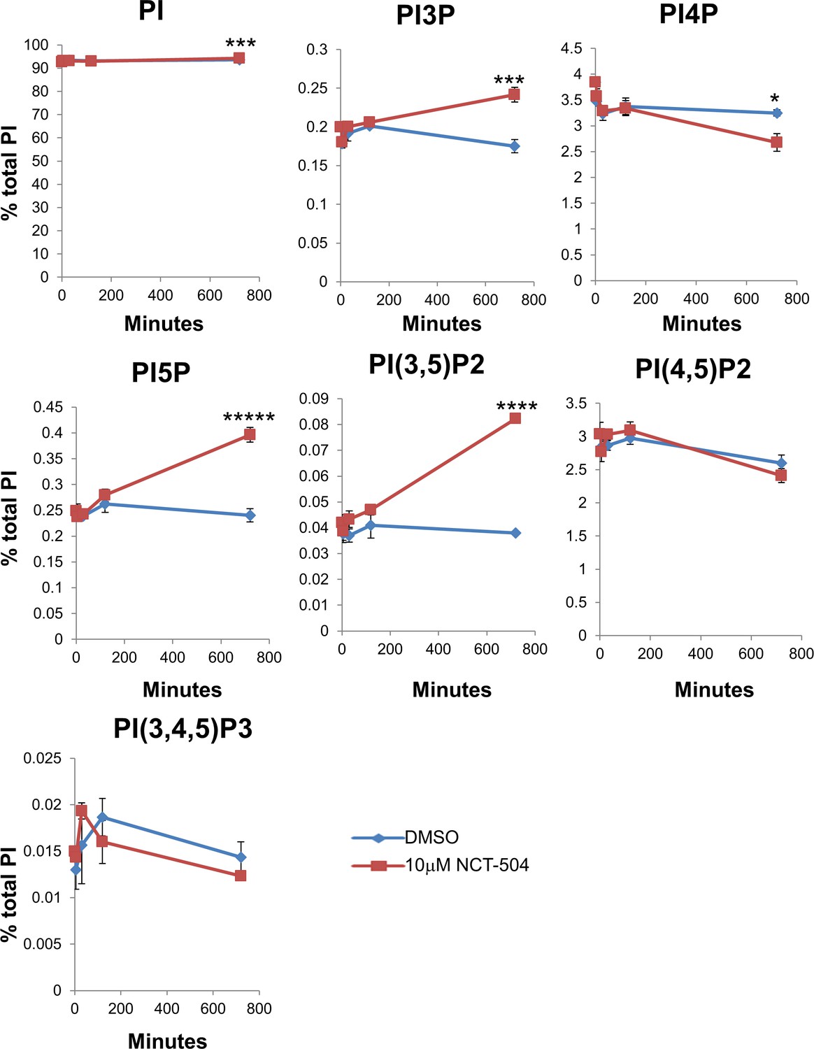
Time course of phosphatidylinositol lipid changes upon NCT-504 treatment (10 μM) in MEFs.
Time shown in minutes: 0, 5, 30, 120, 720 min. Average of three experiments with error bars. Statistical significance between DMSO-treated and NCT-504 treated samples were analyzed using paired one tailed student t-test, *p<0.05, **p<0.01, ***p<0.005, ****p<0.001, *****p<0.0005.
Figure 2—figure supplement 3
Modulation of the levels of phosphatidylinositol lipids by NCT-504 in unaffected human fibroblasts.
Immortalized human fibroblast cells were incubated with media containing 3H-inositol for 48 hr and cells were treated with DMSO or indicated concentrations of NCT-504 for the last 12 hr of labeling.
Figure 3 with 3 supplements
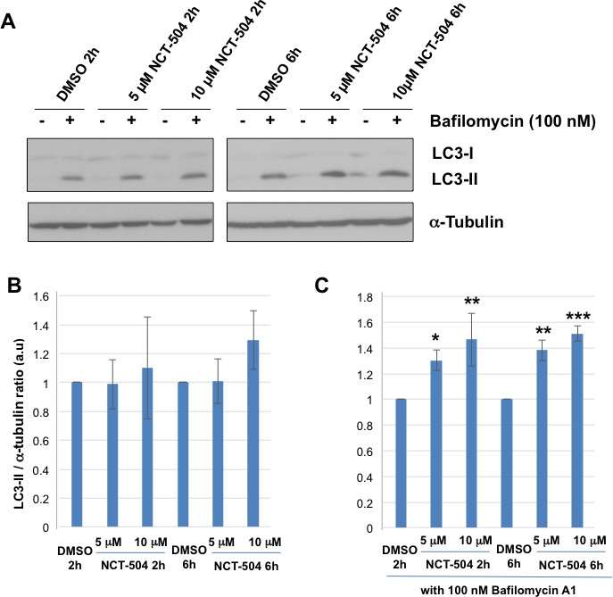
Inhibition of PIP4Kγ increases autophagy flux.
(A) Representative Western blots showing the levels of LC3-I, LC3-II and Tubulin (loading control) in HEK293T cells treated with either 5 or 10 μM NCT-504 or DMSO (control) for two or six hours in the presence or absence of 100 nM bafilomycin. (B–C) Quantification of LC3-II levels detected by western blot normalized to α-tubulin (loading control). Changes in LC3-II with drug treatment alone is presented relative to levels in the DMSO control cell lysates (B) and changes in LC3-II with drug treatment plus bafilomycin is presented relative to DMSO plus bafilomycin (C). Statistical significance was quantified from three independent experiments using Dunnett's multiple comparisons test, *p<0.05, **p<0.01, ***p<0.005.
Figure 3—figure supplement 1
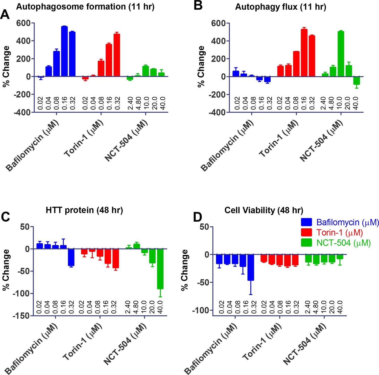
NCT-504 increases autophagy flux and decreases huntingtin protein in 293A cells.
Concentration-response of bafilomycin (20 nM, 40 nM, 80 nM, 160 nM, 320 nM), torin-1 (20 nM, 40 nM, 80 nM, 160 nM, 320 nM), and NCT-504 (2.4 μM, 4.8 μM, 10 μM, 20 μM, 40 μM) as modulators of autophagosome formation and autophagy flux. (A) Autophagosome formation was measured, at 11 hr, as increase of total yellow spots area (GFP spots area overlap with mCherry spots area) normalized with DMSO treatment control from an image taken at each time point using the Opera Phenix (PerkinElmer), data presented here is average of N = 3, one field per well, (B) Autophagy flux at 11 hr was monitored via autolysosome formation as measured by the increase of total red spots area (mCherry spots area) normalized with DMSO treatment control, data presented here is average of N = 3, one field per well, (C) Change in huntingtin protein measured by Homogeneous Time Resolved Fluorescence (HTRF) assay at 48 hr, and (D) change in viability of cells following treatment as measured by ATP concentration using CellTiter-Glo Luminescent Cell Viability Assay (Promega).
Figure 3—figure supplement 2
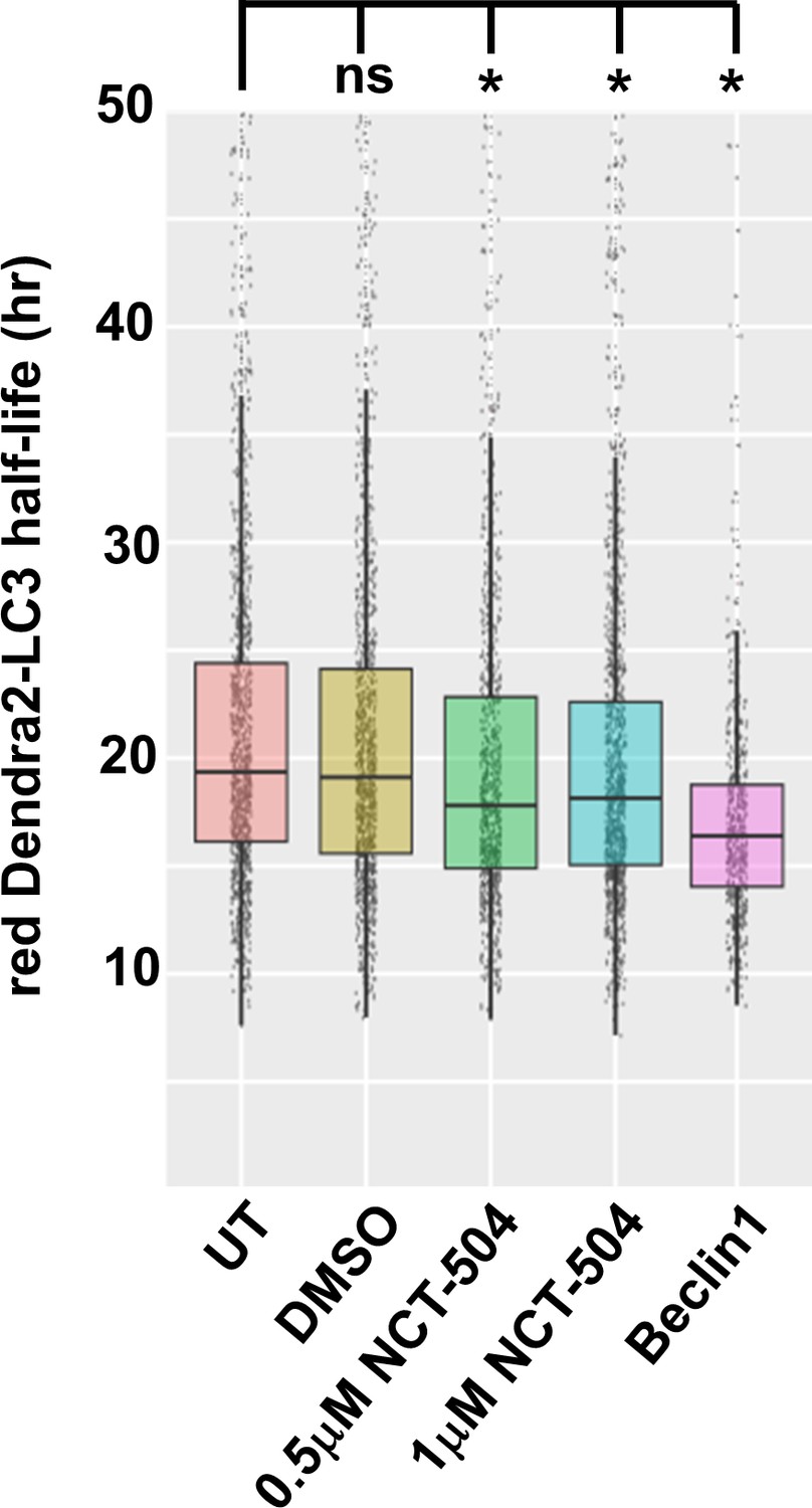
PIP4Kγ inhibition increases the rate of autophagic flux in cortical neurons.
The effect of NCT-504 on autophagic flux was determined in DIV4 rat primary cortical neurons transfected with Dendra2-LC3. Untreated (UT) and DMSO-treated neurons are negative controls; co-transfection with GFP-Beclin serves as a positive control. 30 min post-transfection, neurons were treated with DMSO or NCT-504. After 24 hr, Dendra2-LC3 was photoconverted and the intensity of red Dendra2-LC3 within each neuron was quantified immediately and at four additional time points for a duration of 2 days. A minimum of 750 neurons were quantified for each condition with the exception of 540 for Beclin-transfected neurons. Dendra2-LC3 half-life was calculated from the rate of loss of red-Denra2-LC3 intensity. Kruskal-Wallis (c2 (5, n = 4,743)=177.4, p<0.0001) and Dunnet's post hoc test results indicate that differences between no treatment and each treatment are significant: Beclin (p<0.0001), 0.5 µM NCT-504 (p<0.0001) and 1 µM NCT-504 (p=0.0003). *=P < 0.0005.
Figure 3—figure supplement 3
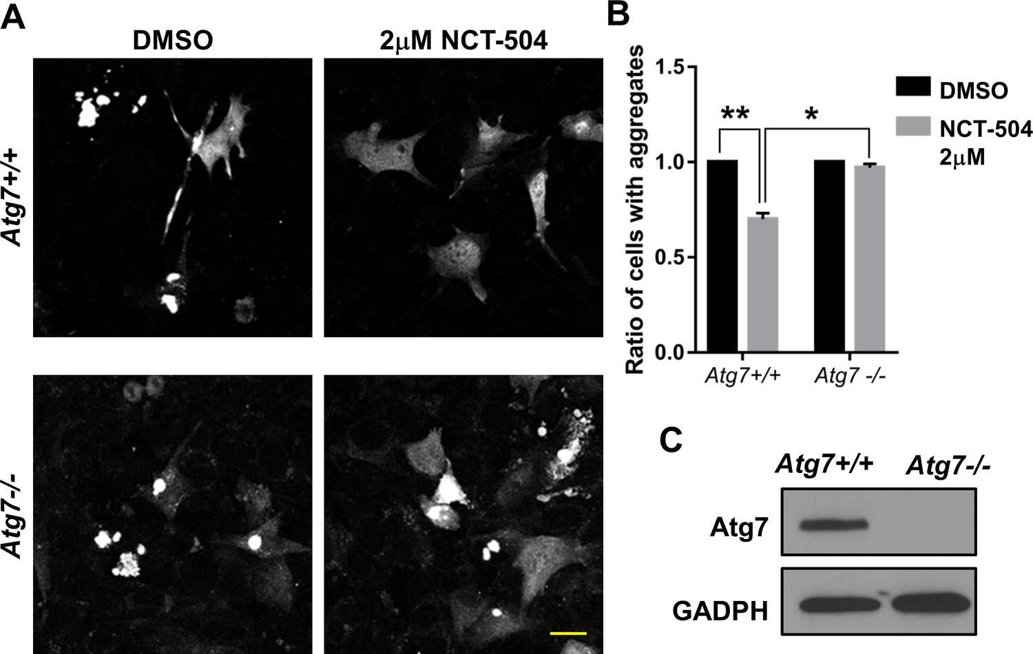
Lowering mHtt aggregates via NCT-504 requires macroautophagy: (A-B) Atg7+/+ and Atg7-/- cells were transfected with GFP-Htt(exon1)-Q74.
Two hours after transfection, cells were treated with either DMSO or 2 μM NCT-504 for 48 hr, fixed, and the percentage of cells with aggregates was quantified. The ratio of aggregates in NCT-504 vs. DMSO treatments were determined separately for wild-type and mutant cells. Statistical significance was analyzed using paired one tailed student t-test from three individual experiments. **p<0.01, *p<0.05. Bar 20 μm. (C) Atg7+/+ and Atg7-/- cells were lysed and immunoblotted with antibodies against Atg7 and GADPH.
Figure 4 with 5 supplements
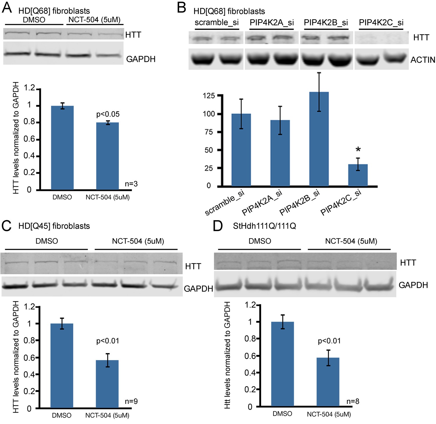
Chemical inhibition of PIP4Kγ or knock-down of the corresponding mRNA, PIP4K2C, lowers mHtt protein levels in cells from HD patients and HD knock-in mice.
(A) Reduction of mHtt protein levels in an HD patient fibroblast cell line (Q68) following exposure for 12 hr to NCT-504 (5 μM) (B) mHtt protein levels in patient fibroblast cell line (Q68) were analyzed following siRNA-mediated silencing of PIP4K2A, PIP4K2B and PIP4K2C genes. Note that only PIP4K2C knockdown lowers mHtt levels. Control experiments showing silencing specificity on PIP4K protein levels are in Figure 4—figure supplement 3. (C) Reduction of mHtt protein levels in an HD patient fibroblast cell line (Q45) following exposure to NCT-504 (5 μM). (D) Reduction of mHtt protein levels in immortalized striatal cells from knock-in HD mice (STHdhQ111) treated for 12 hr with NCT-504 (5 μM).
Figure 4—figure supplement 1

Cell viability of HD patient fibroblasts (Q45) exposed to the indicated doses of NCT-504 for 12 hr as per the CellTiter-Glo Promega assay.
https://doi.org/10.7554/eLife.29123.016
Figure 4—figure supplement 2
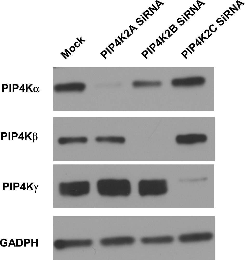
Knock-down efficiency and specificity of small interfering RNA in HD patient fibroblasts (Q45).
Cells grown in 35 mm culture dishes were treated with 800 pmoles of On-target plus smartpool SiRNA for 72 hr. Cells were lysed and immunoblotted with antibodies for PIP4Kα, PIP4Kβ, PIP4Kγ and GADPH.
Figure 4—figure supplement 3
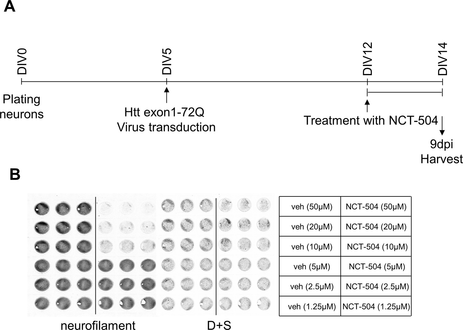
Experimental details and controls for mouse primary cortical neurons transduced with Htt(exon1)-Q72.
(A) Experimental design of PIP4Kγ inhibitor treatment in neurons. Mouse primary cortical neurons transduced with Htt(exon1)-Q72 lentivirus and treated with PIP4Kγ inhibitor NCT-504 for 48 hr. (B) Assessment of baseline toxicity of NCT-504 to find non-toxic concentrations. The indicated concentrations of NCT-504 were applied to neurons for 48 hr and baseline toxicity assessed by in-cell western analysis using neurofilament antibody. To normalize cell number Draq5 and sapphire 700 staining (D + S) was performed. Veh represents vehicle (DMSO) treatment.
Figure 4—figure supplement 4
Reduction of Htt protein levels or aggregate by inhibition of PIP4Kγ or PIP4K2C knockdown.
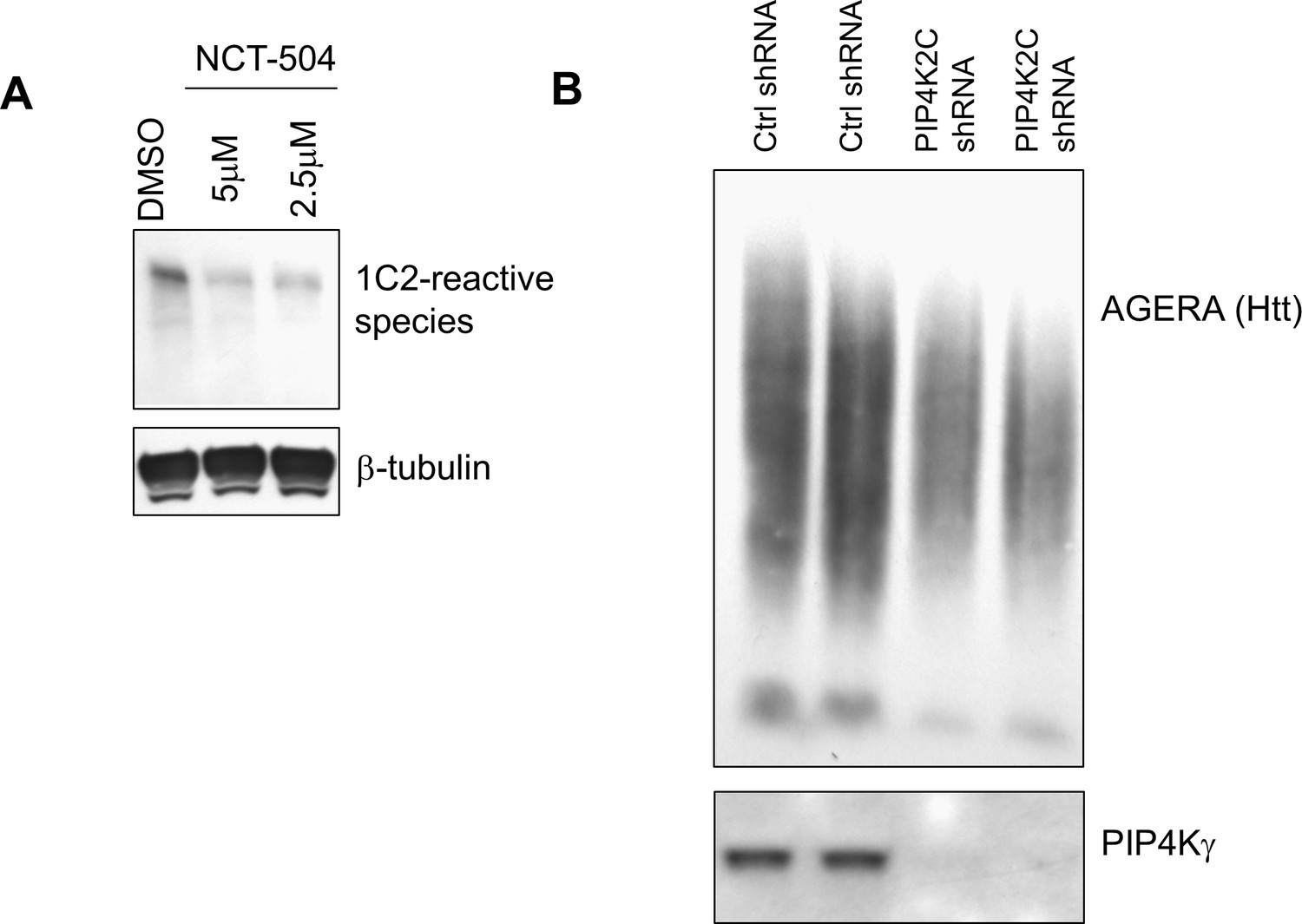
(A) NCT-504 reduces the levels of Htt(exon1)-Q72 in primary cortical mouse neurons as measured by western blot analysis using the monoclonal antibody 5TF-1C2, which recognizes polyQ. β-tubulin was used as a loading control. (B) Effect of knockdown of PIP4K2C on mHtt aggregates in mouse primary cortical neurons, as measured by two-dimensional resolution of high molecular weight species using AGERA.
Figure 4—figure supplement 5
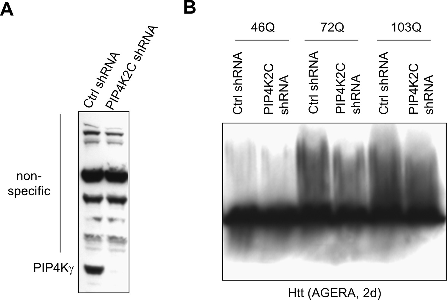
Effect of PIP4K2C knockdown on mHtt aggregates in N2a transfected cells.
(A) Effect of knockdown on PIP4Kγ protein assessed by western of N2a cell lysates. This is a Neuro-2a neuroblastoma cell line purchased from the ATCC (RRID: CVCL_0470) (B) Effect of knockdown of PIP4K2C on formation of mHtt aggregates as measured by two-dimensional resolution of high molecular weight species using AGERA. Htt(exon1) polyglutamine expansion lengths are indicated.
Figure 5 with 1 supplement
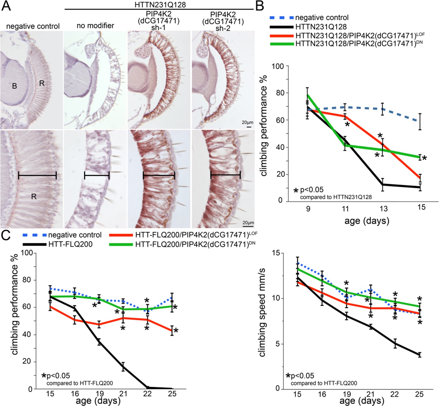
Reduced dPIP4K gene activity ameliorates photoreceptor degeneration and behavioral impairments in a Drosophila HD model.
(A) Sections through the Drosophila retina showing loss of photoreceptor cells and retinal tissue in animals expressing N-terminal mHtt (HTTNT231Q128) in the eye (compare no modifier with negative control panels). The photoreceptor and retinal loss phenotype is ameliorated in HttNT231Q128 animals that also express anyone of two shRNAs targeting dPIP4K. (B) Chart shows motor performance (%) as a function of age in negative controls (dPIP4K+/+, blue dotted line), Drosophila expressing N-terminal mHtt in the CNS (HTTNT231Q128/dPIP4K+/+, black line) or animals expressing N-terminal mHtt in the CNS together with a dPIP4K heterozygous loss of function (HTTNT231Q128/dPIP4K+/-, red continuous line) or a dPIP4K kinase dead isoform (HTTNT231Q128/dPIP4K+/DN, green continuous line). Notice the amelioration of mHtt-induced deficits upon decreasing the activity of dPIP4K. (C) Chart shows motor performance (%) and climbing speed as a function of age in negative controls (dPIP4K+/+, blue dotted line), Drosophila expressing full length mHtt in the CNS (HTT-FLQ200/dPIP4K+/+, black line) or animals expressing FL mHtt in the CNS together with a dPIP4K heterozygous loss of function (HTT-FL200/dPIP4K+/-, red continuous line) or a dPIP4K kinase dead isoform (HTT-FL200/dPIP4K+/DN, green continuous line). Note amelioration of neural HttNT231Q128-induced motor deficits by decreasing the activity of dPIP4K. Genotypes in A: Negative control: GMR-GAL4/+; dPIP4K+/+. No modifier: GMR-GAL4/+; UAS:HTTNT231Q128/+; dPIP4K+/+. PIP4K2 sh1/sh2: GMR-GAL4/+; UAS:HTTNT231Q128/UAS:dPIP4Ksh-1 or sh-2. Genotypes in B: Negative control: elavc155GAL4/+; dPIP4K+/+.HTT231Q128: elavc155GAL4/+; UAS:HttNT231Q128/+; dPIP4K+/+. HTT231Q128/PIP4K2LOF: elavc155GAL4/+; UAS:HttNT231Q128/+; dPIP4K29/+ and HTT231Q128/PIP4K2DN: elavc155GAL4/+; UAS:HttNT231Q128/UAS:dPIP4K29[D271K].. Genotypes in C: Negative control: elavc155GAL4/+; dPIP4K+/+. HTT-FLQ200: elavc155GAL4/+; UAS:HttFLQ200/+; dPIP4K+/+. HTT-FLQ200/PIP4K2LOF: elavc155GAL4/+; UAS:UAS:HttFLQ200/+; dPIP4K29/+ and HTT-FLQ200/PIP4K2DN: elavc155GAL4/+; UAS:UAS:HttFLQ200/UAS:dPIP4K29[D271K]. elavc155GAL4 drives expression of mHtt to all neurons but not other cell types. Means between points at each age were analyzed by ANOVA followed by Dunnet’s post hoc test. Error bars indicate the s.e.m. *p<0.05.
Figure 5—figure supplement 1

Reduced dPIP4K gene activity in wild type Drosophila does not impact motility.
https://doi.org/10.7554/eLife.29123.022
Scheme 1
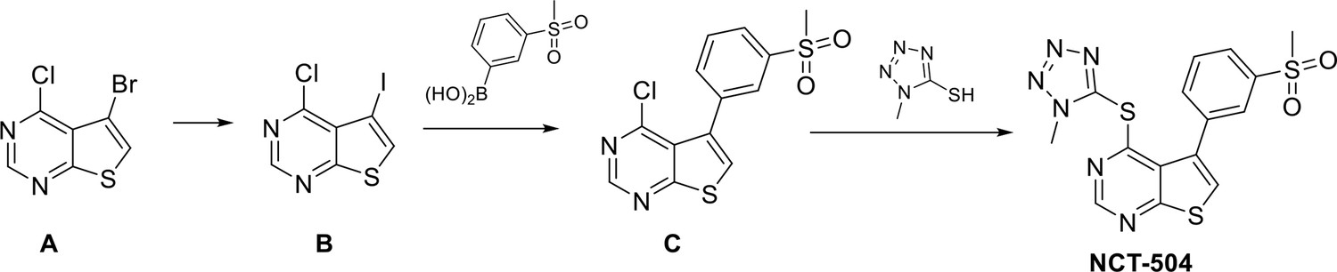
Synthetic scheme to prepare NCT-504.
https://doi.org/10.7554/eLife.29123.023Tables
Table 1
Kinase profiling results for NCT-504 and ML168.
Percent activity remaining at 10 μM exposure of NCT-504 and ML168 in KINOMEscan kinase panel/profiling http://www.discoverx.com/technologies-platforms/competitive-binding-technology/kinomescan-technology-platform. Top 3 NCT-504 inhibited kinases are reported as single replicate data. Full data set is provided in Table 1 – source data file. PIP4K2γ potencies were confirmed in triplicate concentration-response testing (Figure 1D).
| Kinase | ML168 | NCT-504 |
|---|---|---|
| PIP4K2C | 23 | 4.9 |
| RSK1(Kin.Dom.2-C-terminal) | 20 | 40 |
| GAK | 10 | 42 |
-
% Control Legend
0% ≤ x < 10%
-
10% ≤ x < 35%
35% ≤ x
Additional files
-
Source code 1
Custom Software for statistical analysis.
- https://doi.org/10.7554/eLife.29123.024
-
Transparent reporting form
- https://doi.org/10.7554/eLife.29123.025
Download links
A two-part list of links to download the article, or parts of the article, in various formats.
Downloads (link to download the article as PDF)
Open citations (links to open the citations from this article in various online reference manager services)
Cite this article (links to download the citations from this article in formats compatible with various reference manager tools)
Inhibition of PIP4Kγ ameliorates the pathological effects of mutant huntingtin protein
eLife 6:e29123.
https://doi.org/10.7554/eLife.29123


